MTA DOKTORI ÉRTEKEZÉS
|
|
|
- Etelka Halász
- 6 évvel ezelőtt
- Látták:
Átírás

1 MTA DOKTORI ÉRTEKEZÉS Háziállatok neonatális Fc receptorának (FcRn) karakterizálása; az FcRn fokozott kifejeződésén alapuló új transzgénikus technológia az immunválasz jelentős fokozására Dr. Kacskovics Imre Eötvös Loránd Tudományegyetem Természettudományi Kar, Biológiai Intézet Immunológiai Tanszék Budapest, 2012
2 Tartalomjegyzék 1. Bevezetés, irodalmi áttekintés Témaválasztás A maternális IgG transzport A neonatális Fc receptor (FcRn) Az FcRn szerkezeti sajátosságai Az FcRn szerepe a maternális immuntranszportban Az FcRn szerepe az IgG homeosztázisában FcRn mediált kétirányú IgG transzport a nyálkahártya felszíneken: antigén mintavétel Az FcRn szerepe a fagocitózisban és antigénprezentációban Az FcRn kifejeződésének szabályozása Monoklonális ellenanyagok előállítása transzgénikus állatokban Monoklonális ellenanyag fejlesztés hatékonyságának fokozása Tg egerekben Humán monoklonális ellenanyagok fejlesztése humanizált Tg állatokban Célkitűzések Eredmények és diszkusszió A szarvasmarha és a vele rokon juh, illetve teve tejmirigy és egyéb nyálkahártya IgG szekretáló mechanizmusának elemzése A szarvasmarha, juh, sertés, teve és a nyúl FcRn -lánc szekvenciájának meghatározása és elemzése A bfcrn -lánc alternatív RNS hasítással létrejött variánsai A bfcrn -lánc funkcionális elemzése ph függő IgG kötésen keresztül, in vitro sejtes rendszerben A szarvasmarha, juh és teve FcRn -lánc szövettani kifejeződése a tejmirigyben A szarvasmarha és juh FcRn -lánc szövettani kifejeződése a bélcsatornában A szarvasmarha és juh FcRn -lánc szövettani kifejeződése a tüdőben A bfcrn-t laktáló tejmirigyben kifejező Tg egérmodell előállítása és jellemzése A bfcrn-t laktáló tejmirigyben kifejező Tg egerek IgG homeosztázisának elemzése A bfcrn - bigg1 és bigg2 kötések elemzése felületi plazmon rezonanciás (SPR) méréssel A bfcrn IgG katabolizmusban betöltött szerepének elemzése A bfcrn kapcsolódása szarvasmarha és humán IgG molekulákhoz A bfcrn kimutatása a kapilláris endothel sejtekben, vesében A humán IgG felezési idejének meghatározása szarvasmarhában A bfcrn -láncot kifejező BAC Tg egérmodellek előállítása és jellemzése A bfcrn -láncot kifejező BAC Tg egerek előállítása A bfcrn -láncot kifejező BAC Tg egerek kromoszóma elemzése A bfcgrt szabályozásának elemzése A bfcrn -láncot kifejező BAC Tg egerek IgG katabolizmusának elemzése A bfcrn -láncot kifejező BAC Tg egerek humorális immunválaszának elemzése szolubilis fehérje antigén (ovalbumin) immunizálással A bfcrn -láncot kifejező BAC Tg egerek professzionális antigén prezentáló sejtjeinek jellemzése A bfcrn -láncot kifejező BAC Tg egerek humorális immunválaszának diverzitás elemzése A bfcrn -láncot kifejező BAC Tg egerek humorális immunválaszának elemzése konjugált haptén (TNP-KLH) immunizálással
3 Tartalomjegyzék A bfcrn -láncot kifejező BAC Tg egerek humorális immunválaszának elemzése gyengén immunogén antigénekre adott immunválasz A bfcrn -láncot kifejező BAC Tg egerek humorális immunválaszának elemzése FITC-dextrán immunizálással A bfcrn -láncot kifejező BAC Tg egér albumin homeosztázisa A bfcrn -láncot kifejező BAC Tg egerek alkalmazása a monoklonális ellenanyagok előállításában TNP specifikus monoklonális ellenanyagok fejlesztése Humán CXCR4 specifikus monoklonális ellenanyagok fejlesztése A bfcrn túltermeltetése nem jár együtt autoreaktív ellenanyagok képződésével Tg egerekben A nyúl FcRn -láncot kifejező BAC Tg nyulak előállítása és jellemzése A nyúl FcRn szöveti lokalizációja, ph függő IgG kötésének elemzése A nyúl FcRn ph dependens IgG kötésének elemzése A nyúl FcRn BAC Tg nyulak előállítása és immunológiai jellemzése Új eredmények összefoglalása Az eredmények gyakorlati jelentősége Az értekezés alapjául szolgáló közlemények jegyzéke Anyagok és módszerek Köszönetnyilvánítás Irodalomjegyzék Függelék
4 Rövidítések APC β2m bfcrn BAC BAEC BALB/c_Tg5 BCE BCR bfcrn bigg, bigg1, bigg2 CFA BSA DC FCGRT FCS FcRn FcRB FVB/N_Tg4 / Tg5 higg HRP IFA IgM IgD IgG IgA IgE IK KLH LPS migg NF-κB OVA PBS PCR QCM RACE-PCR RT-PCR SPR TBS TD TF Tg TI TLR TNBSA TNP TNP-BSA TRIS vt antigen presenting cell / antigén prezentáló sejt β 2 -mikroglobulin bovine neonatal Fc receptor / szarvasmarha neonatális Fc receptor bacterial artificial chromosome / mesterséges bakteriális kromoszóma bovine aortic endothelial cell / szarvasmarha aorta eredetű endothel sejt a bfcrn α-láncát 5 kópiában hordozó BALB/c genetikai hátterű egértörzs bovine capillary endothel cell / szarvasmarha kapilláris endothel sejt B cell receptor / B sejt receptor szarvasmarha neonatális Fc receptor Szarvasmarha IgG, IgG1, IgG2 complete Freund s adjuvant / komplett Freund adjuváns szarvasmarha szérum albumin dendritic cell / dendritikus sejt FcRn nehézláncát kódoló gén fetal calf serum / magzati borjú savó neonatális Fc receptor Brambell receptor (FcRn korábbi elnevezése a receptor első leírójáról) a bfcrn α-láncát 4 / 5 kópiában hordozó FVB/N genetikai hátterű egértörzs Humán IgG horse radish peroxidase / torma peroxidáz incomplete Freund s adjuvant / inkomplett Freund adjuváns immunglobulin M immunglobulin D immunglobulin G immunglobulin A immunglobulin E immunkomplex keyhole limpet hemocyanin / kulcslyuk csiga (Megathura crenulata) hemocianin lipopoliszacharid egér IgG nuclear factor κb ovalbumin Phosphate buffered saline / foszfát pufferelt fiziológiás sóoldat polymerase chain reaction / polimeráz láncreakció Quartz crystal microbalance Rapid Amplification of cdna Ends / cdns végek PCR alapú gyors amplifikációja reverz transzkripciót követő polimeráz láncreakció surface plasmon resonance / felszíni plazmon rezonancia tris pufferelt fiziológiás sóoldat T cell dependent / T sejt függő transzkripciós faktor transzgénikus T cell independent / T sejttől független Toll-like Receptor 2,4,6-trinitrobenzene sulfonic acid / 2,4,6-trinitrobenzén szulfonsav 2,4,6-trinitrophenol / 2,4,6-trinitrofenil szarvasmarha szérum albuminhoz konjugált TNP tris (hydroxymethyl) aminomethane vad típus 4
5 1. Bevezetés, irodalmi áttekintés 1.1 Témaválasztás A hosszú távú immunitás fenntartásában a plazmasejtek által termelt IgG izotípusú molekulák játsszák a legnagyobb szerepet. A szérumban és a nem-mukozális szövetekben ez az immunglobulin izotípus fordul elő legnagyobb mennyiségben (Waldmann and Strober, 1969). Az IgG hatékony effektor funkcióját biztosítja a komplement C1 komponensével és a különböző Fc gamma receptorokkal kialakuló interakciója. E molekula kiemelten fontos szerepére utal az a tény, hogy valamennyi immunglobulin izotípus közül ennek a leghosszabb a felezési ideje, illetve a maternális immunitásban is elsődleges szerepet tölt be. Korábbi kutatásaim a háziállatok immunglobulinjainak karakterizálására irányultak és ennek kapcsán elemeztem a sertés IgG és IgA izotípusait és a nehézlánc variábilis génjeit (Kacskovics et al., 1994; Sun et al., 1994; Brown et al., 1995; Butler et al., 1996); illetve a szarvasmarha IgG izotípusait (Kacskovics and Butler, 1996; Corbeil et al., 1997; Kelm et al., 1997). Ezeket a kutatásaimat PhD disszertációmban foglaltam össze 1998-ban. A háziállatok immunglobulinjainak karakterizálását ezt követően is folytattam (Zhao et al., 2002; Zhao et al., 2003a; Zhao et al., 2003c; Butler et al., 2009a; Butler et al., 2009b; Butler et al., 2009c); de érdeklődésem 1995-től kezdve mindinkább a szarvasmarha tejmirigy IgG szekretáló mechanizmusának tisztázására irányult és önálló kutatásom is leginkább erre a területre korlátozódott. Feltételezésünk szerint e folyamat befolyásolása nagyban hozzájárult volna olyan transzgénikus (Tg) szarvasmarha előállításához, amely lényegesen nagyobb mennyiségű ellenanyagot (IgG) szekretál a tejbe és ezáltal új, passzív immunterápiás lehetőséget nyújt a humán gastrointestinális fertőzések kezelésében (Kacskovics, 2003; Kacskovics, 2006; Hammarstrom and Weiner, 2008). A szarvasmarha tőgyhámsejteken keresztüli IgG transzport régóta kutatott terület, ám a szekrécióban részt vevő receptort azonosítani, és a mechanizmust tisztázni mindeddig nem sikerült ben egy új típusú IgG kötő receptort, az MHC I-típusú molekulákkal rokon, neonatális Fc receptort (FcRn) karakterizáltak molekuláris szinten (Simister and Mostov, 1989). Minthogy a 90-es évek közepén (ez irányú vizsgálataim kezdetén), az FcRn-ről mutatták ki egyedül, hogy az IgG-t hámsejteken juttatja keresztül, elhatároztuk, hogy karakterizáljuk e receptort a kérődzők és néhány egyéb háziállat esetén. Vizsgálataink során klónoztuk a szarvasmarha, juh, sertés és teve FcRn molekuláit és kimutattuk ezek jelenlétét egyebek mellett a tőgyszöveti hámsejtekben (Kacskovics et al., 2000; Mayer et al., 2002b; Mayer et al., 2002a; Zhao et al., 2003b; Kis et al., 2004; Mayer et al., 2004; Doleschall et al., 2005; Kacskovics et al., 2005; 5
6 Bevezetés, irodalmi áttekintés Témaválasztás Mayer et al., 2005; Kacskovics, 2006; Kacskovics et al., 2006a; Kacskovics et al., 2006b). Eredményeink alapján arra gondoltunk, hogy az FcRn e fajokban részt vesz a maternális immuntranszportban, és a föcstejbe (kolosztrumba) szekretálja az anyai IgG-t (Kacskovics, 2004). Később kimutattuk, hogy ez a receptor, más állatfajokhoz hasonlóan, a szarvasmarhában is fontos szerepet tölt be az IgG homeosztázis szabályozásában (Kacskovics et al., 2006a). Mindezen vizsgálatok kapcsán felmerült, hogy az igen költséges és számos technológiai nehézséget jelentő nagyállat in vivo vizsgálatok mellett, olyan a szarvasmarha FcRn-t (bfcrn) kifejező transzgenikus (Tg) egér modelleket hozzunk létre, amelyek hatékonyabb in vivo génregulációs és funkcionális elemzéseket tesznek lehetővé. Az egyik ilyen Tg modellben a bfcrn-t az egér laktáló tejmirigyében fejeztettük ki egy tejspecifikus promoterrel, és azt találtuk, hogy a tejmirigyben kifejeződő bfcrn az IgG-t a tejmirigyből a vérbe, nem pedig a vérből a tejbe juttatja, amint arra korábban gondoltunk (Lu et al., 2007). Ezt a funkciót támasztja alá egy, a bfcrn-igg kölcsönhatást elemző kutatásunk is (Takimori et al., 2011). A másik egérmodellben a bfcrn-t is kódoló kromoszóma szakaszt integráltuk az egér genomba (BAC transzgenezis) és felismertük, hogy a fokozott mértékű, szövetspecifikus bfcrn kifejeződés hatására az IgG lebomlása csökken (Bender et al., 2007), valamint a humorális immunválaszképesség sokszorosára fokozódik (Cervenak et al., 2011; Kacskovics et al., 2011; Onisk et al., 2011; Schneider et al., 2011; Vegh et al., 2011; Vegh et al., 2012). Az FcRn Tg egérmodellekkel kapcsolatos adataink alapján nyúl FcRn-t nagyobb mértékben kifejező Tg nyulakat is előállítottunk, amelyek az FcRn Tg egerekhez hasonlóan fokozott humorális immunválaszt mutatnak (Catunda Lemos et al., 2012; Duranthon et al., 2012). Kutatásaink hasznosíthatósága: A szarvasmarha tőgyszövetben kimutattuk a bfcrn jelenlétét és igazoltuk, hogy szerepe a tőgyből vérkeringésbe irányuló IgG reciklizáció. Ezek az eredmények az állatbiotechnológiai kutatásokat, illetve állatgyógyászatot segíthetik. Az FcRn-t nagyobb mértékben kifejező Tg egerek és nyulak fokozott immunválaszképessége, azaz 1) a magasabb antigén(ag)-specifikus szérum IgG titer; 2) nagyobb számú Ag-specifikus B-sejt és hibridóma; 3) nagyobb mértékű humorális immundiverzitás; és a 4) hatékony immunválasz gyengén immunogén Ag esetén új lehetőséget kínál a terápiás, diagnosztikai és kutatási monoklonális- és poliklonális ellenanyagok előállítása terén. Ennek kiaknázására szabadalmi bejelentést tettünk 2007-ben, amelyet az értekezés benyújtásáig az Európai és Ausztrál Szabadalmi Hivatalok szabadalomnak nyilvánítottak (EP , ill. AU ; míg a többi régióban a szabadalmi eljárások jelenleg is zajlanak), ill. egy hasznosító vállalatot alapítottunk (ImmunoGenes Kft; Értekezésemben az FcRn el kapcsolatos kutatásainkról adok részletes áttekintést. 6
7 1.2 A maternális IgG transzport Már a jelenkori tudományos, immunológiai ismeretek elterjedése előtt jól ismert volt a gazdasági haszonállatokkal foglalkozók körében az a jelenség, hogy a csikó, bárány vagy borjú, amely születése után nem jutott föcstejhez (kolosztrumhoz), rövid időn belül elpusztult. Először 1892-ben Paul Ehrlich hívta fel arra a figyelmet, hogy a bekövetkező elhullás oka fertőző betegség (Ehrlich, 1892). Az újszülött immunrendszere a születést követő hetekben meglehetősen fejletlen és éppen ezért nem is tud hatékonyan részt venni a fertőzések megakadályozásában. Ezt az időleges védelmi hiányt pótolják az anya immunrendszere által termelt ellenanyagok, amelyek a kórokozók széles spektrumával szemben nyújtanak specifikus védelmet. Ezt a folyamatot anyai, vagy maternális immunitásnak nevezzük, amelynek során az anya jelentős mennyiségű immunglobulin átadásával biztosítja az újszülött életben maradását az élet első időszakában. Ezt a rendszert végső soron egyfajta immunológiai tapasztalat közvetítésének is felfoghatjuk, hiszen az anyában olyan ellenanyagok találhatók, amelyeket a környezetében található kórokozókkal szemben termelt. Minthogy az újszülött természetes élettere megegyezik az anyáéval, az ily módon nyert tapasztalat 1. ábra - Az emlősök az anyai immunglobulin utódba irányuló hatásos az újszülöttet transzportja alapján három csoportba sorolhatók (Butler, 1999). fenyegető kórokozók semlegesítésében is. Az emlősök az anyai immunglobulin utódba irányuló transzportja alapján három csoportba sorolhatók (1. ábra). A főemlősök valamint a nyúl (I. csoport) a magzati élet során kapják meg a maternális immunglobulinok teljes készletét, így az újszülöttek vérében jórészt az anyai immunglobulinok (IgG) találhatók. Emellett e fajok újszülöttjei jelentős mennyiségű IgA molekulához is jutnak az anyatejjel, amely helyileg a béltraktusban vesz részt a kórokozók visszaszorításában (Brandtzaeg, 2003; Labbok et al., 2004). (Az emberi anyatej csupán nyomokban tartalmaz IgG-t, amelynek felszívódása elenyésző (Van de Perre, 2003)). Ezzel szemben a patás emlősök (ló, a sertés és a kérődzők; III. csoport) magzatai a maternális immunglobulin készletet (elsősorban IgG) kizárólag a születést követő néhány óra alatt elfogyasztott föcstej (kolosztrum) révén veszik fel. Ebben az időszakban az újszülött állatok rendhagyó bélhámsejt szerkezete biztosítja, hogy a béltraktusba 7

8 Bevezetés, irodalmi áttekintés Maternális IgG transzport került immunglobulinok intakt formában felszívódhassanak, és a vérpályába kerülhessenek. E folyamat a születést követő egy-két napon belül lezárul, azaz a bélben levő immunglobulinok ezután már nem képesek ilyen formában a vérbe kerülni. A rágcsálók valamint a ragadozók (II. csoport) újszülöttjei mind magzati élet során, mind pedig a kolosztrum révén részesülnek a maternális IgG transzportban (Butler, 1999). Ehrlich korai sejtését, amely szerint a tehéntej protektív ellenanyagokat tartalmaz, csak 1946-ban igazolta Emil Smith, aki a szarvasmarha kolosztrum fő összetevőjét immun laktoglobulinként jelölte meg (ma IgG1) (Smith, 1946). Míg az IgA epithel sejteken keresztüli transzportjáról tudjuk, hogy a szekretoros IgA a polimer immunglobulin receptor (pigr) közvetítésével kerül a nyálkahártya felszínére (Mostov and Deitcher, 1986), addig az IgG epithel sejteken keresztül megvalósuló transzportjáról nagyon keveset tudtunk, bár régóta receptor mediált transzporttal magyarázzák. A szarvasmarha IgG1 (bigg1) szérum-kolosztrum irányú transzportját először 1961-ben Dixon és mtsai írta le (Dixon, 1961), majd mások kimutatták, hogy ez a szekréció igen jelentős mértékű (mintegy 500g bigg1 szekretálódik az ellést megelőző 3 hét során) (Butler, 1974; Newby and Bourne, 1977), és hogy ez idő alatt a szérum bigg1 szintje hasonló mértékű csökkenést mutat (Sasaki et al., 1976). A tehén szempontjából drámai mértékű IgG vesztés csupán az ellés idejére korlátozódik, azt követően a tej IgG tartalma mintegy két nagyságrenddel alacsonyabb, mint a föcstejé (2. ábra). A transzport ellés körüli időzítése és nagyfokú szelektivitása (a vér bigg1 és bigg2 koncentrációja közel azonos) specifikus receptor közvetített folyamatot feltételez, amely az ellés előtti illetve azt közvetlenül követő időszakban jelentős mennyiségű bigg1-et juttat a tőgy acinus sejtjein keresztül a kolosztrumba. E hipotézist valószínűsítik azok a vizsgálatok, amelyek bigg1 specifikus kötést mutattak ki tőgy sejteken (Kemler et al., 1975; Sasaki et al., 1977; Leary et al., 1982; Barrington et al., 2. ábra - a szarvasmarha IgG1 és IgG2 koncentrációja az 1997), bár e receptort azonosítani ontogenezis során (sematikus ábra), illetve a szarvasmarha IgG alosztályok koncentrációja (mg/ml) a kolosztrumban, tejben és a mindeddig nem sikerült. vérben (Butler, 1983) 8
9 Bevezetés, irodalmi áttekintés Maternális IgG transzport A maternális (kolosztrális) immunglobulinok felszívódása az újszülött borjak vékonybeléből nem-specifikus folyamat. Mégis kiemelten fontos, hogy a fiatal kérődző állatok esetén a már vérpályába került bigg1 egy része a vékonybél crypta sejtjeinek aktív transzportáló tevékenysége révén ismét a lumenbe kerül, és ott hozzájárul az emésztőtraktus specifikus immunvédelméhez (Newby and Bourne, 1976a; Besser et al., 1987; Besser et al., 1988). A korábbi vizsgálatok alapján ezek a molekulák intakt módon jutnak a lumenbe és ott képesek a kórokozók semlegesítésében részt venni (Besser et al., 1988). Ismert továbbá, hogy a kérődzők esetén a bigg1, az IgA molekulához hasonlóan, más nyálkahártya felületekre - szájnyálkahártya, orrnyálkahártya, alsó légutak, nemi utak - is aktívan szekretálódik (Duncan et al., 1972; Corbeil et al., 1976; Newby and Bourne, 1976a; Wilkie, 1982; Butler, 1983), illetve, hogy a bigg1 az IgA izotípushoz hasonlóan rezisztens a bakteriális proteázokkal szemben (Newby and Bourne, 1976b). Munkatársaimmal ebbe a kutatási irányba kapcsolódtunk be 1995-ben azzal, hogy klónoztuk és számos szövetben így a tőgyszövetben is - lokalizáltuk a szarvasmarha (és más rokon háziállat) neonatális Fc receptorát (FcRn). 9
10 1.3 A neonatális Fc receptor (FcRn) Az FcRn szerkezeti sajátosságai F. W. Rogers Brambell és munkatársai számos kísérletüket követve írták le először, hogy az anyai immunglobulin G (IgG) molekulák utódba kerülését egy telíthető receptor biztosítja (Brambell et al., 1958); majd nem sokkal később felvetették, hogy egy hasonló vagy azonos receptor védi meg az IgG molekulákat a gyors lebomlástól (Brambell et al., 1964). Ezeket az anyai IgG transzportban résztvevő, illetve a szérum IgG felezési idejét meghatározó receptorokat nevezték első leírójáról Brambell receptornak (FcRB) ben Jones és Waldman újszülött patkányok vékonybeléből izolált egy fehérjét (innen kapta a neonatális receptor nevet, ám elnevezése ellenére az FcRn kifejeződése nem korlátozódik az újszülöttkori időszakra, hanem felnőttkorban is kimutatható különböző sejt- és szövettípusokban, ld. később), amely az anyatejben található IgG transzportját biztosítja (Jones and Waldmann, 1972); és amelyről ezt követően kimutatták, hogy egy 12 és egy kd-os fehérjéből álló heterodimer (Simister and Rees, 1985), valamint azt, hogy az IgG erősen ph-függő módon kötődik a receptorhoz. A kötődés enyhén savas közegben (ph ) jön létre, míg semleges-enyhén bázikus közegben (ph ) az IgG/FcRn komplex disszociál (Rodewald, 1976; Rodewald and Kraehenbuhl, 1984). A patkány FcRn-t 1989-ben klónozták és megállapították, hogy egy nehéz-láncból (α-lánc) és a β2- mikroglobulinból (β2m) épül fel. Az α-lánc az MHC I molekula α-láncával homológ, 3 extracelluláris doménből (α1, α2 és α3), a sejtmembránban horgonyzó transzmembrán régióból és citoplazmikus farokrészből áll (Simister and Mostov, 1989). Ha a két lánc kapcsolódása elmarad, az α-láncok diszulfid hidakkal összekötött oligomereket képeznek és az endoplazmatikus retikulumban maradnak, majd valószínűleg lebomlásra kerülnek (Claypool et al., 2002; Praetor and Hunziker, 2002; Zhu et al., 2002). Időközben, 2m génkiütött (knock-out, KO) egereken bizonyították, hogy az IgG felezési idejét ugyanez a receptor szabályozza (Ghetie et al., 1996; Israel et al., 1996; Junghans and Anderson, 1996), ezzel Brambell sejtését formálisan is igazolták, azaz ugyanaz a molekula vesz részt az anyai IgG transzportban, és a keringő IgG lebontásának szabályozásában (Junghans, 1997). Ezt később az FcRn -lánc KO egereken is igazolták (Roopenian et al., 2003). Röntgen-krisztallográfiás elemzések kimutatták (3. ábra), hogy bár az FcRn α-lánca szerkezetileg szoros rokonságot mutat az MHC I molekulával (Burmeister et al., 1994), a peptidkötő zseb, amelyhez a klasszikus és nem klasszikus MHC I molekuláknál peptid vagy egyes esetekben glikolipid ligandum kapcsolódik (Bjorkman et al., 1987; Zeng et al., 1997) sztérikusan nem hozzáférhető. A sztérikus gátlást az α2 domén helikális részében, a polipeptidlánc 162. pozíciójában lévő prolin okozza (West and Bjorkman, 2000). A peptidkötő 10

 3D számítógépes modellje (Kacskovics, 2003); B.")
11 Bevezetés, irodalmi áttekintés A neonatális Fc receptor (FcRn) zseb záródását a 164. pozícióban lévő arginin és az α1-α2 hélixek között kialakuló ionos és hidrofób kölcsönhatások is elősegítik. Ezért az FcRn, az MHC I molekulákkal ellentétben nem képes antigén fragmentumok bemutatására. Az FcRn IgG kötés középpontjában lévő aminosav maradványok egy része hidrofób kölcsönhatást alakít ki (FcRn Trp-133 :Fc Ile-253 és 2m Ile- 1:Fc Pro-307), amelyet ionos kölcsönhatás kialakítására képes aminosav-maradványok vesznek körül (patkány FcRn Glu-117, Glu- 118, Glu-132, és Asp- 3. ábra - A. a szarvasmarha FcRn és a kötődött IgG(CH2) 3D számítógépes modellje (Kacskovics, 2003); B. egy intakt IgG - nyíl jelöli az FcRn-hez 137 ill. patkány IgG Fc kapcsolódási pontját; C. az FcRn IgG kötés középpontjában lévő aminosav His-310, Arg-311, Hismaradványok egy része hidrofób más részük ionos kölcsönhatást alakít ki enyhén acidikus közegben (Martin et al., 2001; Kacskovics et al., 2006a). 435, és His-436). Enyhén acidikus közegben a hisztidinek protonálódnak, míg a glutaminsav és aszparaginsav maradványok negatív töltésűvé válnak. Az így kialakuló erős ionos kapcsolat magyarázatul szolgál az FcRn-IgG interakció szigorú ph-függésére, ugyanis semleges kémhatáson a hisztidinek deprotonálódnak és ennek következtében az IgG disszociál a receptorról (Martin et al., 2001). Ezek az aminosav-maradványok az IgG Fc His-436 kivételével - nagymértékben konzerválódtak az evolúció során. Az FcRn molekulának két ismert liganduma van ban Chaudhury és munkatársai bizonyították, hogy az FcRn az albumin molekulákat is megköti és megvédi a gyors lebomlástól. Ez a kötés is ph-függő, amely az FcRn His-166 és az albumin Glu- 54 és Tyr-60 aminosav maradványok között jön létre. Az FcRn 166. pozíciójában lévő hisztidint 9 emlős fajban mutatták ki, 4. ábra az FcRn két különböző régióban ph dependens kötéssel kapcsolódik az IgG, ill. albumint valószínű tehát, hogy ez az erősen molekulákhoz (Andersen et al., 2012). konzerválódott aminosav a legkritikusabb 11
, azaz a két ligandum nem befolyásolja egymás kötődését a receptorhoz (Chaudhury et al., 2003;")
12 Bevezetés, irodalmi áttekintés A neonatális Fc receptor (FcRn) ebben az interakcióban. Az IgG és az albumin kötőhelyei különböznek az FcRn molekulán (4. ábra), azaz a két ligandum nem befolyásolja egymás kötődését a receptorhoz (Chaudhury et al., 2003; Chaudhury et al., 2006; Andersen et al., 2012) Az FcRn szerepe a maternális immuntranszportban Story és mtsai az emberi placenta syntitiotrophoblast sejtjeiben mutatták ki az FcRn jelenlétét, és bizonyították annak szerepét az IgG transzportjában (Story et al., 1994). Simister és mtsai a receptort az IgG molekulával együtt a syncytiotrophoblast sejtek savas endoszómáiban lokalizálta. Kimutatták, hogy az IgG folyadék fázisú pinocitózist követően a savas kémhatású endoszómákban kötődik az FcRn molekulákhoz, átszállítódik a sejten a bazolaterális oldalra, majd a fiziológiás közegben kiszabadul a komplexből és a magzati keringésbe jut (5. ábra) (Simister, 2003). Az FcRn-t humán emlő tejmirigyből is kimutatták. Mivel az emberi tej nagyon kis mennyiségű IgG-t tartalmaz, 5. ábra FcRn közvetített anyai IgG feltételezték, hogy a tejmirigyben expresszálódó FcRn az transzport az emberi placentában (Roopenian and Akilesh, 2007). egérhez hasonlóan az IgG molekulákat a tejből nagy hatékonysággal visszajuttatja a szérumba (Cianga et al., 2003). A rágcsálókban az anyai IgG egyrészt a szikzacskón keresztül jut a magzatba, másrészt az újszülött állatok vékonybelében a tejből is felszívódik (Simister and Rees, 1985). Az FcRn szerepét az anyai IgG szikzacskón keresztül zajló transzportjában az utóbbi években igazolták (Kim et al., 2009; Mohanty et al., 2010). A tejmirigyben zajló IgG transzporttal összefüggésben laktáló egér tejmirigyében az acinusok epithel sejtjeiben lokalizálták az FcRn molekulát, és úgy találták, hogy az IgG alosztályok tejbe történő transzportja fordított korrelációt mutat az FcRn kötési képességükkel, azaz az FcRn-hez nagyobb affinitással kötődő IgG izotípusok kisebb koncentrációban kerülnek a tejbe. Feltételezésük szerint a laktáló tejmirigyben lévő FcRn a szekréció helyett inkább a visszaforgatásban (recycling) játszik szerepet, vagyis a tejmirigyből a keringésbe juttatja vissza a hozzákapcsolódott IgG molekulákat és az FcRn-hez kevésbé kötődő IgG vélhetően diffúzóval kerül a tejbe (Cianga et al., 1999). 12


13 Bevezetés, irodalmi áttekintés A neonatális Fc receptor (FcRn) 6. ábra - FcRn közvetített anyai IgG transzport az újszülött rágcsáló vékonybélsejtjeiben (Roopenian and Akilesh, 2007). Az IgG az újszülött állatok vékonybél enterocitáinak lumenális felszínén, enyhén savas közegben, az FcRn molekulához kötődik, és a sejt az FcRn-IgG komplexet receptor-mediált endocitózissal veszi fel. A komplexek transzcitózis révén keresztüljutnak a sejten, ezt követően exocitózissal a bazolaterális oldalra kerülnek, ahol a fiziológiás kémhatáson szétválik a receptor és a ligandum (6. ábra). Az FcRn szerepét a rágcsálók maternális IgG transzportjában az a vizsgálat is alátámasztotta, amelyben kimutatták, hogy az FcRn -lánc vagy 2m KO egerekben az anyai eredetű IgG töredéke volt annak, amit vad típusú alomtársaikban mértek (Israel et al., 1995; Roopenian et al., 2003) Az FcRn szerepe az IgG homeosztázisában 7. ábra Az IgG farmakokinetikájának és farmakodinamikájának sematikus ábrázolása. Az IgG áramlással és esetleg az FcRn közvetítette transzport révén a keringésből a szövetközi térbe kerül, majd onnan ugyanezekkel a mechnizmusokkal, valamint a nyirokkeringéssel újra a vérbe jut. A vérpályán belül, és a szövetközi térben számos sejt veszi fel az IgG-t nem-specifikusan. Amennyiben a felvett IgG FcRn-hez kapcsolódik, akkor az IgG-FcRn komplex a sejtfelszínre kerül, ahol az IgG leválik és újra intakt molekulaként vesz részt a szervezet immun-homeosztázisában. Amennyiben az IgG nem kapcsolódik FcRn-hez, akkor a lizoszómában lebomlik. Az IgG akár a vérpályán belül, akár a szövetek között specifikusan, másodlagos kötőerőkkel (a kötésre jellemző asszociációval (kon) és disszociációval (koff) kapcsolódhat a célmolekulával. Egy kis része a szérum IgG-nek a vese filtrációval ürül. Az IgG sorsát az is befolyásolja, hogy az antigén-igg immunkomplex milyen sejtekhez kötődik és ott milyen reakciókat (szignál-transzdukciót) gerjeszt (Lobo et al., 2004). 13
14 Bevezetés, irodalmi áttekintés A neonatális Fc receptor (FcRn) A plazmasejtek által termelt (vagy befecskendezett - exogén) IgG a vérbe kerül, majd azt követően nagyon hamar az extracelluláris térbe jut, ill. onnan diffúzióval és a nyirokkeringéssel újra visszajut a vérbe. Ezzel a szérum szövetközi tér között órák alatt kialakul az IgG dinamikus egyensúlyi állapota. Az IgG a szervezetből folyamatosan ürül, amely egyfelől az IgG-t nemspecifikusan felvevő sejtekben zajló lebomlás miatt, másfelől a vese filtráció következtében, illetve az antigénnel történt kapcsolódás, majd az így keletkezett immunkomplexek eliminálása révén következik be (7. ábra) (Lobo et al., 2004). A legtöbb szérumfehérjéhez hasonlóan a nem-igg izotípusú ellenanyagok felezési ideje viszonylag rövid (~1-2 nap), az IgG izotípusoké ezeknél lényegesen hosszabb, alosztálytól és fajtól függően: egerekben az IgG1, IgG2a és IgG3 felezési ideje 6-8 nap, az IgG2b felezési ideje valamivel rövidebb, 4-6 nap (Vieira and Rajewsky, 1988); az emberi IgG felezési ideje nap, ettől csak az IgG3 tér el (7 nap) (Spiegelberg et al., 1968). A szarvasmarhában az IgG termelődése, immunfolyamatokban betöltött szerepe alapvetően azonos a többi emlősállatokkal összehasonlítva. A bigg1 és bigg2 alosztályokat elemezve megállapítható, hogy vérbeli koncentrációjuk hasonló (2. ábra), és együttes szérumkoncentrációjuk átlagosan 20 mg/ml. A szarvasmarha IgG vérpályán belüli feleződési üteme mintegy nap, és ezzel lényegesen meghaladja a vérbeli IgA, IgM molekulák (3,5-4 nap) fél-életidejét. Több publikációban is megerősítették, hogy a bigg2 hosszabb fél-életidővel rendelkezik, mint a bigg1 (Butler, 1983). A hasonlóságok mellett a kérődző állatok IgG metabolizmusának jellegzetes különbsége, hogy a bigg1 molekula számos nyálkahártya felszínére (tőgy, vékonybél, alsó légutak, uterus) szekretálódik és ott - az IgA protektív hatását kiegészítve - hatékonyan részt vesz az immunvédelemben (Butler, 1983). Brambell és munkatársai az általuk már 1964-ben feltételezett, az anyai IgG transzporttal kapcsolatba hozható és a szérum IgG felezési idejét meghatározó receptor egyik feladatát abban látták, hogy az IgG molekulákat megóvja a lizoszómális lebontástól (Brambell et al., 1964). Korábbi vizsgálatok kapcsán jól ismert, hogy magasabb szérum IgG koncentráció mellett az IgG molekulák frakcionált katabolikus rátája (időegység alatt lebomló IgG mennyiség) is megnövekszik (8. ábra) (Brambell et al., 1964; Morell et al., 1970). Ilyen jellegű összefüggés a többi Ig izotípus esetében 8. ábra - összefüggés az IgG szérum koncentráció és a felezési nem fordul elő (Waldmann and Strober, 1969). A koncentráció idő között egérben (Brambell et al., 1964). és a frakcionált katabolikus ráta közötti pozitív korreláció, 14

15 Bevezetés, irodalmi áttekintés A neonatális Fc receptor (FcRn) illetve a kiemelkedően magas szérum szint az albumin molekulára is jellemző (Freeman and Gordon, 1965). Az ezerkilencszáz ötvenes-hatvanas években végzett farmakokinetikai vizsgálatok arra az eredményre vezettek, hogy a legtöbb plazmafehérje, így az IgG is, a vérpálya mentén bomlik le, és egyetlen szerv sem játszik kitüntetett szerepet ebben a folyamatban. Mindezek alapján Thomas Waldman és Warren Strober 1969-ben arra a következtetésre jutottak, hogy a plazmafehérjék lebomlása, illetve az IgG védelme a kapillárisok endothel sejtjeiben zajlik (Waldmann and Strober, 1969). FcRn α-lánc, vagy β2m génkiütött egerekben, normális B-sejt állomány mellett az IgG rövid fél-életidejét ill. gyors lebomlását (9. ábra) és abnormálisan alacsony szérum IgG szintet mutattak ki. Mindez arra utalt, hogy az FcRn fontos szerepet játszik a szérum IgG szint szabályozásában (Ghetie et 9. ábra IgG lebomlás vadtípusú (WT) és 2m génkiütött al., 1996; Israel et al., 1996; Junghans and Anderson, 1996; ( 2m-) egérben (Junghans and Roopenian et al., 2003). Humán kapilláris endotheliumban is Anderson, 1996). megfigyelték az FcRn funkcionális expresszióját, ami újabb bizonyítékkal szolgált arra, hogy ezek a sejtek részt vesznek az IgG homeosztázisban (Ward et al., 2003). A vérben keringő IgG más makromolekulákkal együtt nem-specifikus pinocitózissal az endothel sejtekbe kerül, majd az endocitotikus vezikulumok összeolvadnak az FcRn-t tartalmazó, enyhén savas kémhatású korai endoszómákkal. Itt megtörténik az FcRn-IgG interakció és az IgG recirkulációval újra a szérumba jut, vagy transzcitózissal a szövetek közötti térbe, ahonnan a diffúzió, ill. nyirokkeringés juttatja vissza a véráramba (10. ábra) (Roopenian and Akilesh, 2007). Ez a mechanizmus magyarázatot ad Brambell korai hipotézisére, amelyet az IgG koncentrációja és frakcionált katabolikus rátája között fennálló direkt összefüggés ismeretében javasolt, azaz az endothel sejtekbe nem-specifikus pinocitózissal felvételre került makromolekulák 10. ábra - FcRn közvetített IgG védelem a kapilláris endothel sejtekben (Roopenian and Akilesh, 2007). 15
16 Bevezetés, irodalmi áttekintés A neonatális Fc receptor (FcRn) közül az IgG-t egy receptor (Brambell Fc receptor, FcRB) specifikusan visszajuttatja a véráramba, míg a többi fehérje a sejtben degradálódik. Ez a receptor fiziológiás IgG koncentrációnál hatékonyan megvédi az IgG-t a lebomlástól, de magas IgG koncentrációnál telítődik és a receptorhoz nem kötődő IgG molekulák lebomlanak (10. ábra) (Brambell, 1970a) ben Simister, Ghetie és Junghans egymástól függetlenül kimutatták, hogy az FcRn azonos a korábban feltételezett FcRB molekulával, és egyfelől az IgG homeosztázisában, másfelől az IgG transzepithelialis transzportjában vesz részt (Ghetie and Ward, 1997; Junghans, 1997; Simister and Story, 1997). Humán vese glomerulus epithel sejtjeiben és a proximális tubulusok sejtjeiben is mutattak ki FcRn expressziót. A glomerulusok epithel sejtjeiben található FcRn valószínűleg immunkomplexek kiürülésében játszik szerepet, míg a proximális tubulusok sejtjeinek apikális részén elhelyezkedő receptor feltehetően a primer szűrletbe kis mennyiségben bekerült IgG felvételét/visszaszívását végzi (Haymann et al., 2000). Az FcRn expresszióját felnőtt patkány és egér májsejtekben is leírták (Blumberg et al., 1995; Borvak et al., 1998). Funkciójáról valószínűsítik, hogy a májban is az IgG vérkeringés felé irányuló visszaforgatásában (recycling) vesz részt (Telleman and Junghans, 2000). Humán monocitákban, makrofágokban és dendritikus sejtekben is kimutatták az FcRn expresszióját. Ezek, az ún. professzionális antigén prezentáló sejtek (APC) nagymértékű fagocitózisra és makropinocitózisra képesek, ezért felmerült, hogy hasonlóan az endothel sejtekhez, az APC-k is szerepet játszanak a szérum IgG homeosztázisban (Zhu et al., 2001). Vad típusú és FcRn génkiütött egerekben végzett, csontvelői kiméra kísérletek segítségével sikerült igazolni, hogy a hemopoetikus eredetű sejtek az endothel sejtekhez mérhető mennyiségben képesek megvédeni a monomer IgG molekulákat a lebomlástól (Akilesh et al., 2007). Az FcRn IgG katabolizmusban betöltött szerepe miatt alkalmas célpont a humán terápia számára is. Jól ismert, hogy egyes autoimmun betegségekben, amikor a szervezet saját antigénjei ellen termelődik ellenanyag (főleg IgG), a nagy mennyiségben beinjektált IgG (IVIG terápia) jótékony hatású lehet. Ennek egyik magyarázata, hogy a nagy mennyiségű exogén IgG telíti az FcRn molekulákat és általánosan csökkenti valamennyi IgG - így a kóros IgG-k védelmét is (Li et al., 2005). Hasonló hatásúak azok az ellenanyagok, vagy egyéb molekulák, amelyek az FcRnhez specifikusan kötődnek és megakadályozzák az FcRn-IgG interakciót (Mezo et al., 2008; Patel et al., 2011). Másfelől, a terápiás monoklonális ellenanyagok IgG-Fc részének célzott mutációjával fokozható az IgG kötődésének affinitása az FcRn-hez, és ezáltal hosszabb életidejű immunglobulin molekulákat lehet előállítani, amelyeket például a daganatok immunterápiájára hatékonyabban lehet használni (Kuo et al., 2010; Zalevsky et al., 2010). Ismeretesek továbbá 16
17 Bevezetés, irodalmi áttekintés A neonatális Fc receptor (FcRn) olyan preparátumok is, amelyben a hatóanyagot az IgG-Fc régióhoz konjugálják a hasznos élettartam megnövelése érdekében (Dumont et al., 2012) FcRn mediált kétirányú IgG transzport a nyálkahártya felszíneken: antigén mintavétel A nyálkahártyák felszínén főleg az IgA molekulák vannak jelen nagy mennyiségben és látják el a szervezet védelmét. A dimer IgA molekulákat a lamina propriában lévő, helyileg aktiválódott B limfociták szekretálják, és azok a polimer Ig receptor (pigr) által közvetített, egyirányú aktív transzporttal jutnak át az epithel sejteken (Rojas and Apodaca, 2002). Az IgA molekulák mellett azonban IgG is szekretálódik a nyálkahártyák felszínére, így pl. a humán orrnyálkahártyában 300 μg/ml, vastagbélben mintegy 800 μg/ml IgG-t mutattak ki (Kozlowski et al., 1997). Míg a rágcsálóknál az FcRn expressziója a bélhámsejtekben a szoptatás idejére korlátozódik (Martin et al., 1997) (ld. még fejezet), addig a humán receptort mind magzati, mind felnőtt bélhámsejtekben ki tudták mutatni (Israel et al., 1997). Humán T84 sejtvonalon végzett in vitro kísérletek FcRn közvetített kétirányú IgG transzportot mutattak (Dickinson et al., 1999). A kétirányú transzport révén a humán FcRn az IgG molekulákat az epithel sejteken keresztül a bél lumenébe szállítja, ahol azok a jelenlévő antigénekhez kötődnek. Ezt követően az immunkomplexeket a receptor visszaforgatja a lamina propriába a dendritikus sejtek számára, amelyek az immunkomplexeket feldolgozzák, és bemutatják a CD4 + T-sejteknek (Yoshida et al., 2004; Yoshida et al., 2006). Hasonló folyamat játszódhat le a gyomorban is, amelynek epithel sejtjeiben kifejeződő FcRn részt vesz a bakteriális infekciók (Helicobacter) szabályozásában (Ben Suleiman et al., 2012). A gastrointestinális traktustól eltérően, a humán és rágcsálók alsó légútjaiban, illetve a női nemi szervek nyálkahártya felületén az IgG koncentrációja meghaladja az IgA szintet (Merrill et al., 1985; Johansson and Lycke, 2003). Spiekermann és munkatársai mutatták ki először, hogy az ember, a makákó és az egér légutak bronchus epithel sejtjei expresszálják az FcRn-t (Spiekermann et al., 2002). A közelmúltban igazolták, hogy a női nemi utakban is az epithel sejtekben kifejeződő FcRn szekretálja ezen sejtek felszínére az IgG-t (Li et al., 2011; Ye et al., 2011). Mindezekben a szervekben igazolták azt is, hogy az FcRn aktívan részt vesz a nyálkahártyák felületének védelmében, azaz a lumenális antigének immunkomplex formájában történő neutralizációjában, valamint folyamatosan hozzájárul az antigén mintavételéhez ( immune surveillance ) (Kuo et al., 2010; Roopenian and Sun, 2010; Kuo and Aveson, 2011). Az FcRn nyálkahártya IgG transzportban betöltött szerepe miatt több munkacsoport is olyan megoldásokkal kísérletezik, amelyekben az IgG-Fc régiójához terápiás makromolekulákat 17
18 Bevezetés, irodalmi áttekintés A neonatális Fc receptor (FcRn) kapcsolnak és ezeket főképp a légutakon keresztül FcRn közvetített transzporttal juttatják a szervezetbe (Low et al., 2005; Vllasaliu et al., 2012) Az FcRn szerepe a fagocitózisban és antigénprezentációban A neutrofil granulociták hatékonyan fagocitálják az antigén-igg immunkomplexeket és azok eliminálása mellett jelentős szereppel bírnak az immunválasz befolyásolásában. Ismeretes, hogy a neutrofil granulociták IgG-mediált fagocitózisának kiinduló pontja az FcγRIIa és az FcγRIII típusú Fc receptorokhoz történő kötődés, amelyek elsősorban a sejtfelszínen vannak, de nagyon gyorsan internalizálódnak, ha IgG-vel fedett antigén kötődik hozzájuk. Kimutatták, hogy az IgG-mediált fagocitózis során ezekben a sejtekben az FcRn az IgG-vel borított (opszonizált) baktériumokat tartalmazó fagolizoszómák köré csoportosult és a hatékony fagocitózishoz az FcRn szerepe nélkülözhetetlen (Vidarsson et al., 2006). Bár az FcRn önmaga nem képes antigént prezentálni (Simister and Ahouse, 1996), expressziója mégis kimutatható az összes hivatásos antigénprezentáló sejtben (APC), azaz a monocitákban, makrofágokban, dendritikus sejtekben (Zhu et al., 2001), illetve a lép B sejtekben is (Mi et al., 2008; Montoyo et al., 2009). Qiao és mtsai egér dendritikus sejtekhez OVA-IgG-t tartalmazó multimer immunkomplexet adva erős, dózis-függő OVA-specifikus T-sejt proliferációt figyeltek meg. Abban az esetben, ha FcRn génkiütött állatból származó dendritikus sejteket töltöttek fel multimer immunkomplexekkel, vagy vad típusú állat dendritikus sejtjeihez olyan immunkomplexet adtak, amiben az IgG Fc részén kimutálták az FcRn-kötő helyet, a T-sejt stimuláció drasztikusan lecsökkent. Ugyanakkor kimutatták azt is, hogy a monomer IgG-vel ellentétben, ami egy reciklizációs útvonalat követ, a multimer immunkomplexek a dendritikus sejtekben degradációs útra, azaz a lizoszómába kerülnek. Tehát az FcRn elősegítette az immunkomplexek lizoszómális degradációját, ami az MHC II molekulák antigénnel való feltöltődéséhez, majd intenzív T-sejt proliferációhoz vezetett (Qiao et al., 2008; Liu et al., 2011). Legújabban azt is kimutatták, hogy a dendritikus sejtekben kifejeződő FcRn részt vesz az antigén- IgG kereszt-prezentációjában is, amellyel a citotoxikus T-sejteket (CD8 + ) aktivál (Baker et al., 2011). Megjegyzendő, hogy az FcRn antigén-prezentációban és az immunválaszban betöltött szerepének fentiekben történő pontos karakterizálását saját eredményeink megelőzték, amelyekben az FcRn fokozott kifejeződését mutató Tg egerekben lényegesen nagyobb számú antigén-specifikus B-sejtet és jelentősen magasabb antigén-specifikus ellenanyag titert mértünk már a es években (részletesen ld. később). Ezek szabadalmi bejelentése is ekkor történt (Kacskovics et al., 2007) és ennek megfelelően kaptunk erre az eljárásra szabadalmi védettséget (Bosze et al., 2011; Kacskovics et al., 2012). 18

19 Bevezetés, irodalmi áttekintés A neonatális Fc receptor (FcRn) Az FcRn kifejeződésének szabályozása Bár a génexpresszió szabályozása több szinten valósul meg, ezek egyik legfontosabb része a transzkripciós szabályozás. A transzkripciós faktorok (TF) közül néhány, mint pl. az Sp1, az állandó szintű transzkripció fenntartásáért felelős oly módon, hogy erősíti a promoterhez gyengén kapcsolódó transzkripciós komplex aktivitását, különösen a TATA-box nélküli gének esetén (Lania et al., 1997; Emami et al., 1998), mint amilyen az FcRn -láncot kódoló FCGRT is. Korábbi vizsgálatok igazolták az egér, patkány és humán FCGRT esetén az Sp1 aktiváló hatását (Mikulska and Simister, 2000; Jiang et al., 2004; Tiwari and Junghans, 2005), ill. az egér FCGRT promoter esetén az Ets és NF1 családba tartozó TF-ok jelenlétét is igazolták (Tiwari and Junghans, 2005). Liu és mtsi a humán FcRn expresszió fokozódását figyelték meg humán makrofág jellegű sejtvonalban (THP-1) és frissen izolált monocitákban TNF-α, IL-1β, CpG és LPS hatására. Kimutatták, hogy az expresszió 11. ábra - Az NF-κB aktivációja (Li and Verma, 2002) fokozódást az nuclear factor-κb (NF-κB) okozza, amely a humán FCGRT 2. és 4. intronjának transzkripciós kötőhelyeihez kapcsolódik (11. ábra) (Liu et al., 2007). Ismert továbbá az is, hogy a TNF-α az NF-κB aktivációján keresztül a β2m expresszióját is fokozza (Gobin et al., 2003). A két gén kiegyensúlyozott regulációja funkcionális szempontból rendkívül fontos, ugyanis az FcRn csak a β2m-nal együtt tud kijutni az endoplazmatikus retikulumból (Zhu et al., 2002). Meg kell jegyezni azonban, hogy a β2m, az IgG metabolizmus mellett, a többi MHC I típusú gén chaperone molekulája is, így részt vesz az antigén prezentációban (HLA-A-B-C, CD1), valamint a vas metabolizmusban is (HFE). Éppen ezért e funkciók szempontjából is alapvető a többi gén szempontjából is kielégítő kifejeződése, így az nem köthető kizárólag az FcRn szabályozásához (Gobin et al., 2003). Bár az INF-γ, a TNFα-val együtt az MHC I gének átírását általában aktiválja, egy további vizsgálat szerint a humán FcRn génkifejeződést csökkentheti (Liu et al., 2008). 19
20 1.4 Monoklonális ellenanyagok előállítása transzgénikus állatokban A terápiában, diagnosztikában és kutatásban egyre nagyobb igény van a monoklonális ellenanyagokra. Számos előnyük mellett előállításuk sok esetben nehézségekbe ütközik. Óriási problémát jelentenek ugyanis a gyengén immunogén antigének, amik ellen nagyon ritkán, vagy soha nem sikerül monoklonális ellenanyagot előállítani. Ezért számos kísérlet és fejlesztés irányul ennek megoldására Monoklonális ellenanyag fejlesztés hatékonyságának fokozása Tg egerekben A monoklonális ellenanyagok fejlesztésének növekvő igényével már korábban előtérbe került olyan genetikailag módosított egértörzsek alkalmazásának lehetősége, melyek használata több hibridóma kialakulását eredményezi. Ilyenek azok az egértörzsek, amelyekben a B-sejt apoptózist gátolják, például az MRL/lpr egértörzs, melynek egyedei spontán kialakult Fas molekula hiánnyal jellemezhetők (Takahashi et al., 2000); vagy az, amelyik fokozott mértékben fejezi ki a Bcl-2 molekulát (Strasser et al., 1990). Bár ezekben az állatokban fokozott mértékű az antigén-specifikus B-sejtek kialakulása, erősen autoimmun tulajdonságaik jelentősen korlátozzák használatukat. A nagy mennyiségű immunkomplex jelenlétében életbe lépő, immunválaszt szabályozó mechanizmusok blokkolása, vagyis a B-sejt aktiválást gátló FcγRIIB receptorok kiütése újabb ígéretes lehetőséget jelentett az immunválasz fokozás és nagyobb számú antigén specifikus B-sejt képződés irányába. Az FcγRIIB hiányos egerek valóban fokozott ellenanyag termeléssel, azonban egyúttal megnövekedett anafilaktikus reakcióval is jellemezhetők (Takai et al., 1996), ezenkívül spontán sejtmag ellenes ellenanyagokat (antinuclear antibodies, ANA) termelnek és végzetes glomerulonefritisz alakul ki szervezetükben (Bolland et al., 2002; Tiller et al., 2010). Kiderült az is, hogy a follikuláris dendritikus sejteken kifejeződő FcγRIIb fontos szerepet tölt be az antigén prezentációjában, azaz a receptor hiánya ezt az alapvetően fontos folyamatot is károsítja (Qin et al., 2000). Mindezek alapján ez a Tg modell sem váltotta be a hatékonyabb monoklonális ellenanyag előállításhoz fűzött reményeket. Egy másik Tg egérmodellben a csíraközpont asszociált DNS primáz (GANP) kifejeződését fokozták és értek el nagyobb affinitású, antigén-specifikus ellenanyagok termelését (Sakaguchi et al., 2005; Ono et al., 2009); ill. egy további modellben is a szomatikus hipermutációt fokozták a DNS polimeráz expresszió fokozásával (Daly et al., 2012). 20
21 Bevezetés, irodalmi áttekintés Monoklonális ellenanyagok előállítása Tg állatokban Humán monoklonális ellenanyagok fejlesztése humanizált Tg állatokban Napjaink egyik legdinamikusabban fejlődő terápiája a monoklonális ellenanyagok alkalmazása. Míg a korábbi, egérben előállított monoklonális ellenanyagokat humanizálni kellett annak érdekében, hogy az emberi szervezetben ne alakuljon ki nagymértékű egér IgG elleni immunválasz (HAMA) és az ne csökkentse a terápia hatékonyságát (egér-humán kiméra monoklonális ellenanyagok létrehozása, ill. egér CDR graftok humán IgG Fab vázba illesztése: humanizált ellenanyagok), addig az elmúlt több mint két évtizedben számos új, humán immunglobulin génszekvenciákat hordozó, genetikailag módosított állatot fejlesztettek ki humán terápiás monoklonális ellenanyagok előállítására. Ezek közül is azok, amelyekben a nehéz-lánc konstans régiója is humán molekula (HuMab, XenoMouse, Kirin TC Mouse) viszonylag gyenge humorális immunválasszal rendelkeznek feltehetőleg azért, mert a B-sejt receptoraként hibrid komplexet hoznak létre, amelyben a humán BCR-ek kapcsolódnak a szignalizációban részt vevő egér Ig - és Ig -láncokkal (Lonberg, 2005). A humanizált állatok humorális immunválaszának fokozása érdekében az újabb generációjú Tg egereket, sőt újabban patkányokat úgy hozzák létre, hogy a könnyű láncot teljesen humánra cserélik, míg a nehézlánc esetén csupán annak variábilis részét helyettesítik humán genetikai elemekkel, és a nehézlánc konstans részét az eredeti egér szekvenciák kódolják (VelocImmune Mouse, OMT rat) (Dechiara et al., 2009; Menoret et al., 2010). (A hibridóma előállítást követően az egér, ill. patkány konstans régiókat a későbbiekben humánra cserélik.) Bár az egér sokáig egyeduralkodónak számított a monoklonális technológia szempontjából, a nyúl hibridóma előállításához szükséges fúziós partner kifejlesztését követően megnyílt a lehetőség arra, hogy ebben a fajban is előállítsanak monoklonális ellenanyagot (Spieker-Polet et al., 1995). Jelenleg humanizált (CDR grafting) nyúl monoklonális ellenanyagot terápiás célra az Apexigen, Inc. fejleszt, de a humanizált nyúl előállítása is megkezdődött, a nyúl immunglobulin gének célzott kiiktatásával (Flisikowska et al., 2011). 21
22 2. Célkitűzések I. A szarvasmarha és a vele rokon juh, illetve teve tejmirigy és egyéb nyálkahártya IgGszekretáló mechanizmusának elemzése a. A szarvasmarha, juh, sertés és teve FcRn -lánc karakterizálása. b. Az FcRn jelenlétének kimutatása a tőgyszöveti, bélcsatorna és légúti hámsejtekben. c. Az IgG metabolizmusának elemzése a bfcrn-t a laktáló tejmirigyben kifejező Tg egerekben. d. A bfcrn - bigg1 és bfcrn - bigg2 interakcióinak elemzése felületi plazmon rezonancia (SPR) rendszerben. II. A szarvasmarha FcRn IgG katabolizmusban betöltött szerepének elemzése a. Az FcRn jelenlétének kimutatása a szarvasmarha kapilláris endothel sejtekben. b. A bfcrn - humán IgG (higg) interakciók elemzése SPR és sejtes rendszerekben. c. A higg kiürülésének vizsgálata normál és higg-t termelő transzkromoszómális szarvasmarhákban. III. A szarvasmarha FcRn gént (bfcgrt) kifejező Tg egerek előállítása e receptor in vivo génregulációs és funkcionális sajátosságának tanulmányozására a. A szarvasmarha FcRn-t kódoló FCGRT (bfcgrt) gén szabályozásának elemzése. b. Az IgG- és albumin-homeosztázis, és a humorális immunválasz elemzése a bfcrn-t fokozott mértékben kifejező BAC Tg egerekben. IV. A bfcrn-t fokozott mértékben kifejező BAC Tg egerek alkalmazhatóságának elemzése a monoklonális ellenanyag termelés során a. TNP-specifikus monoklonális ellenanyagok előállítása bfcrn BAC Tg egerekben. b. Humán CXCR4 specifikus monoklonális ellenanyagok előállítása bfcrn BAC Tg egerekben (független validálás). c. Autoreaktív ellenanyagok jelenlétének elemzése bfcrn BAC Tg egerekben. V. A nyúl FcRn klónozása és karakterizálása, illetve a nyúl FcRn-t fokozott mértékben kifejező Tg nyulak immunválaszának elemzése 22
23 3. Eredmények és diszkusszió 3.1 A szarvasmarha és a vele rokon juh, illetve teve tejmirigy és egyéb nyálkahártya IgG szekretáló mechanizmusának elemzése A szarvasmarha, juh, sertés, teve és a nyúl FcRn -lánc szekvenciájának meghatározása és elemzése Mivel az epithel sejtek IgG transzportáló képességét FcRn közvetített folyamat, kiindulási hipotézisünk során Dr. Lennart Hammarströmmel (Division of Clinical Immunology, Karolinska Institutet, Huddinge, Sweden) együtt feltételeztük, hogy a kérődzők és más patás emlősfajok esetén ez a receptor biztosítja a tőgy acinus epithel sejtjeinek bigg1 transzportját is. E fajok FcRn elemzésének első lépéseként a már ismert egér, patkány és humán FcRn -lánc szekvenciák alapján, a konzervatív cdns szakaszok figyelembevételével és a cdns végek PCR alapú gyors amplifikációs módszerével (Rapid Amplification of cdna Ends; RACE-PCR) határoztuk meg a szarvasmarha, juh és teve FcRn α-láncának teljes cdns-ét (a sertés FcRn cdns-t a velünk párhuzamosan végzett kutatási eredményeket alkalmazva klónoztuk és elemeztük), amelyek magukban foglalták a 5 - és 3 - nem-transzlálódó, ill. a köztes teljes transzlálódó régiót, azaz a szignál szekvenciát, az 1-3 doméneket, transzmembrán és citoplazmikus régiókat. A későbbiekben az FcRn humorális immunválaszra gyakorolt hatásának fokozása érdekében elemeztük a nyúl FcRn -lánc szekvenciáját, amelyhez szintén a RACE-PCR eljárást alkalmaztuk. A 12. ábra az általunk klónozott szarvasmarha (Kacskovics et al., 2000), juh (Mayer et al., 2002b), sertés (Zhao et al., 2003b), teve (Kacskovics et al., 2006b) és nyúl (Catunda Lemos et al., 2012) FcRn -lánc cdns szekvenciáiból származtatott aminosav szekvenciákat mutatja, összehasonlítva a humán (Story et al., 1994), egér (Ahouse et al., 1993), és a filogenetikailag mindegyiktől távoli közönséges rókakuzu (Adamski et al., 2000) FcRn szekvenciákkal. Megállapítható, hogy a közönséges rókakuzu kivételével, valamennyi vizsgált faj FcRn aminosav szekvenciája nagymértékű (>60%) hasonlóságot mutat. Az FcRn 2 doménjében minden fajra jellemzően megtalálhatók azok az aminosav maradványok, amelyek az FcRn IgG ph függő kapcsolatában vesznek részt: Glu-117, Glu-118/Asp-118, Glu-132/Asp-132, Glu-135 (a számozás alapjául az egér/patkány FcRn szekvencia szolgált) (Burmeister et al., 1994; Martin et al., 2001). Az IgG Fc régióban a Gly-191, Ile-253, His-310 és His-435 aminosav-maradványok kapcsolódnak az FcRn-el, és ezek valamennyi emlős faj IgG molekuláiban megtalálhatók (konzerváltak). Bár az Asp-137 fontos a rágcsálók FcRn IgG kapcsolódásában, ez az aminosavmaradvány nem konzervált, hiszen az ember, nyúl, rókakuzu esetén leucint, a juh, szarvasmarha, 23
24 Eredmények és diszkusszió A háziállatok tejmirigy IgG szekretáló mechanizmusának elemzése sertés, teve esetén pedig arginint találunk ebben a pozícióban. A patkány/egér Glu-137 az IgG His-436 aminosav-maradványával kapcsolódik (Burmeister et al., 1994; Martin et al., 2001), ez szignál humán : MGVPRPQPWALGLLLFLLPGSLG-- : juh : MRLPRPQPWGLGLFLVLLPGALS-- : szarvasm. : MRLPRPQPWGLGLLLVLLPGALS-- : sertés : MRVPRPQPWWLALLFLLLPGTPS-- : teve : MRVPRSQPWGLALLLLLLPGTLRA- : nyúl : MGRPRLLPGALRLLLCLLPGTLA-- : egér : MGMPLPWALSLLLVLLPQTWG---- : rókakuzu : MCQALGLSMGRPSPNPQGLPFLLLLILPDLRVLS : 1 domén humán : AESHLSLLYHLTAVSSPAPGTPAFWVSGWLGPQQYLSYNSLRGEAEPCGAWVWENQVSWYWEKETTDLRIKEKLFLEAFKALGGK-- : 85 juh : AENHRSLQYHFTAVSAPAAGTPAFWVSGWLGPQQYLSYNNLRAQAEPYGAWVWESQVSWYWEKETTDLRNQEKLFLQALQVLGE--- : 84 szarvasm. : AENYRSLQYHFTAVSAPAAGTPAFWVSGWLGPQQYLSYNNLRAQAEPYGAWVWESQVSWYWEKETMDLRNQETLFLEALQALGE--- : 84 sertés : ADNHRSLLYHLTAVSAPTPGAPAFWVSGWLGPQQYLSYNNLRAQAEPYGAWVWESQVSWYWEKETADLRNKQKLFLEALKTLEEG-- : 85 teve : AESHRSLLYHFTAVSNPASGTPAFSLSGWLGPQQYLSYNNLRAQAEPYGAWVWESQVSWYWEKETTDLRDKEKLFLEALKVFGDR-- : 85 nyúl : AGSHLSLLYHITAVTDPHGGTPSFFVSGWLGPQQYLSYSNRRSEAEPYGAWIWESQVSWYWEKETVDLKNKQQLFLEALEVLGE--- : 84 egér : SETRPPLMYHLTAVSNPSTGLPSFWATGWLGPQQYLTYNSLRQEADPCGAWMWENQVSWYWEKETTDLKSKEQLFLEALKTLEKILN : 87 rókakuzu : AEA-PSLFYQLTAVSVAPKGTPRFWASGWLGPQLFLTYSSG-GNAEPWGAWRWEPQEPWFWEKETWYLKTQERLLQEALKLSKKE-- : 83 2 domén ** ** * * + humán : GPYTLQGLLGCELGPDNTSVPTAKFALNGEEFMNFDLKQGTWGGDWPEALAISQRWQQQDKAANKELTFLLFSCPHRLREHLERGRGNLEWK : 177 juh : GPFTLQGLLGCELGPDNVSVPVAKFALNGEEFMMFDPKLGIWDGDWPESRTVSIQWTKQPEAVNKEKTFLLYSCPHRLLGHLERGRGNLEWK : 176 szarvasm. : GPFTMQGLLGCELGPDNVSVPVAKFALNGEEFMMFDPKLGIWDGDWPESRTVSIKWTQQPEAVNKEKTFLLYSCPHRLLGHLERGRGNLEWK : 176 sertés : GPFTLQGLLGCELGPDNVSVPVATFALNGEEFMKFDTKLGTWDGEWPEARTIGSKWMQEPDAVNKEKTFLLYSCPHRLLGHLERGRGNLEWK : 177 teve : DSYTLQGLLGCELGPDNVSVPMAKYALNGEEFMEFDPKLGIWDGDWPEARTIGIKWMKHPEAVNKEKTFLLYSCPHRLLGHLERGRGNLEWK : 177 nyúl : GSYTLQGLLGCELGPDNASVPTAKFALNGEDFMSFDPKQGSWAGEWPEALRISTRWQQQAEAVSKERTFLLTSCPQRLLGHLERGRGNLEWK : 176 egér : GTYTLQGLLGCELASDNSSVPTAVFALNGEEFMKFNPRIGNWTGEWPETEIVANLWMKQPDAARKESEFLLNSCPERLLGHLERGRRNLEWK : 179 rókakuzu : GAHTFQGLVGCQLNPDNSSQHTARYALDGLDLLTFDPVSREWFGDTAEALNVKKGWANESQRAEKDAEFLLTTCPQKLKSHLQKGQGNFHWK : domén humán : EPPSMRLKARP-SSPGFSVLTCSAFSFYPPELQLRFLRNGLAAGTGQGDFGPNSDGSFHASSSLTVKSGDEHHYCCIVQHAGLAQPLRVEL : 267 juh : EPPSMRLKARP-SSPGLSVLTCSAFSFYPPELKLHFLRNGLAIGSGEIDMGPNGDGSFYAWSSLTVKSGDEHHYRCVVQHAGLAQPLTVEL : 266 szarvasm. : EPPSMRLKARP-GSPGFSVLTCSAFSFYPPELKLRFLRNGLAIGSGEIDMGPNGDGSFYAWSSLTVKSGDEHHYRCVVQHAGLAQPLTVEL : 266 sertés : EPPSMRMKARPGTAPGFSVLTCIAFSFYPPELQLRFLRNGLAAGSGESDIGPNGDGSFHAWSSLTVKSGDEHHYCCVVQHAGLAQPLTVEL : 268 teve : EPPSMRLKARP-GNPGFSVLTCSAFSFYPPELQLRFLRNGLAAGSGEGDVVPNGDGSFYAWSSLTVKSGDEHQYRCWVQHVGPAQPLTVEL : 267 nyúl : EPPSMRLKARP-GSPGFSVLTCSAFSFYPPELQFGFLRNGMAAGSGEGGFGPNGDGSFYAWASLSVRSGDEYRYSCVVRHAGLPQPLSVAL : 266 egér : EPPSMRLKARP-GNSGSSVLTCAAFSFYPPELKFRFLRNGLASGSGNCSTGPNGDGSFHAWSLLEVKRGDEHHYQCQVEHEGLAQPLTVDL : 269 rókakuzu : EAPEVRAGGHVEPGSAWSTLTCQAFSFFPPELELTFLREGQPQLVKGVEPWPNRDGAFYSRGTLLVPFGDEAFYSCLVQHPALASSITVNF : 265 TM CYT # ## humán : ESPAKSSVLVVGIVIG---VLLLTAAAVGGALLWRRMRSGLPAPWISLR--GDDTGVLLPTPGEAQDADLKDVNVIPATA : 342 juh : ESPARTSMPVVGIVIG---FFLLLTVAAGGALLWRRMRKGLPASWISFR--GEDVGALLPTPGLSKDGES : 331 szarvasm. : ESPARTSVPVVGIVIG---LFLLLTVAAGGALLWRRMRKGLPAPWISFR--GEDVGALLPTPGLSKDGES : 331 sertés : ESPAKSSMPVVGIMVG---FLLLLIVAGGGALLWRRMRKGLPAPWISFH--GDDVGALLPTPDLAKDAES : 333 teve : ESPAKSSVPVIGISIG---FLLLMTVAAGGALLWR-RRKGLPAPWIAFR--GDDIGALLPTPGLSKDAES : 332 nyúl : DWPAQSSVTVAGIVVG---VLLLVAVAAGAPLAWRR-R-GLPAPWVFLR -GDDIRTLLP-----QDEGPQDVSAFPATA : 336 egér : DSSARSSVPVVGIVLG---LLLVVVAIAGGVLLWGRMRSGLPAPWLSLS--GDDSGDLLPGGNLPPEAEPQGANAFPATS : 344 rókakuzu : EASGGLPLPIRVSLVAGSLLFFACLVGVVAWVVISRKRGARPAPWIFRRRAGDDVGSLLSAPASAQDSSP : ábra - a humán (Story et al., 1994), juh (Mayer et al., 2002b), szarvasmarha (Kacskovics et al., 2000), sertés (Zhao et al., 2003b), teve (Kacskovics et al., 2006b), nyúl (Catunda Lemos et al., 2012), egér (Ahouse et al., 1993) és oposszum (Adamski et al., 2000) FcRn -lánc aminosav szekvenciák összehasonlítása. A strukturális és funkcionális szempontból fontos aminosav-maradványok jelölése: rágcsálókban, illetve minden eddig vizsgált fajban jelöli a potenciális N-kapcsolt glikolázációs helyeket (N-X-S, vagy N-X-T, ahol az X a prolint kivéve bármely aminosav-maradvány lehet); a * jelöli a patkány IgG FcRn komplex röntgendiffrakcióval meghatározott aminosav-maradványait (Burmeister et al., 1994); a + az albumin kötésért felelős hisztidint a jelöli. A citoplazmikus doménban két endocitózist közvetítő szignált mutattak ki, amelyeket a # jelöl. Az egyes aminosav-maradékok konzerváltsági fokát az egy oszlopban előforduló azonos aminosav-maradékok száma alapján becsültük; minél magasabb a konzerváltság foka, annál sötétebb az adott betűjel háttere (Nicholas and Nicholas, 1997). CYT, citoplazmikus; TM, transzmembrán regiókat jelöli; az általunk karakterizált FcRn szekvenciák fajneveit vastagított karakter jelöli. azonban nem konzervált aminosav-maradvány az emlősök IgG molekuláiban. Mindez arra utal, hogy az FcRn IgG ph függő interakciójában kulcsfontosságú szereppel bíró két ionos-kötés kialakulása - a rókakuzu kivételével - minden eddig vizsgált emlősfaj esetén megvalósulhat. Az IgG kötésben résztvevő aminosav maradványok magas konzerváltságához hasonlóan, minden eddigi emlősfajban megtalálható az a hisztidin (His-166), amelyik az albumin 24
25 Eredmények és diszkusszió A háziállatok tejmirigy IgG szekretáló mechanizmusának elemzése megkötésében játszik szerepet (12. ábra). Megjegyzendő, hogy ebben a vonatkozásban a rókakuzu FcRn szekvenciája is konzervált, ami arra utalhat, hogy ebben a fajban az FcRn még csak az albumin homeosztázisban vesz részt, és csak az evolúció későbbi fázisában alakult ki az FcRn IgG kötése, illetve az IgG homeosztázisban betöltött sokoldalú szerepe. Ez a rókakuzu és más alacsonyabb rendű emlős (pl. kacsacsőrű emlős) FcRn IgG, illetve albumin interakciók elemzésével derülhetne ki. Az FcRn molekula szénhidrát oldallánca általánosan az Asn-104 aminosav maradványhoz kapcsolódik (a rágcsálókban ezen kívül az Asn-87, Asn-128, és Asn-225 aminosav maradványokon is kialakulhat N-kapcsolt glikolizáció) (12. ábra). A legnagyobb eltérések a citoplazmikus doménben mutatkoznak. A citoplazmikus régió szerepét az endocitózisban, illetve a bazolaterális irányú transzportban a patkány FcRn esetén elemezték legmélyrehatóbban, mutált FcRn variánsok segítségével. Ezzel a módszerrel két jelentős szignált, a triptofán (Trp-311) és a di-leucine (Leu-322, Leu-322) motívumot azonosították (Wu and Simister, 2001; Newton et al., 2005; Wernick et al., 2005), amelyek valamennyi vizsgált emlős fajban kimutathatók. Mindazonáltal a citoplazmikus régió az általunk karakterizált szarvasmarha, juh, sertés és teve fajokban 10 aminosavval rövidebb, mint a humán (és főemlősök), ill. rágcsálók esetén. A nyúl esetén a citoplazmikus régióban egy 5 aminosav hosszú régió delécióját azonosítottuk (Catunda Lemos et al., 2012) (12. ábra). Az NCBI és Ensembl adatbázisaiban számos FcRn -lánc ortológot találtunk, amelyek elemzése kapcsán megállapíthattuk, hogy a citoplazmikus régió eltérései tükrözik leginkább az egyes fajok filogenetikai pozícióját (13. ábra). Az erszényeseknek (rókakuzu, oposszum, és wallaby) viszonylag rövid, aminosavból álló citoplazmikus régiójuk van. A korai emlős filogenezis az Atlantogenata és Boreoeutheria kládokat hozta létre. Az egyetlen Atlantogenatához tartozó predikciós szekvencia az afrikai elefánté, amely 7-8 aminosav maradvánnyal hosszabb citoplazmikus régióval rendelkezik, mint az erszényesek. A Boreoeutheria a Laurasiatheria és Euarchontoglires öregrendekre oszlik. Az Euarchontoglires öregrendbe tartozó fajok, amelyek esetén FcRn szekvenciát találtunk (ember, csimpánz, orángután, gibbon, makákó, selyemmajom, maki, nyúl, pika, mókus, hörcsög, patkány és egér) megőrizték az FcRn C-terminálisán lévő extra aminosav maradványokat, kivétel a tengerimalac (a feltételezett aminosav szekvenciája alapján). A nyúl esetén egy 5 aminosav deléciót azonosítottunk a citoplazmikus régió középső szakaszán. Tekintettel arra, hogy a pika (szintén a Nyúlalakúak rendjébe tartozó faj) esetén ez a deléció nem mutatható ki, ez a sajátosság vélhetően a nyulakra vagy a Nyúlfélékre (Leporidae) jellemző. A Laurasiatheria öregrendbe tartozó fajok (szarvasmarha, juh, sertés, ló, denevér, kutya és panda) elveszítették a citoplazmikus régiójuk utolsó 10 aminosavból álló szakaszát. Ennek okát a szarvasmarha esetén egy pont-mutációban azonosítottuk, amely a leolvasási keretben a többi 25
26 Eredmények és diszkusszió A háziállatok tejmirigy IgG szekretáló mechanizmusának elemzése fajhoz képest 10 aminosavval korábban eredményez stop-kódont (Kacskovics et al., 2000). Mivel nemcsak a szarvasmarhában, de a többi eddig vizsgált patás emlősfajban is hasonlóan rövid a citoplazmikus régió, feltételezzük, hogy ez a pont mutáció egy közös ősben alakult ki, illetve, hogy ez a változás nem okoz alapvető funkcionális hiányosságot (Catunda Lemos et al., 2012). 13. ábra Az FcRn -lánc citoplazmikus szekvenciái tükrözik leginkább az egyes szekvenciák filogenetikai pozicióját. A filogenetikai dendrogramot Prasad és mtsai tanulmánya alapján hoztuk létre (Prasad et al., 2008), úgy hogy néhány helyen az ágak hosszát az ábra méretéhez optimalizáltuk. A szürkével jelzett citoplazmikus régiók predikciós szekvenciák (Catunda Lemos et al., 2012). 26

27 Eredmények és diszkusszió A háziállatok tejmirigy IgG szekretáló mechanizmusának elemzése A bfcrn -lánc alternatív RNS hasítással létrejött variánsai A bfcrn -lánc szekvencia elemzésekor a teljes hosszúságú szekvencia mellett, egy rövidebb szekvenciát is azonosítottunk. Ez utóbbi szekvenciából teljes hosszában hiányzott a transzmembrán régió, amelyet a 6. exon kódol. Az FcRn -láncot kódoló genomikus DNS (FCGRT) elemzésekor kiderült, hogy a szarvasmarha FCGRT exon-intron struktúrája, RNS hasítási pontjai megegyeznek az ortológ gének szerkezetével. Míg az 5. intron 5 hasítási szekvenciája (donor szekvencia) konzervált, addig a 3 hasítási pont (akceptor szekvencia) eltér a konszenzusos polipirimidin szekvenciától (PPyT) (14. ábra). Ennek eredménye lehet, hogy az 14. ábra a bfcrn genomikus DNS szekvencia, amely az 3 domént (5.exon), a teljes 5. intront, a transzmembrán régiót (6. exont), a 6. intront, és a citoplazmikus régió (7. exon) egy részét mutatja. A pontozottan aláhúzott szekvencia az 5. intron RNS hasítás nem konzervált 3 akceptor pontja, míg a szaggatott vonallal aláhúzott szekvencia a 6. intron RNS hasítás konzervált 3 akceptor pontja (PPyT) (Kacskovics et al., 2000). 15. ábra a teljes (A) és deletált (B) szarvasmarha FcRn mrns variánsok szöveti megoszlása Northern blot (A) és reverz-transzkriptáz-pcr (B) módszerekkel elemezve. M tőgyszövet, P nyálmirigy, L máj, J vékonybél (jejunum), K vese, S lép, C sejtvonal (MDBK) (Kacskovics et al., 2000). RNS hasítás során esetenként (akár szabályozott módon) az RNS hasítási mechanizmus nem ismeri fel pontosan az 5. intron végét (akceptor szekvenciát), ill. a 6. intron megfelelő szekvenciájában azonosíthatja azt. Ezáltal a transzmembrán régiót kódoló 6. exon kivágódik a nukleáris RNS-ből. További elemzéseink azt is kimutatták, hogy a nyálmirigy kivételével valamennyi vizsgált szövetben - a tőgyszövetben is - jelentős mennyiségben ki tudtuk mutatni a teljes hosszúságú FcRn mrns-t (Northern blot elemzéssel), addig a transzmembrán régiót nem tartalmazó variánst csak reverz-transzkriptáz-pcr módszerrel sikerült kimutatni, ami az utóbbi jóval kisebb mértékű kifejeződésére utal (15. ábra) (Kacskovics et al., 2000). Hasonló alternatív RNS hasítással létrejött FcRn variánst sertés esetén írtak le, bár abban az esetben a deletált mrns szakasz nem a transzmembrán régió, hanem az IgG-vel kapcsolódó 2-domén (Ye et al., 2008b). Az alternatív hasítással létrejött formák funkcióját, esetleg szabályozással összefüggő megjelenését további vizsgálatok kell, hogy felderítsék. 27

28 Eredmények és diszkusszió A háziállatok tejmirigy IgG szekretáló mechanizmusának elemzése A bfcrn -lánc funkcionális elemzése ph függő IgG kötésen keresztül, in vitro sejtes rendszerben Az általunk izolált bfcrn cdns molekula funkcionális épségéről transzfektált sejtvonalon végzett kísérletsorozatunkkal győződtünk meg. Ennek során a cdns molekulát egy eukarióta expressziós vektorba integráltuk, amelyet egy FcRn expressziót nem mutató patkány sejtvonalba (IMCD) (McCarthy et al., 2000) transzfektáltuk. Ezt követően szelekciós marker (geneticin) segítségével, stabil bfcrn expresszáló sejtvonalat hoztunk létre. A bfcrn -lánc fehérjeszintű 16. ábra a bfcrn fehérje szintű kifejeződésének elemzése transz- kifejeződését Western blottal detektáltuk, amelyhez egy korábban előállított, FcRn peptid (N -LEWKEPPSMRLKARP-C ) specifikus, és fektált sejtvonal fehérje kivonatból Western blot a patkány, humán és szarvasmarha FcRn -lánccal keresztreakciót technikával, amellyel a bfcrn -lánc molekula mutató, nyúlban termelt poliklonális ellenanyagot (McCarthy et al., tömege kb. 38 kda-nak 2000) használtunk. Vizsgálatunk szerint a bfcrn -lánc molekula bizonyult (Kacskovics et al., 2000). tömege kb. 38 kda, azaz kisebb, mint a humán és egér FcRn molekulatömege, ami egyrészt a rövidebb citoplazmikus régióval, másrészt az eltérő mértékű glikolizációval magyarázható (16. ábra) (Kacskovics et al., 2000). In vitro kísérleteinkben az általunk izolált cdns molekulát funkcionálisan is elemeztük és kimutattuk, hogy a korábban vizsgált patkány és humán molekulához hasonlóan a szarvasmarha FcRn is ph függő módon köti az bigg molekulát (17. ábra), sőt képes azt a sejteken keresztül is juttatni (Kacskovics et al., 2000). 17. ábra I-bIgG phdependens kötése bfcrn transzfektált és nem-transzfektált (kontroll) IMCD sejteken; kompetitív, jelöletlen bigg-vel, vagy anélkül (Kacskovics et al., 2000). Mindezek az eredmények arra utaltak, hogy az általunk klónozott bfcrn egy valódi IgG kötő FcRn és feltételezhetően a többi emlős fajhoz hasonlóan vesz részt az IgG homeosztázisában. 28

 kiindulva olyan Hibridizáció bfcrn -lánc specifikus anti-sense próbával, a képbetéten a nyilak digoxigenin-jelölt az interstitium pontozott jelölődésére mutatnak, b) hibridizáció sense próbával")
29 Eredmények és diszkusszió A háziállatok tejmirigy IgG szekretáló mechanizmusának elemzése A szarvasmarha, juh és teve FcRn -lánc szövettani kifejeződése a tejmirigyben Az FcRn -lánc mrns tőgybioptátumokból és a tüdőszöveti metszetekből történő kimutatásához a cdns szekvenciából 18. ábra - In situ hibridizáció szárazonálló tehéntőgy metszeteken. a) kiindulva olyan Hibridizáció bfcrn -lánc specifikus anti-sense próbával, a képbetéten a nyilak digoxigenin-jelölt az interstitium pontozott jelölődésére mutatnak, b) hibridizáció sense próbával (negatív kontroll), L, lumen; a léptékvonalak 50 µm-t jelölnek (Mayer et al., DNS próbát készítettünk, amely a 2005). transzmembrán régiót, a citoplazmikus régiót és a 3 -nem-transzlálódó régió egy részéhez képes megfelelő körülmények mellett kapcsolódni. Következő lépésként az in situ hibridizációs módszert szárazon álló tehén tőgyszöveti metszeten optimalizáltuk. Az FcRn -lánc mrns jelenlétét az acinus és ductus epithel sejtekből mutattuk ki. Az FcRn -lánc mrns-hez kötődni nem képes, sense próbával (negatív kontroll) hibridizált metszeteken gyenge, nem-specifikus jelet tapasztaltunk (18. ábra) (Mayer et al., 2005). Az FcRn expresszió lokalizációját a szarvasmarha tőgyben megfigyelt lokalizációval azonosnak találtuk azokban a tőgybioptátumokban is, amelyeket a szarvasmarhához közeli rokonságban anyajuhoktól vettünk ellés körüli időpontokban (Mayer et al., 2002a; Mayer et al., 2002b). Ellés előtt egy héttel álló szarvasmarha tőgybioptátumából készített metszeteken kimutattuk a bigg ph függő jelölődését, azaz a tőgybioptátum acinus epithel sejtjei ph 6.0 környezetben megkötötték a jelölt bigg-t, míg ph 7.4 értéken az epithel sejtek 19. ábra - Cy 2-vel jelölt bovin IgG ph-függő kötődése egy héttel az bigg kötése alig volt ellés előtt álló szarvasmarha tőgy metszethez. Az acinus epithel sejtek ph 6.0 kémhatáson kötik a jelölt bovin IgG molekulákat, ph 7.4 kémhatáson kimutatható (19. ábra) alig kimutatható az epithel sejtek IgG kötése, léptékvonal 50 µm (Kis et (Kis et al., 2004). al., 2004). 29

30 Eredmények és diszkusszió A háziállatok tejmirigy IgG szekretáló mechanizmusának elemzése A szarvasmarha és vele rokon fajok FcRn -láncának fehérje szintű kimutatásához nyúlban poliklonális ellenanyagot termeltettünk, amelyhez az irodalomból ismert, és korábban már használt antigént használtuk (KLH-hoz konjugált oligopeptid, amely a rókakuzu kivételével az összes eddig ismert fajban azonos, így a szarvasmarha, juh és teve FcRn esetén is: N - LEWKEPPSMRLKARP-C ; (McCarthy et al., 2000)). Az oligopeptid ellen termeltetett ellenanyagot affinitásoszlopon tisztítottuk, majd szarvasmarha FcRn-t stabilan kifejező IMCD sejteken teszteltük (B1, (Kacskovics et al., 2000)). A B1 sejtek kivonata az FcRn -láncának megfelelő méretű, 20. ábra - Az FcRn specifikus ellenanyag elemzése Western körülbelül 38 kda moláris tömegű fehérjét tartalmazott, blot módszerrel: A) affinitás tisztítás előtt; B) affinitás amelyet az anti-fcrn szérum a Western blottokon felismert, tisztítást követően szarvasmarha ill. nem detektáltunk FcRn -láncot a nem-transzfektált FcRn -láncot stabilan kifejező IMCD sejtek (B1), és nemtranszfektált IMCD sejtek IMCD sejtek esetén, így a későbbiekben az alkalmazásával. A nyíl a bfcrn immunhisztokémiai vizsgálatokban ezt az ellenanyagot nehézláncot jelzi (kb. 38 kda) használtuk (20. ábra). (Mayer et al., 2002b). Az affinitástisztított anti-fcrn nyúl szérummal kivitelezett immunhisztokémia megerősítette in situ hibridizációs eredményeinket. Az ellés környékén vett szarvasmarha és juh tőgybioptátumok acinus epithel sejtjei jelölődtek, azonban jelentős különbség mutatkozott az ellés előtti és utáni festődési mintázatban. Az ellés előtti mintáknál az FcRn fehérje diffúz megoszlást mutatott az acinus epithel sejtekben, az ellést követően (ellés napján, ellés után 1 hét és 2 hét) pedig az acinus epithel sejtek apikális része festődött. Involúcióban lévő tőgyet nem vizsgáltunk, de a szárazonálló vágóhídi minta szintén diffúz festődést adott az acinus epithel sejtekben. Az ellés előtt 1 nappal a két festődési mintázat (diffúz és apikális) közötti átmeneti állapotot figyeltünk meg (a szarvasmarha metszeteken megfigyelt jelölődés: 21. ábra, megegyezett a juh, sőt a egy másik patás, a teve tőgymetszetein tapasztaltakkal) (Mayer et al., 2002a; Mayer et al., 2002b; Mayer et al., 2005; Kacskovics et al., 2006b). Eredményeink rávilágítottak arra, hogy kérődzők esetén az FcRn szöveti lokalizációja a tejmirigy élettani állapotától függ. Hasonló összefüggést mindeddig sem a humán, sem az egér tejmirigy FcRn lokalizációja esetén nem mutattak ki. Mivel a kérődzők esetén az ellés előtt és azt követően az IgG1 jelentős mértékben szekretálódik, azonban azt követően töredékére csökken az IgG1 tejbe irányuló szekréciója feltételeztük, hogy a tőgyhámsejtekben kifejeződő FcRn jelentős lokalizációs változása összefüggésben van ezekkel a folyamatokkal. 30

31 Eredmények és diszkusszió A háziállatok tejmirigy IgG szekretáló mechanizmusának elemzése A kérődzők tőgyének elemzése idején már ismert volt, hogy az FcRn az IgG felezési idejét is szabályozza és az, hogy minél nagyobb az IgG FcRn kötés erőssége, annál hosszabb az adott IgG izotípus felezési ideje. Korábbi vizsgálatok alapján általános volt az a vélemény, hogy a bigg2 felezési ideje hosszabb, mint a bigg1 felezési ideje (Butler, 1983). Bár ez a humán és egér modellek alapján arra utal, hogy a bfcrn - bigg2 interakció erősebb, mint a bfcrn bigg1 kötés, azonban erről kísérletes bizonyítékunk ebben az időszakban még nem állt rendelkezésre. Figyelembe kellett vennünk azt is, hogy a rágcsálók és a higg homeosztázisától eltérően a kérődzők esetén az IgG1 jelentős mértékben szekretálódik a nyálkahártyák felszínére, azaz a bigg1 rövidebb 21. ábra - Az ellés körüli időben vett szarvasmarha tőgybioptátumok immunhisztokémiai elemzése. Diffúz FcRn expressziót detektáltunk a tőgy acinus epithel szérum felezési idejét az FcRn sejtjeiben ellés előtt 14 (a) és 7 (b) nappal. A diffúz és esetlegesen csekélyebb védelme mellett apikális festődés közötti átmeneti állapotot figyeltünk meg az ellés előtt 1 nappal (c). Apikális festődés a fokozottabb kiürülés is magyarázhatja. mutatkozott az acinus epithel sejtekben az ellés napján, ellés után (d), valamint 7 (e) és 14 (f) nappal az ellés Az egerek tejmirigyében kifejeződő FcRn szerepével kapcsolatban azt után. L, az acinusok lumene. Léptékvonal 20 µm (Mayer et al., 2005). találták, hogy a receptorhoz nagy affinitással kötődő IgG izotípusok a véráramba kerülnek vissza, és nem a tejbe szekretálódnak (Cianga et al., 1999), azonban a kérődzők jellegzetes IgG homeosztázis alapján, továbbá a patások (és egyes ragadozók) lényegesen rövidebb citoplazmikus régiója (13. ábra) miatt nem zárhattunk ki egy alternatív lehetőséget sem, azaz a szarvasmarhában az FcRn nem a bigg2 visszatartásában, hanem a bigg1 szekréciójában vesz részt. A kérődző FcRn nyálkahártya IgG1 szekréciós folyamataiban betöltött szerepének pontosabb jellemzését további szövetek bélcsatorna, tüdő immunhisztokémiai elemzésével egészítettük ki. 31


32 Eredmények és diszkusszió A háziállatok tejmirigy IgG szekretáló mechanizmusának elemzése A szarvasmarha és juh FcRn -lánc szövettani kifejeződése a bélcsatornában Az IgG újszülött vékonybélből történő felszívódása nem receptor mediált folyamat, ugyanakkor a már felszívódott bigg1 az újszülöttben, illetve azt követően is jelentős mértékben szekretálódik (ld fejezet). Mivel a humán és rágcsálók esetén az FcRn lokalizációját és funkcióját számos nyálkahártya epithel sejtben kimutatták és jellemezték (ld fejezet), az FcRn expresszióját és lokalizációját újszülött bárány duodenumából származó mintán is vizsgáltuk. Az affinitástisztított FcRn specifikus ellenanyagot használva erős jelölődést láttunk a crypta epithel sejtek apikális részén. Ezekben a sejtekben a bazális oldalon gyengébb jelet, míg a citoplazmában pontozott festődést figyelhettünk meg. Tekintettel arra, hogy a bigg1 szekréció ezekben a sejtekben zajlik, az FcRn specifikus megjelenése arra utalhat, hogy szerepe van a 22. ábra - Immunhisztokémiai elemzéssel erős apikális és gyenge bazális (nyilak) FcRn bigg1 szekréciójában. A korábbi elképzelésnek jelenlétét mutattuk ki az újszülött bárány megfelelően, amely nem-specifikusnak jellemzi a duodenumának cryptasejtjeiben. A duodenalis enterocytákban azonban nem detektáltuk a kolosztrális IgG enterocytákban végbemenő receptort. Léptékvonal 20 µm (Mayer et al., felszívódását, ezekben a sejtekben nem tudtuk 2004). kimutatni az FcRn jelenlétét. A vékonybél lamina propria elszórt festődését vélhetően az intestinalis makrofágok FcRn expressziója okozta (Zhu et al., 2001) (22. ábra). Az előbbi eredményeket alátámasztja a felnőtt szarvasmarha vékonybél metszeteinek immunhisztológiai elemzése is, amely során a crypta epithel sejtekből kimutattuk az FcRn expressziót és az enterociták itt sem festődtek (23. ábra). Rágcsálókban az intestinalis epithel sejtekben (enterocitákban) az FcRn expressziós szintje magas és a kolosztrális IgG transzcitózisában vesz részt. A bélben az FcRn expresszió az újszülött rágcsáló fejlődése során lecsökken, majd az elválasztás idejére majdnem teljesen eltűnik (Berryman and 23. ábra - Az FcRn nehézlánc Rodewald, 1995; Ghetie et al., 1996; Martin et al., 1997). Az expresszó kimutatása a duodenalis crypta (c) sejtekben, FcRn-t humán intestinalis epithel sejteken immunhisztokémiával detektálták, amely erős jelet mutatott a sejtek amelyekben apikális lokalizációt detektáltunk. A léptékvonal 20 µm-t jelölnek, apikális (lumenális) részén (Israel et al., 1997). Dickinson és Mayer féle hematoxylin kontrasztfestés (Mayer, 2005). munkatársai leírták, hogy az FcRn felnőtt emberi vékonybélben 32
. Igazolták továbbá azt is, hogy a humán FcRn az IgG molekulákat az epithel sejteken keresztül a bél lumenébe szállítja, ahol azok a jelenlévő antigénekhez kötődnek.")
33 Eredmények és diszkusszió A háziállatok tejmirigy IgG szekretáló mechanizmusának elemzése nemcsak az enterocitákban, hanem crypta epithel sejtekben is kifejeződik és kétirányú IgG transzportot közvetít (Dickinson et al., 1999). Igazolták továbbá azt is, hogy a humán FcRn az IgG molekulákat az epithel sejteken keresztül a bél lumenébe szállítja, ahol azok a jelenlévő antigénekhez kötődnek. Ezt követően az immunkomplexeket a receptor visszaforgatja a lamina propriába a dendritikus sejtek számára, amelyek az immunkomplexeket feldolgozzák, és a CD4 + T sejteknek bemutatják (Yoshida et al., 2004). Tekintettel arra, hogy saját vizsgálataink nem igazolták az FcRn kifejeződését a kérődzők enterocitáiban, feltételezzük, hogy az ember esetén megismert immun-felismerési mechanizmus a kérődzőkre nem jellemző. Mindazonáltal ezt a kérdést is további funkcionális elemzések deríthetik fel A szarvasmarha és juh FcRn -lánc szövettani kifejeződése a tüdőben In situ hibridizációs vizsgálatokkal a szarvasmarha tüdő metszeteken, a bronchiolus epithel sejtekben erős FcRn α-lánc expressziót detektáltunk. Az alveolusok szintén pozitívnak bizonyultak, de nem lehetett pontosan meghatározni, hogy az endothel vagy epithel 24. ábra - Digoxigeninnel jelölt, bfcrn α-lánc specifikus DNS próbával végzett in situ sejtek festődtek. Ezen felül elszórt, pontozott hibridizáció bika tüdő metszeteken. a) antisense próbával hibridizált bronchiolus a tüdő festődést figyeltünk meg az egész szöveten (24. metszeten, b) ugyanarról a területről készített ábra) (Mayer et al., 2004). metszet hibridizációja sense próbával, mint Az in situ hibridizációs eredményekkel negatív kontrollal. Léptékvonal 20 µm (Mayer et al., 2004). megegyezően véletlenszerű pontozott festődést kaptunk a szarvasmarha tüdő alveolusokban, erős jelet a bronchiolus epithel sejtekben és valamivel gyengébb festődést a bronchus epithel sejtekben. A tracheát borító epithel sejtekben sem in situ hibridizációval, sem immunhisztokémiai módszerrel nem tudtunk FcRn expressziót kimutatni (25. ábra) (Mayer et al., 2004). 33
.")
34 Eredmények és diszkusszió A háziállatok tejmirigy IgG szekretáló mechanizmusának elemzése A vékonybél mellett, az újszülött borjakban az anyai immunglobulinok a légutakban is megjelennek, és hozzájárulnak a nyálkahártya helyi immunvédelméhez (Belknap et al., 1991). Az IgG1 aránya az életkorral fokozatosan csökken a szekrétumban az IgA javára, de még a kifejlett állatoknál is az IgG1 a meghatározó az alsó bronchoalveoláris régióban (Wilkie, 1982). Vizsgálatainkban más fajoknál tapasztaltakhoz (Spiekermann et al., 2002) hasonlóan megállapítottuk, hogy az FcRn az alsó légutak és a tüdő alveolusok hámsejtjeiben expresszálódik, ahol a szekrétumban IgG túlsúly a jellemző. Az alveoláris régióban tapasztalt pontozott festődés valószínűleg ebben a szövetben is a makrofágok FcRn-t jelöli (Zhu et al., 2001). A légutak felső szakaszában az IgA a meghatározó izotípus, és ennek megfelelően a trachea epithel rétegéből 25. ábra - Az FcRn expresszió immunhisztokémiai lokalizációja tehén tüdő metszeteken. a) az alveolaris terület immunfestése, A az alveolus nem tudtunk FcRn lumenét jelöli, a nyilak és a képbetét az alveolusok pontozott festődését expressziót kimutatni (25. mutatják; b) bronchiolus; c) bronchus; d) trachea (bikából származó metszet), E az epithel sejtréteget jelöli, L pedig a trachea lumenét, a ábra). Feltételezésünk léptékvonalak 20 µm-t jelölnek (Mayer et al., 2004). szerint a szarvasmarha FcRn biztosítja az alsó bronchoalveoláris régióban az IgG dominanciáját. Szállító receptorként szerepe lehet az immunvédelemben vagy az immunfelismerésben, esetleg mindkettőben. 34

35 Eredmények és diszkusszió A háziállatok tejmirigy IgG szekretáló mechanizmusának elemzése A bfcrn-t laktáló tejmirigyben kifejező Tg egérmodell előállítása és jellemzése Az előző fejezetekben bemutatott eredményeink - FcRn kimutatása tőgyszöveti epithel sejtekben in situ hibridizációs és immunhisztokémiai vizsgálatokkal - pusztán közvetett bizonyítékokkal szolgáltak arra nézve, hogy az FcRn részt vesz a tejmirigy IgG szekréciójában. A konkrét bizonyítékokat szolgáltató funkcionális vizsgálatok során szerettük volna elemezni jelölt IgG molekulák kolosztrum/tej irányú szekrécióját. Kezdeti, ellés előtt álló tehenekben végzett kísérleteink azonban jelentős technikai, pénzügyi és etikai problémákat vetettek fel, amelyek miatt kénytelenek voltunk azokat leállítani. A szarvasmarha FcRn tejmirigyben betöltött szerepének funkcionális elemzésére végül Dr. Ning Li (The State Key Laboratory for Agrobiotechnology, College of Biological Sciences, China Agricultural University, Beijing, China) munkacsoportjával közösen olyan Tg egereket hoztunk létre, amelyek a bfcrn -láncot és szarvasmarha béta 2-mikroglobulint (b 2m) kizárólag a tejmirigyben, a laktáció idején fejezik ki. Ezekben az állatokban különböző típusú IgG molekulák szekrécióját és katabolizmusát elemeztük annak érdekében, hogy a bfcrn tejmirigyben betöltött szerepéről további információt nyerjünk. A bfcrn-t laktáló tejmirigyben kifejező transzgénikus egerek előállítása - Kísérleteinkhez expressziós konstrukciókat készítettünk, amelyekben a bfcrn -láncot, vagy a b 2m cnds-t integráltuk a pbc1 vektorba (pbc1-bfcrn és pbc1- b 2m). A pbc1 vektorban a kecske - kazein gén promoter szabályozza az integrált cdns molekulák kifejeződését szövet-specifikusan, úgy hogy a rekombináns fehérje szinte kizárólag a laktáló tejmirigyben fejeződik ki (csupán kismértékű vázizom és bőrkifejeződést írtak le) (Roberts et al., 1992). A Tg egereket (Kunming White genetikai hátterű) mindkét 26. ábra a transzgénikus állatok genomikus DNS linearizált konstrukció együttes elemzése Southern blot eljárással. P - pbc1-bfcrn és pbc1- b 2m plazmidok, CK nemtranszgénikus egér DNS, a számok a különböző mikroinjektálásával hoztuk létre, majd a genotipizálást PCR és Southern-blot transzgénikus egér vonalakat jelölik. A jelerősséget vettük alapul az integrálódott vektorok számának elemzésekkel végeztük. Eljárásunkkal 11 Tg becsléséhez (Lu et al., 2007). egérvonalat hoztunk létre, amelyek közül 10 vonalban (14, 13, 11, 5, 33, 28, 9, 2, 1, 6) mindkét gén (pbc1-bfcrn és pbc1- b 2m) integrálódott, egyben (19) pedig csak a pbc1-bfcrn. Az integrálódott transzgének kópiaszámát (1-15) a Southern-blot jelerőssége alapján számítottuk ki (26. ábra). Laktáló Tg egerek különböző szövetmintáit (szív, máj, lép, tüdő és tejmirigy) Northern-blot eljárással elemeztük, és 35
. 3.1.")
36 Eredmények és diszkusszió A háziállatok tejmirigy IgG szekretáló mechanizmusának elemzése megállapítottuk, hogy a várakozásoknak megfelelően csak a tejmirigy expresszálja a transzgént. A továbbiakban azt is kimutattuk, hogy a bfcrn -lánc és b 2m kifejeződésének mértéke jól korrelált az integrálódott transzgének számával (Lu et al., 2007) A bfcrn-t laktáló tejmirigyben kifejező Tg egerek IgG homeosztázisának elemzése Kópia szám IgG szint 2 A bfcrn és b 2m molekulákat Egér bfcrn b 2m RNS Tej Szérum különböző mértékben kifejező Tg vonal szint 1 ( g/ml) (mg/ml) vt ,7 0,67 vonalak, ill. vad típusú (vt) egerek ,8 0,91 esetén kimutattuk, hogy a 2-es Tg ,1 2,43 vonalhoz tartozó egerek enyhén ,2 1,01 magasabb, míg a 11-es, 14-es és 19-es ,7 1,41 Tg vonalakhoz tartozó tejelő ,3 2,51 nőstények esetén a szérum és a tej IgG ,3 2,64 koncentrációja is szignifikánsan 1. táblázat Transzgén kifejeződés és a tej, szérum IgG szintek alakulása 6 bfcrn transzgénikus vonal egyedeiben. A nőstény transzgénikus egerek tejmirigyében kifejeződő RNS szintek egymáshoz viszonyított értéke: + alacsony; ++ közepes; +++ magas és ++++ nagyon magas. 2 A szérum és a tej IgG szintek 3-3 nőstény egér mintája alapján számítva (Lu et al., 2007). magasabb volt a vt egerekhez képest (1. táblázat). Az emelkedett IgG értékek arra utaltak, hogy a bfcrn képes az egér IgG-t is megkötni, amit időközben egy másik vizsgálatunkban is igazoltunk (Bender et al., 2007). Ugyancsak megvizsgáltuk a receptort nem expresszáló hím Tg egerek szérum IgG koncentrációit, és várakozásunknak megfelelően ezekben az állatokban nem találtunk magasabb szérum IgG szinteket a kontrollokhoz képest. Ezek az adatok arra utaltak tehát, hogy a bfcrn fokozott kifejeződése a tejmirigyben emeli a szérum és a tej IgG koncentrációját (Lu et al., 2007). Meghatároztuk továbbá a szérum IgG és a tej IgG arányát több Tg egérvonal és a vt kontroll állatok esetén. Elemzésünk azt mutatta, hogy a bfcrn-t kismértékben kifejező 2-es Tg vonal esetén nem volt különbség ezekben a paraméterekben, azaz a 27. ábra A szérum és tej IgG koncentrációinak aránya az egyes transzgénikus egérvonalakban és a kontroll szérumban és a tejben hasonló mértékben emelkedik az IgG. Ugyanakkor a 19-, 6- és 11-es Tg vonalakhoz tartozó egerek esetén a szérumban nagyobb mértékben emelkedik az IgG szint, egerekben (vt) (Lu et al., mint a tejben (27. ábra). Ez arra utal, hogy a bfcrn-t a laktáló 2007). tejmirigyben kifejező nőstényekben nincs a receptortól függő 36
.")
37 Eredmények és diszkusszió A háziállatok tejmirigy IgG szekretáló mechanizmusának elemzése szekréció, hanem éppen ellenkezőleg a bfcrn megakadályozza az IgG tejbe irányuló ürülését, hasonlóképpen ahhoz, amit korábban az egér FcRn esetén leírtak (Cianga et al., 1999). Egér genotípus Felezési idő Egy további kísérletünkben a humán vonal -fázis -fázis IgG kiürülését vizsgáltuk Tg és kontroll vt - 0,233±0,03 2,931±0,105 laktáló egerekben. Elemzéseink azt mutatták, 19 bfcrn 0,220±0,121 8,547±0,304 hogy a Tg egerekben lényegesen hosszabb a 9 bfcrn+b 2m 0,201±0,07 7,935±0,415 befecskendezett humán IgG felezési ideje, 2. táblázat A humán IgG farmakokinetikai paraméterei bfcrn transzgénikus és vad típusú (vt) egerekben (Lu et al., 2007). mint a kontroll állatokban (2. táblázat). Ez a kísérleti adatsor alátámasztja korábbi megfigyelésünket, miszerint a humán IgG jól kötődik a bfcrn-hez (Kacskovics et al., 2006a). Megerősíti továbbá az egér IgG szérum / tej arányából levont következtetésünket, azaz a laktáló Tg egerek szérumában jelentősen megnő a humán IgG koncentrációja, ami ismételten arra utal, hogy az FcRn megakadályozza a hozzá erősen kötődő IgG molekulák tejbe irányuló ürülését (Lu et al., 2007). Végül, a bigg1 és bigg2 befecskendezésével meghatároztuk e két IgG izotípus szérum és tej IgG koncentrációját Tg és kontroll laktáló egerekben. Amint a 28. ábrán látható, a bigg1 szérum/tej arányában nincs különbség a Tg és kontroll csoport egyedei között, ami arra utal, hogy a bfcrn nem vesz részt a bigg1 szekréciójának szabályozásában. Ezzel szemben, a bigg2 szérum / tej aránya a Tg állatokban lényegesen magasabb a kontrollokhoz 28. ábra - A szérum és tej bigg1 és bigg2 koncentrációinak aránya az egyes transzgénikus egérvonalakban és a kontroll képest, ami a bfcrn bigg2 nagyfokú visszatartását jelzi, hasonlóképpen ahhoz a jelenséghez, amit a egerekben (vt). A tehén szérum és tej bigg1 tehén kolosztrum, ill. később a tej termelés során és bigg2 koncentrációinak arányát egy korábbi publikáció (Farrell et al., 2004) megfigyelhetünk (Lu et al., 2007). alapján tüntettük fel (Lu et al., 2007). 37
38 Eredmények és diszkusszió A háziállatok tejmirigy IgG szekretáló mechanizmusának elemzése A bfcrn - bigg1 és bigg2 kötések elemzése felületi plazmon rezonanciás (SPR) méréssel A szarvasmarha kolosztrumban megjelenő bigg1 és bigg2 glikolizáltsága jelentősen eltér a szérum és a tej bigg1 és bigg2 glikolizáltságától, mivel a kolosztrum jelentős mértékben tartalmaz sziálsavban gazdag IgG molekulát. Ismert, hogy az IgG N-glikolizáltsága, illetve a szénhidrátlánc összetétele alapvetően befolyásolja az IgG effektor funkcióját. A közelmúltban számos tanulmány született arról, hogy a szénhidrát-láncvégi sziálsavtartalma miatt az ilyen IgG molekulák kevéssé kötődnek a leukociták sejtfelszíni Fc receptoraihoz és ezért citotoxikus hatásuk jelentős mértékben csökkent (Kaneko et al., 2006). Kimutatták továbbá azt is, hogy az ilyen IgG molekulák gyulladáscsökkentő hatással bírnak egérben (Anthony et al., 2008). 29. ábra Az IgG N-glikolizációjának hatása a bfcrn kötődésére. Az A. inzert négy bigg preparátum tömegspektroszkópiás elemzését mutatja, amelyet a szarvasmarha kolosztrumból izolált bigg különböző enzimekkel történő, egymást követő emésztése révén állítottunk elő. B. A négy különböző mértékben N-gikolizált bigg molekula FcRn kötődésének szenzorgramjait (SPR elemzés) a B. inzert mutatja (Takimori et al., 2011). Az eddig vizsgált rágcsáló és humán FcRn IgG interakciók során az IgG N- glikolizáltságáról nem mutatták ki, hogy az befolyásolná az FcRn-IgG kötődést (Simmons et al., 2002; Mi et al., 2008). Ismert ugyanakkor, hogy az FcRn-IgG kapcsolatok sok esetben jelentős mértékű fajspecifikusságot mutatnak (Ober et al., 2001) és ezért nem tartottuk kizártnak, hogy a szarvasmarha esetében az ellés környéki jelentős N-glikolizációs változás befolyásolja az FcRn- IgG kötődést, ami valamiképpen magyarázza a kolosztrumba irányuló jelentős mértékű IgG1 szekréciót. Dr. Shin-Ichiro Nishimura (Laboratory of Advanced Chemical Biology, Graduate School of Advanced Life Science, Hokkaido University, Japan) és munkatársaival közösen, elsőként szarvasmarha kolosztrumból izolált bigg különböző enzimekkel történő, egymást követő emésztése révén állítottunk elő négy bigg változatot és azok bfcrn (a felhasznált rekombináns 38
39 Eredmények és diszkusszió A háziállatok tejmirigy IgG szekretáló mechanizmusának elemzése szolubilis bfcrn fehérjét egy korábbi vizsgálatunk kapcsán állítottuk elő: (Kacskovics et al., 2006a)) kötődését vizsgáltuk SPR rendszerben. Megállapítottuk, hogy a négy eltérő N- glikolizáltságú IgG molekula kötődése a bfcrn-hez lényegében azonos, azaz a többi állatfajhoz hasonlóan az IgG N-glikolizáltsága ebben a fajban sem befolyásolja az FcRn IgG kötődést (29. ábra) (Takimori et al., 2011). Ezt követően elemeztük a bigg1 és bigg2 kapcsolódását a bfcrn molekulával és azt találtuk, hogy a bigg2 jelentősen nagyobb affinitással (6-7-szeres különbség a K D értékben) kapcsolódik a bfcrn-hez, mint a bigg1 (30. ábra). 30. ábra A bfcrn és bigg1, bigg2 kapcsolódás kinetikai és Scatchard plot elemzése. Hasonló N- glikolizációs profilú, különböző koncentrációjú bigg1-t (A) és bigg2-t (B) fecskendeztünk bfcrn kötött felületre (SPR). C. Stachard plot elemzés a bigg1 (teli kör) / bigg2 (üres kör) és bfcrn kapcsolódásáról. Az egyenes meredeksége és metszéspontja a K A, ill. K A R max értékeknek felel meg. D, E. A kinetikai adatokat heterogén modellt (D), ill. a Scatchard számítást (E) alapul véve határoztuk meg (Takimori et al., 2011). Konklúzió: In situ és immunhisztokémiai vizsgálataink kimutatták, hogy a kérődző FcRn minden olyan epithel sejtben kifejeződik, amelyről korábban igazolták, hogy IgG1-et szekretál. Kimutattuk továbbá, hogy kérődzők esetén az FcRn szöveti lokalizációja a tejmirigy élettani állapotától függ. Hasonló összefüggést mindeddig sem a humán, sem az egér tejmirigy FcRn lokalizációja esetén nem találtak. Mivel a kérődzők esetén az ellés előtt és azt követően az IgG1 jelentős mértékben szekretálódik, azonban azt követően töredékére csökken az IgG1 tejbe irányuló 39
40 Eredmények és diszkusszió A háziállatok tejmirigy IgG szekretáló mechanizmusának elemzése szekréciója, feltételezzük, hogy a tőgyhámsejtekben kifejeződő FcRn jelentős lokalizációs változása összefüggésben van ezekkel a folyamatokkal. A bfcrn-t laktáló tejmirigyben kifejező Tg egerek IgG homeosztázisának elemzése bizonyította, hogy a bfcrn az egér tejmirigyben megakadályozza a hozzá jól kötődő molekulák (egér IgG, higg, bigg2) tejbe történő ürülését, ahogy ezt korábban az egér és humán FcRn tejmirigybeli szerepével kapcsolatban leírták (Cianga et al., 1999; Cianga et al., 2003), ill. ezzel egy időben fokozta a szérum IgG koncentrációját. Vizsgálataink azt is kimutatták, hogy a bfcrn csak -láncát kifejező Tg egerek (19-es vonal) nem mutatnak jelentős különbséget a bfcrn - láncot és b 2m is kifejező Tg egerekhez képest. Ez arra utal, hogy a bfcrn -lánc és az egér 2m molekula funkcionális receptort alkot. Azt is mutatja, hogy az egér 2m molekula endogén expressziója elegendő mértékű ahhoz, hogy a Tg egerekben fokozott mértékben kifejeződő FcRn (egér és szarvasmarha FcRn -lánc együttese) esetén se legyen korlátozó tényező (Lu et al., 2007). Felületi plazmon rezonancia alapú méréseink alapján a bfcrn-bigg2 kölcsönhatás sokkal nagyobb affinitású, mint az FcRn-bIgG1 kötés, ami egyértelműen arra utal, hogy a szarvasmarha tejmirigyben a bigg1 nem az FcRn révén szekretálódik a kolosztrumba, hanem a receptor feladata a hozzá erősebben kötődő bigg2-t keringésbe irányuló visszajuttatása (Takimori et al., 2011). Lényegében ez a modell az egérben és emberben már korábban leírt mechanizmussal azonos (Cianga et al., 1999; Cianga et al., 2003) és a bfcrn-t laktáló tejmirigyben kifejező Tg egerekben tapasztalt jelenségekkel (Lu et al., 2007) teljes egyezést mutat. A szarvasmarha maternális immuntranszportjának sajátossága ugyanakkor, hogy az újszülött bélcsatornába került bigg1 nem-specifikusan kerül a vérkeringésbe, szemben az egérrel, ahol ez a folyamat FcRn-től függ. Ugyanakkor, a már felszívódott IgG1 ismételten a nyálkahártyára kerül. Mivel mind a bélcsatornában, mind pedig a tüdőben kimutattuk az FcRn jelenlétét elképzelhető, hogy ezeken a helyszíneken is elsősorban a bigg2 visszatartásában, nem pedig a bigg1 szekréciójában vesz részt e receptor. Kérdéses ugyanakkor, hogy ha nem az FcRn, akkor milyen receptor biztosítja a bigg1 szekrécióját a kolosztrumba, ill. különféle nyálkahártya felszínekre. 40
41 3.2 A bfcrn IgG katabolizmusban betöltött szerepének elemzése A humán poliklonális ellenanyagokat számos terápiás célra alkalmazzák, mint pl. antibiotikum-rezisztens fertőzések, immunhiányos betegségek és különféle autoimmun kórképek kezelésére. Tekintettel azonban arra, hogy ezt humán donorok szérumából állítják elő, akiknek hiperimmunizálása etikai okok miatt nem megengedett, a termék mennyiségi és minőségi szempontból nem mindig éri el a kívánt mértéket. Az 1990-es években több olyan transzgénikus humanizált egeret is előállítottak, amelyek humán monoklonális ellenanyagokat termelnek terápiás célokra. Ezekből az állatokból azonban nem lehet nagy mennyiségű humán poliklonális ellenanyagot kinyerni. E kérdés megoldására állítottak elő olyan transzkromoszómális szarvasmarhát, amely a humán immunglobulin nehéz- és könnyű-lánc géneket hordozza, és amelyek vérében az emberi immunglobulinok kimutathatók (Kuroiwa et al., 2004; Kuroiwa et al., 2009). Mivel a humán terápiás ellenanyagok többsége IgG izotípusú, különösen fontos a humán IgG homeosztázisának elemzése a szarvasmarhában. Amint a bevezetőben is tárgyaltuk, az IgG homeosztázisának szabályozásában az FcRn szerepe kulcsfontosságú, mivel a hozzákapcsolódó IgG lebomlását megakadályozza. E kérdés tanulmányozására elemeztük a szarvasmarha és humán IgG szarvasmarha FcRn kapcsolódásainak sajátosságait, a receptor kifejeződését az IgG katabolizmus fő színtereként jellemzett kapilláris endothel sejtekben és egyéb szövetekben pl. vesében, valamint meghatároztuk a humán IgG felezési idejét normál és transzkromoszómális borjakban A bfcrn kapcsolódása szarvasmarha és humán IgG molekulákhoz Kísérleteink kezdetén először bfcrn-t stabilan kifejező sejtvonalat hoztunk létre (B4) egy szarvasmarha tőgy epithel sejtvonal (MAC-T; (Boudjellab et al., 2000)) stabil transzfekciójával. A sejtek bfcrn -lánc kifejeződését Western blottal ellenőriztük, amelyhez a korábbiakban előállított affinitás tisztított univerzális FcRn oligopeptid-specifikus nyúl poliklonális ellenanyagot (Mayer et al., 2002b) használtuk; míg a bfcrn funkciót 125 I-jelölt bigg ph függő felvételével ellenőriztük. Miután a receptor működéséről megbizonyosodtunk, a sejtekhez 125 I- jelölt szarvasmarha és humán IgG-t adtunk és elemeztük azok ph függő felvételét, úgy hogy a rendszerhez egyes esetekben, feleslegben adtunk jelöletlen szarvasmarha, humán IgG-t is. Néhány beállításban a sejtekhez jelöletlen csirke IgY-t adtunk, amelyről korábban leírták, hogy az FcRnhez nem kötődik (Israel et al., 1996). Vizsgálatainkkal kimutattuk, hogy a 125 I-jelölt szarvasmarha és emberi IgG hatékonyan kapcsolódik a szarvasmarha FcRn receptorhoz, míg a csirke IgY a bfcrn-hez nem kötődik (31. ábra) (Kacskovics et al., 2006a). 41
42 Eredmények és diszkusszió A szarvasmarha FcRn IgG katabolizmusban betöltött szerepének elemzése 31. ábra In vitro radioreceptor assay a bfcrn és a szarvasmarha, humán IgG, illetve a csirke IgY kapcsolódásának vizsgálatára. A. Western blot elemzéssel kimutattuk, hogy a bfcrn -láncot kifejező vektorral stabilan transzfektált MAC- T sejtek (B4) jelentős mértékben kifejezik a receptort; B. 125 I-jelölt bigg ph függő felvételével igazoltuk a B4 sejtekben kifejeződő bfcrn funkcionális intaktságát; C. 125 I-jelölt bigg és higg ph függő felvételét elemeztük. Jól látható, hogy a felvételt ph 6 kémhatáson teljesen meggátolta a sejtekhez nagy mennyiségben (1000-szeres moláris koncentráció) adott jelöletlen bigg, higg (kompetitív gátlás), míg ez a gátlás nem volt megfigyelhető, ha nagy mennyiségű, FcRn-hez nem kötődő, csirke IgY-t adtunk, amelyről korábban leírták, hogy nem kötődik az FcRn-hez. ph 7.5 kémhatáson a sejtek nem vették fel (azaz a bfcrn nem kötötte specifikusan) a bigg és higg molekulákat (Kacskovics et al., 2006a). Ezt követően azt vizsgáltuk, hogy a bigg, vagy a higg kötődik-e erősebben a bfcrn-hez. Ehhez a B4 sejtekhez ph 6 kémhatáson 125 I-jelölt szarvasmarha, vagy humán IgG-t adtunk, illetve emelkedő moláris feleslegben, kompetítorként jelöletlen IgG molekulákat. A kompetíció mértékéből meghatároztuk az IC 50 értékeket (a gátláshoz szükséges maximális koncentráció érték fele), amelyek a bigg esetén 6.6 x 10⁻⁸ M, a higg esetén pedig 1.52 x 10⁻⁹ M volt. Ennek alapján megállapítottuk, hogy a higg lényegesen nagyobb (~43-szor) affinitással kapcsolódik a bfcrnhez, mint a bigg (32. ábra) (Kacskovics et al., 2006a). 42
43 Eredmények és diszkusszió A szarvasmarha FcRn IgG katabolizmusban betöltött szerepének elemzése A szarvasmarha és humán IgG FcRn kötődésének elemzését egy sokkal kvantitatívabb eljárással, a felületi plazmon rezonancia (SPR) módszerével is elvégeztük, Dr. Pamela J. Bjorkman (Division of Biology and Howard Hughes Medical Institute, California Institute of Technology, Pasadena, California, USA) segítségével. Ehhez elsőként olyan rekombináns bfcrn fehérjét állítottunk elő, amelyben a szarvasmarha FcRn -lánc 1-3 doménjei (azaz a transzmembrán és citoplazmikus domének hiányoztak a molekulából), valamint a szarvasmarha 32. ábra A. Kompetítív bigg és higg felvételi vizsgálat B4 sejteken ph 6 kémhatáson, amelynek során egyre fokozódó moláris feleslegben adtunk jelöletlen IgG-t, mint kompetítor molekulát (3 független mérés eredménye). B. A görbék alapján a Prism for Windows 4.0 program számítása alapján megállapítottuk a jelölt IgG-k felvételét gátló jelöletlen IgG-k koncentrációjának a felét (IC 50 ) (GraphPAd Software) (Kacskovics et al., 2006a). béta 2-mikroglobulin (b 2m) együttes kifejezésével hoztuk létre. Az elemzéshez a rekombináns bfcrn-b 2m, ill. hfcrn-h 2m (West and Bjorkman, 2000) molekulákat kötöttük a felületre és a szarvasmarha, ill. humán IgG-t tartalmazó oldatokat (2 nm - 10 M koncentrációban) injektáltuk ph 6 kémhatású oldatban a chipre. Az adatelemzés során olyan modellt alkalmaztunk, amelyben két független kötőhely létezését vettük figyelembe, amelyeket két disszociációs konstanssal (K D1 és K D2 ) és frakcionális telítéssel (f 1 és f 2 ) jellemeztünk, a korábbi vizsgálatoknak megfelelően (West and Bjorkman, 2000). A kötések többsége nagy affinitással (K D1 ) volt jellemezhető, amely a bfcrn-higg esetén 0.45 nm, a bfcrn-bigg esetén 2 nm, a hfcrn-higg esetén 4.9 nm volt. 43

44 Eredmények és diszkusszió A szarvasmarha FcRn IgG katabolizmusban betöltött szerepének elemzése Egyedül a hfcrn-bigg esetén kaptunk alacsony affinitású kötést (430 nm) (33. ábra). SPR mérésünk igazolta tehát, hogy a bfcrn nagyobb 33. ábra a bfcrn és a hfcrn kapcsolódása a szarvasmarha affinitással kapcsolódik a humán, és humán IgG molekulákkal, ph 6 kémhatáson, felületi mint a szarvasmarha IgG plazmon rezonancia elemzéssel (SPR) (Kacskovics et al., 2006a). molekulához, illetve hogy a hfcrn gyakorlatilag elenyésző affinitással kötődik a bigg-hez (Kacskovics et al., 2006a) A bfcrn kimutatása a kapilláris endothel sejtekben, vesében 34. ábra - Az FcRn α-lánc immunhisztokémiai detekciója szarvasmarha duodénum (a) és bőralatti kötőszövet (b) kapilláris endothel sejtekben; illetve a vesében, ahol a receptor a proximális tubulusok (PT) bazális részén lokalizálódott, míg a glomerulus (G) és a distális tubulusok (DT) nem festődtek. Mayer féle kontrasztfestés, léptékvonal 50 µm (Kacskovics et al., 2006a) A bfcrn specifikus jelenlétét tudtuk kimutatni a duodénum, ill. a bőralatti kötőszövet kapilláris endothel sejtjeiben, amelyeket más állatfajokban az FcRn függő IgG protekció helyszíneként írtak le (Borvak et al., 1998; Ward et al., 2003; Montoyo et al., 2009). A vese filtráció során, bár csekély mértékben, IgG is ürül. A vesében több munkacsoport is vizsgálta az FcRn kifejeződését, és kapcsolatát az IgG ürüléssel, ill. annak megakadályozásával (Kobayashi et al., 2002; Akilesh et al., 2008). A szarvasmarha vesemintákban, a proximális tubulusok epithel sejtjeinek bazális oldalán lokalizáltuk az FcRn molekulát. A vese glomerulusokban elszórtan gyenge nemspecifikus jelet tapasztaltunk, a medulla, az interstitium és a distalis tubulusok epithel sejtjei nem festődtek (34. ábra). A szarvasmarha FcRn lokalizációja alapján tehát feltételeztük, hogy a rágcsálókhoz, emberhez hasonlóan ebben az állatfajban is részt vesz az FcRn az IgG katabolizmus szabályozásában. Ennek igazolására borjakban humán IgG kiürülési vizsgálatokat végeztünk. 44
45 Eredmények és diszkusszió A szarvasmarha FcRn IgG katabolizmusban betöltött szerepének elemzése A humán IgG felezési idejének meghatározása szarvasmarhában Ismert, hogy az IgG-FcRn kötés affinitása és az IgG felezési ideje között szoros az összefüggés, azaz minél - erősebb a kapcsolat, annál hosszabb a felezési idő (Ghetie and Ward, 2002; Zalevsky et al., 2010). Mivel vizsgálataink alapján a bfcrn-higg interakció nagyobb affinitású volt, mint a bfcrn-bigg kapcsolódás, feltételeztük, hogy az emberi IgG felezési ideje szarvasmarhában meghaladja a szarvasmarha IgG fél-életidejét, amennyiben az FcRn ebben a fajban is szabályozza az IgG homeosztázist. A B higg konc. ( g/ml) Alpha_HL= 0.35 nap Beta_HL= 32.9 nap mért higg számított higg mért anti-higg anti-higg (OD) 1000 higg konc ( g/ml) 100 Alpha_HL= 0.32 nap Beta_HL= nap számított higg mért higg C Dózis mg/ttkg idő (nap) Elemzett időtartam (nap) Cmax g/ml CL ml/nap AUC day* g/ml idő (nap) t1/2 (nap) n WT-hIgG ± ± ± ± WT-cIgY (28-49) 6892 ( ) ( ) 2.39 ( ) 2 TC-hIgG ( ) ( ) ( ) ( ) ábra A. HIgG kiürülésének elemzése Holstein-Fríz (WT) borjakban; a fekete vonallal az első 6,8 nap kiürülésének kinetikáját ábrázoltuk, ezt követően az anti-humán IgG megjelenése (szaggatott vonal) megakadályozta a farmakokinetika (PK) hosszabb ideig tartó elemzését. B. >40 napig tartó higg kiürülés elemzése transzkromoszómális (TC) borjakban, amelyek kismértékben higg-t termelnek és és ezért nem alakul ki bennük anti-humán IgG. C. A higg PK elemzése WT borjakban (WT-hIgG), TC borjakban (TC-hIgG); valamint a csirke IgY PK elemzése WT borjakban (WT-cIgY) (Kacskovics et al., 2006a). Vizsgálataink során először négy kb. 200 kg testtömegű borjúnak fecskendeztünk intravénásan (i.v.) 2 g higg-t (10 mg/ttkg). Ezt követően az állatokból rendszeres időközönként vért vettünk, és a vérükben keringő higg koncentrációját meghatároztuk (ELISA). A higg a szarvasmarhában immunogén hatású és az anti-higg specifikus ellenanyagok jelentősen befolyásolják a vizsgálandó higg kiürülését. Ezt a tényezőt úgy vettük figyelembe, hogy meghatároztuk az anti-higg specifikus ellenanyagok jelenlétét, és a higg felezési idejét abban az időintervallumban elemeztük, amikor ezek koncentrációja még nem volt számottevő. Az idegen IgG ellen termelődő saját ellenanyagok IgG kiürülési vizsgálatot torzító hatását elkerülendő, a higg felezési idejét olyan klónozott borjakban is meghatároztuk, amelyek kis mennyiségben maguk is emberi immunglobulint termelnek (vagyis az emberi immunglobulinok számukra nem számítanak idegen fehérjének), mivel az emberi nehéz- és könnyűláncokat tartalmazó 45
46 Eredmények és diszkusszió A szarvasmarha FcRn IgG katabolizmusban betöltött szerepének elemzése kromoszóma régiókat hordozzák (Kuroiwa et al., 2002). Vizsgálatainkat Dr. Richard A. Goldsby (Department of Biology, Amherst College, Amherst, USA) segítségével végeztük el. Elemzéseink alapján (két-kompartmentes modell) megállapítottuk, hogy a higg felezési ideje a szarvasmarhában kb. 33 nap, ami lényegesen hosszabb, mint a bigg felezési ideje (10-22 nap (Brandon and Lascelles, 1971; Nielsen et al., 1978) azzal, hogy a bigg2 felezési idejét hosszabbnak találták, mint a bigg1 felezési idejét (Nansen, 1970; Husband et al., 1972)), sőt mint a higg felezési ideje emberben (IgG1, IgG2 és IgG4: ~21-23 nap, IgG3: ~7 nap; (Spiegelberg and Fishkin, 1972)). A csirke IgG felezési ideje ~2.4 nap ami jól jelzi, hogy ez a molekula nem tud a szarvasmarha FcRn-hez kapcsolódni és bár molekula mérete hasonló az emlős IgG molekula méretéhez, megfelelő védelem nélkül nagyon hamar kiürül a szarvasmarha szervezetéből (35. ábra) (Kacskovics et al., 2006a). Konklúzió: A bfcrn a többi emlőshöz hasonlóan kifejeződik a kapilláris endothel sejtekben és a vesében, illetve hatékonyan védi a keringő IgG molekulákat a lebomlástól. Érdekes és fontos megállapításunk, hogy a bfcrn nemcsak a fajspecifikus, de a higg-t is megköti. Ennek fontos következménye az, hogy a higg-t nagy mennyiségben termelő transzkromoszómális borjak megvédik a termelt higg molekulákat a gyors lebomlástól (Kacskovics et al., 2006a). Korábbi elemzések szerint a bigg2 felezési ideje hosszabb, ami alátámasztja egy másik vizsgálatunk eredményét, azaz ez az izotípus nagyobb affinitással kötődik a bfcrn-hez, mint a bigg1 (ld fejezet). 46
47 3.3 A bfcrn -láncot kifejező BAC Tg egérmodellek előállítása és jellemzése A bfcrn -láncot kifejező BAC Tg egerek előállítása A bfcrn-t laktáló tejmirigyben kifejező Tg egerek (ld fejezet és (Lu et al., 2007)) előállításával egy időben egy másik Tg egérmodellt is létrehoztunk Dr. Bősze Zsuzsanna munkacsoportjával (Mezőgazdasági Biotechnológiai Kutatóközpont) együttműködve. A kisméretű genetikai elemekkel létrehozott Tg állatok esetén a véletlenszerűen integrálódó transzgén kromoszómális környezete által okozott változások a transzgén kifejeződésében pozíció effektust eredményezhetnek. Az ebből fakadó kedvezőtlen hatások elkerülése érdekében a bfcrn Tg egereket ebben az esetben egy olyan konstrukcióval hoztuk létre, amely a bfcrn -lánc genomikus DNS-t (bfcgrt) és azt körülvevő jelentős méretű géndarabot együttesen tartalmazta egy mesterséges bakteriális kromoszómába (BAC) integráltan. Ennek előnyei, hogy a vizsgálni kívánt gén kifejeződése kópiaszám függő, ill. az integrálódó gén regulációs szakaszai szabályozzák annak kifejeződését (ellentétben a nagy hatékonyságú virális promoterek, pl. CMV, SV40 alkalmazásával) és ezért szövet és egyedfejlődés specifikus. (A módszer hátránya, hogy kis hatékonyságú, ezért jelentős pénz- és idő-ráfordítást igényel az ilyen típusú Tg állatok előállítása.) Esetünkben szarvasmarha BAC genomi könyvtárakból PCR módszerrel három klónt azonosítottunk, amelyek közül végül az INRA BAC könyvtár (Eggen et al., 2001) 128E04 BAC klónját használtuk. Ez a klón kb. 100 kb hosszúságú szarvasmarha genomi génszakaszt tartalmaz, és a bfcgrt-től mindkét irányban több mint 25 kb hosszúságú szakaszok találhatók, amelyek biztosítják, hogy a bfcgrt a teljes szabályozó szakaszaival együtt integrálódhat a recipiens állat kromoszómájába. A linearizált BAC transzgént FVB/N genetikai hátterű egerek embrióiba injektáltuk, és az így létrejött 3 alapítóvonalba tartozó egyedekben karakterizáltuk az integrálódott génszakaszt. Kiderült, hogy az egyik alapítóban a beépült gén rövidebb, vagyis az integráció során letört belőle egy szakasz, ezért ezt a vonalat kizártuk a további elemzésből. További elemzéseinkkel azt is megállapítottuk, hogy a #14-es alapítóban két kópia integrálódott (heterozigóta állapotban), míg a #19-es alapítóban öt kópia épült be a kromoszómába. Az egerek mintáinak Northern blot és Western blot elemzésével megállapítottuk, hogy az integrálódott transzgének kópiaszámuknak megfelelően fejeződtek ki (36. ábra) (Bender et al., 2007). 47
 a #14 és #19 vonalak esetén, heterozigóta (hemizigóta; he) és homozigóta (ho) állatok májszövetét")
 állatok tüdőszövetét elemezve. Szarvasmarha májszövet (bov); illetve bfcrn-t stabilan expresszáló sejtvonal (B4; (Kacskovics et al.")
 esetén az integrált transzgének kromoszóma szintű detekcióját")
48 Eredmények és diszkusszió A bfcrn BAC Tg egerek előállítása és jellemzése 36. ábra Transzgén kópiaszám függő bfcrn kifejeződés: a bfcrn -lánc expressziója RNS (Northern blot) a #14 és #19 vonalak esetén, heterozigóta (hemizigóta; he) és homozigóta (ho) állatok májszövetét elemezve; illetve fehérjeszintű detekciója (~40 kda; a szaggatott nyíl a kisebb méretű, valószínűleg nem-glikolizálódott fehérjét jelöli) 2 kópiás #14 hemizigóta (TG2) és 5 kópiás #19 hemizigóta (TG5) állatok tüdőszövetét elemezve. Szarvasmarha májszövet (bov); illetve bfcrn-t stabilan expresszáló sejtvonal (B4; (Kacskovics et al., 2006a)) mint pozitív kontroll minták; illetve vad típusú egerek (WT) mintái, mint negatív kontroll minták (Bender et al., 2007) A bfcrn -láncot kifejező BAC Tg egerek kromoszóma elemzése #14 #19 A két különböző bfcrn Tg egérvonal (FVB/N_#14, FVB/N_#19) esetén az integrált transzgének kromoszóma szintű detekcióját floureszcens in situ hibridizációval (FISH) elemeztük mitótikus kromoszómával rendelkező fibroblaszt sejteken. Megállapítottuk, hogy a két Tg vonalban a BAC integrációja teljesen eltérő kromoszómákba történt; ill. azt is kimutattuk, hogy a BAC integrációk ún. tandem ismétlődéssel, azaz a #14 vonal esetén kettő, a #19 vonal esetén öt transzgén épült be egymást követően (37. ábra). Ezzel az elemzéssel ki tudtuk zárni annak az (inkább elméleti) lehetőségét, hogy a két 37. ábra A BAC 128E04 különböző bfcrn Tg egérvonal (#14, #19) hasonló integrációja két különböző kromoszómába történt a FVB/N_#14, FVB/N_#19 bfcrn Tg egerek esetén (Cervenak et al., 2011). immunfenotípusa és az albumin katabolizmusra gyakorolt hatása (ld. később) abból adódik, hogy mindkettőben azonos kromoszóma helyre integrálódott a transzgén és ott esetleg a két mechanizmusban egyaránt fontos szabályozó szekvenciát semmisített meg (Cervenak et al., 2011). 48
49 Eredmények és diszkusszió A bfcrn BAC Tg egerek előállítása és jellemzése A bfcgrt szabályozásának elemzése Munkacsoportunk izolálta és meghatározta a bfcrn -lánc 5 -végét határoló 1800 nukleotid hosszúságú szakaszának szekvenciáját (Bender et al., 2007). A promoter szakasz in silico elemzése azt mutatta, hogy az egér és humán homológokhoz hasonlóan, a bfcrn -lánc 5 -végét határoló régió sem tartalmaz TATA boxot, de Sp1, C/EBPβ, prolactin response factor MGF/STAT5, interferon response elements IRF1/IRF2 és AP-1 TF kötő helyek mellett, 3 NF-κB kötő helyet mutat (Doleschall et al., 2005). Közülük a legígéretesebb transzkripciós faktor kötőhelyek - NF-κB - funkcionális elemzéséhez különböző hosszúságú bfcrn promoter-luciferáz riportergén expressziós vektort állítottunk elő, amelyeket kezdetben humán NF-κB p65 alegységét expresszáló konstrukciókkal együtt különböző *** sejtvonalakba transzfektáltunk. Dual-luciferase 45 -p65 40 Reporter Assay System (Promega) technikával 35 +p65 30 kimutattuk, hogy a humán p65 luciferáz *** aktivitást vált ki a bfcrn promoter 15 *** *** 10 konstrukciókat is tartalmazó sejtekben. A 5 0 pgl3 - basic bfp3 M1 M2 M3 M1,2,3 promoter szakaszok rövidítésével meghatároztuk a lehetséges NF-κB kötőhelyeket, amelyeknek 38. ábra - Normál (bfp3) és mutált (M1, M2 és kötőképességét PCR alapú mutagenezissel M3) bfcrn promoter luciferáz riporter gén konstrukciók indukálhatósága humán p65 töröltük. Ennek hatására a promoterek overexpresszióval HC11 sejtekben. Ha mindhárom NF-κB kötőhelyet egy konstrukción indukálhatósága csökkent. Ha mindhárom belül töröljük, a promoter indukálhatósága kötőhelyet egyszerre mutáltuk, az megszűnik (Doleschall, 2007). indukálhatóság megszűnt (38. ábra). Az in vitro luciferáz riporter gén kísérleteket EMSA (electromobility shift assay) vizsgálatokkal is megerősítettük és igazoltuk, hogy mind a három NF-κB kötőhely érzékeny az NF-κB-aktivációra (Doleschall, 2007). Ezeket a vizsgálatokat ezt követően az általunk izolált és karakterizált szarvasmarha p65 transzkripciós faktorral (Doleschall et al., 2007) is elvégeztük és hasonló eredményt kaptunk. Relative luciferase activity Ezt követően a bfcgrt NF- B indukálhatóságát egy primer szarvasmarha endothel sejtvonalon (BAEC) teszteltük, amelynek bfcrn kifejezését előzőleg reverz-transzkriptáz PCR módszerrel ellenőriztük. A sejtekben az NF- B hatását lipopoliszacharid (LPS) stimulussal váltottuk ki, majd a bfcrn és bβ2m gének expresszióját valós idejű (real-time) PCR technikával határoztuk meg 1, 2, 4, 6, 8 és 24 óra elteltével. Míg a bfcrn esetében a mrns expresszió az alapszinthez képest kétszeres emelkedéssel a kezelést követő második órában érte el maximumát, a β2m mrns expresszió a kezelést követő 8. óráig fokozatosan emelkedett és ekkor az 49
50 Eredmények és diszkusszió A bfcrn BAC Tg egerek előállítása és jellemzése alapszinthez képest 2.8-szoros emelkedést mutatott. A kezelést követő 24. órára mindkét gén mrns expressziója alapszintre tért vissza (39. ábra) (Cervenak, 2012). Relatív FcRn expresszió A FcRn kezeletlen 2.5 LPS kezelés után * * Kont LPS kezelés után eltelt idő (óra) B Relatív 2m expresszió 2m kezeletlen 4 LPS kezelés után *** 3 * * Kont LPS kezelés után eltelt idő (óra) 39. ábra - LPS kezelés hatása a bfcrn, szarvasmarha β2-mikroglobulin (bβ2m) génexpresszióra BAEC sejtekben. A mrns szinteket real-time PCR segítségével határoztuk meg kezeletlen és LPS-sel kezelt BAEC sejtekből származó mrns kivonatokból. Az LPS kezelést 1, 2, 4, 6, 8 illetve 24 óráig alkalmaztuk. (A) A bfcrn expresszió a kezelést követő második, a (B) bβ2m gén expresszió pedig a 8. órában érte el maximumát. Ezekben az időpontokban a bfcrn esetében 2-szeres, a β2m esetében 2,8-szoros expressziós szint emelkedést tapasztaltunk. A grafikonokon a ubiquitin génre normalizált és a kezeletlen sejtek mintáiban mért értékekhez viszonyított relatív génexpressziót ábrázoltuk. Az oszlopok három párhuzamos minta átlagát mutatják SEM (*, p < 0.5; **, p < 0.01; *** p < 0.001). Következő kísérleteinkben arra kerestük a választ, vajon LPS kezelés hatására fokozódik-e a bfcrn expresszió in vivo, a bfcrn FVB/N_Tg_4 egerekben. Az egerek hasüregébe LPS-t fecskendeztünk, majd a lépükből és a májukból kivont mrns-ből, bfcrn specifikus Northern blot segítségével meghatároztuk a bfcrn gén expresszió mértékét. Az LPS-sel kezelt Tg egerek lépében a bfcrn gén gyors indukcióját figyeltük meg, amely a kezelést követő 4. órában érte el maximumát és mintegy 4-szeres emelkedést mutatott az alapállapothoz képest, majd a 8. órában visszaállt a kiindulási értéket megközelítő szintre (40. ábra). A léppel párhuzamosan az állatok májából is meghatároztuk a bfcrn expressziós szinteket, de ebben a szervben nem tapasztaltunk markáns különbségeket. Az LPS oltás hatékonyságát az állatok testhőmérséklet mérésével monitoroztuk. Rektális hőmérsékletet mértünk a kontroll és LPS kezelt állatokból minden mintagyűjtési időpontban. Az LPS indukálta testhőmérséklet változás az irodalmi adatokkal egybevágóan (Kozak et al., 1995) folyamatos csökkenést mutatott, majd 24 óra elteltével lassan normalizálódott (40. ábra). A bfcrn génexpresszió LPS hatására bekövetkezett dinamikája a lépben jó egyezést mutatott a BAEC sejtekben megfigyelt génexpressziós változással. 50
51 Eredmények és diszkusszió A A bfcrn BAC Tg egerek előállítása és jellemzése B Relatív bfcrn expresszió ** kezeletlen * LPS kezelés után 0 Kont LPS kezelés után eltelt idő (óra) Testhõmérséklet ( o C) ** ** ** *** *** * LPS kezelés után eltelt idő (óra) Kezeletlen LPS kezelés után 40. ábra - A bfcrn gén expressziója LPS hatására fokozódik a bfcrn tg egerek lépsejtjeiben. - mfcrn -/- /bfcrn +/+ transzgenikus egereket 250 μg/100 g LPS-sel oltottunk i.p. (A) 1, 2, 4, 6, 8, 12 és 24 órával az LPS oltás után Northern blottal meghatároztuk a bfcrn gén expressziós szinteket az állatok lépsejtjeiből nyert mrns kivonatokból. A legmagasabb bfcrn expressziót (4x) a kezelést követő 4. órában tapasztaltunk. A grafikonon a mrns betöltési mennyiségre normalizált és a kezeletlen egerek mintáiban mért értékekhez viszonyított relatív génexpressziót ábrázoltuk. Az oszlopok három párhuzamos minta átlagát mutatják SEM (*, p < 0.5; **, p < 0.01). (B) Minden mintagyűjtési időpontban mértük a kontroll és LPS-sel kezelt állatok testhőmérsékletét. Az értékek 3 állatból mért adatok átlagát mutatják ± SEM (*, p<0.05; **, p < 0.01; *** p < 0.001) (Cervenak, 2012). A kísérlet során vizsgáltuk a lép sejtösszetételét, vajon az LPS kezelés okoz-e változást a sejtek arányában és ez esetleg befolyásolhatja-e a teljes lépszövetből mért génexpressziós szinteket. (Azaz a kezelés hatására a lépben megemelkedett bfcrn mrns szint valóban génexpresszió fokozódására, vagy esetleg a lép sejtösszetételének változására utal.) Áramlási citofluorimetriával a B (B220 + ) és T limfociták (CD3 + ), a neutrofil granulociták (CD11b + /Gr1 + ) és a dendritikus sejtek (CD11b + /CD11c + ) arányát követtük nyomon. Az LPS kezelés 8. órájáig nem tapasztaltunk lényegi eltérést a sejtek arányában. A kezelést követő első órában megfigyelhető volt a neutrofil granulociták CD11b expressziójának növekedése, a 12. órában pedig megjelent egy újabb granulocita populáció, amely normál szinten expresszálta a CD11b-t. Megállapíthattuk tehát, hogy a vizsgálati időszak első felében, amikor jelentős bfcrn expresszió emelkedést detektáltunk, nem tapasztaltunk szignifikáns eltérést a lép sejtösszetételében, azaz a bfcrn mrns fokozódását valóban génexpresszós változás okozta (Cervenak, 2012). 51
52 Eredmények és diszkusszió A bfcrn BAC Tg egerek előállítása és jellemzése A bfcrn -láncot kifejező BAC Tg egerek IgG katabolizmusának elemzése A bfcrn BAC Tg egerek különböző szöveteiből kimutatható volt a bfcrn α-lánc jelenléte DNS, mrns és fehérje szinten is, így a receptor funkcionális működőképességét az IgG védelemben betöltött szerepe alapján teszteltük. A kísérlet során 10 mg/ttkg mennyiségű ovalbumin specifikus monoklonális egér IgG1 ellenanyagot fecskendeztünk intravénásan vt és 4 extra kópia bfcrn-t hordozó Tg egerekbe, majd lemértük annak szérumbeli koncentrációját a meghatározott időközönként vett vérmintákból. A kiürülési görbe matematikai modellezése jól korrelált az FcRn mediált IgG farmakokinetika általános sémájával (Lobo et al., 2004), így meghatároztuk az α- (megoszlási) és a β- (kiürülési) fázisra vonatkozó felezési időt. Az α-fázis felezési ideje mind a vt, mind a Tg egerekben hasonló, 5 óra közeli értéket adott. A β-fázisra vonatkozó értékek vt egerek esetén megfeleltek az irodalmi adatoknak (125.4 ± 3.2 óra), míg a bfcrn Tg egerekben szignifikánsan magasabbnak bizonyultak (vt ± 3.2 óra, tg ± 7.8 óra) (41. ábra; (Bender et al., 2007)). Egér IgG kiürülés bfcrn Tg és vt egerekben Humán IgG kiürülés bfcrn Tg és vt egerekben 10Tg 10vt 20Tg Tg vt egér IgG1 koncentráció ( g/ml) 10 felezési idő (óra) * 10vt higg koncentráció ( g/ml) 10 felezési idő (óra) * ** tg vt idő (óra) 0 10tg 10vt 20tg 20vt idő (óra) 41. ábra - Egér IgG1 és higg farmakokinetikai elemzése vt és 4 kópiás bfcrn Tg (FVB/N) egerekben Az ábrán 10 mg/ttkg ovalbumin specifikus egér IgG1 (Bender et al., 2007), illetve 10 mg/ttkg és 20 mg/ttkg higg intravénás injektálását követően az ellenanyag átlagos koncentrációja ± SEM került feltüntetésre, az oszlopdiagram a WinNonLin program kétkompartmentes modelljének alkalmazásával kiszámolt felezési időt jelzi (* - p<0,05; ** - p<0,01, Student-féle t-próba). Annak eldöntésére, hogy a Tg egerekben kifejeződő bfcrn molekula képes-e nemcsak az egér, hanem az emberi IgG molekulát is nagyobb hatékonysággal megóvni a lebomlástól, az előző kísérlethez hasonló módon vt és 4 kópiás Tg egerekbe 10, illetve 20 mg/ttkg higg-t injektáltunk intravénásan, és az állatokból vett vérmintákból meghatároztuk a befecskendezett higg koncentrációját, illetve a felezési időt. A higg felezési ideje ezek alapján 10 mg/ttkg befecskendezett higg esetén Tg állatokban ± 14.6 óra, vt társaikban ± 17.8 óra, míg 52
53 Eredmények és diszkusszió A bfcrn BAC Tg egerek előállítása és jellemzése 20 mg/ttkg emberi ellenanyag intravénás injektálását követően Tg egerekben 181 ± 7.7 óra, vt kontrollokban ± 3.7 óra. A két különböző koncentrációban adott higg felezési ideje között lényegi különbség nem mutatkozott, ami arra utal, hogy még a magasabb dózisban injektált higg sem befolyásolta számottevően a szérum összes IgG szintjét és ezáltal az IgG felezési idejét. Összességében elmondható, hogy a bfcrn Tg állatokban a higg felezési ideje mindkét esetben szignifikánsan meghosszabbodott a kontroll állatokhoz képest (41. ábra) A bfcrn -láncot kifejező BAC Tg egerek humorális immunválaszának elemzése szolubilis fehérje antigén (ovalbumin) immunizálással A következőkben azt vizsgáltuk, hogy a bfcrn fokozott termelődése az állatok immunizálása során anti-ova IgG szérum IgG szint keletkező IgG molekulákat is fokozottan FVB/N_vt *** *** *** *** FVB/N_Tg4 *** 40 *** *** FVB/N_Tg * 30 *** *** képes-e megvédeni, illetve hogyan hat a *** *** *** *** *** ** *** ** *** 20 *** * *** *** 10 ** bfcrn kópiaszámától függő IgG védő hatás az idő (nap) idő (nap) állatok humorális immunizálás immunizálás 42. ábra - bfcrn transzgenikus egerekben ovalbumin immunizálás hatására immunválaszára. Ennek erőteljesebb immunválasz alakul ki, mint a vad típusú állatokban. FVB/N_Tg4, FVB/N_Tg5 (Tg állatok, amelyek 4 illetve 5 kópiában tisztázására ovalbuminnal (OVA) immunizál- hordozzák a bfcrn gént) illetve vad típusú (FVB/N_vt) egereket ovalbuminnal immunizáltunk. Az ábrán az OVA-specifikus IgG titer és a totál IgG mennyisége (mg/ml) látható; az egyes adatpontok 5-5 állatból mért tunk két, a bfcrn-t érték átlagát mutatják SEM (*, p < 0.05; **, p < 0.01; ***, p < 0.001) (Cervenak et al., 2011). eltérő kópiaszámban hordozó Tg (Tg4 és Tg5), illetve egy vt egércsoportot. A második immunizálás után a Tg egerek mindkét csoportjában jóval nagyobb mértékű OVA-specifikus IgG titer alakult ki, mint a vad típusú állatokban. Továbbá, a Tg állatok szérumából mérhető totál IgG mennyisége is jelentősen meghaladta a vt állatokban mérhető szérum IgG mennyiségét (42. ábra). Az antigén-ellenanyag kapcsolódás kimutatására használt, indirekt ELISA módszerrel az ellenanyagok összesített antigén kötő képességét tudtuk csak meghatározni, ami az ellenanyagok mennyiségétől, és az antigénhez való affinitásától is függ. Így ezzel a módszerrel még nem kaptunk választ arra a kérdésre, hogy az antigén(ag)-specifikus ellenanyagok nagyobb koncentrációban vannak-e jelen a Tg állatok szérumában, vagy az ellenanyagok OVA szembeni affinitása nőtt-e meg. További kompetitív ELISA elemzéseink kimutatták, hogy hogy nincs különbség a vt és Tg állatok szérumában termelődött OVA-specifikus IgG molekulák OVA-kötő képességében, tehát minden bizonnyal az titer mg/ml 53
 (Cervenak et al., 2011). 43.")
54 Eredmények és diszkusszió A bfcrn BAC Tg egerek előállítása és jellemzése Ag-specikus IgG molekulák mennyisége nőtt meg a szérumban. Mindebből először arra következtettünk, hogy a fokozott FcRn expresszió nagyobb mértékben képes megvédeni az immunizálás során termelődő IgG molekulákat (43. ábra) (Cervenak et al., 2011). 43. ábra A bfcrn Tg egerekben fokozott mértékben kifejeződő FcRn hatékonyabban védi meg az immunizálás hatására termelődő antigén-specifikus IgG molekulákat. titer FVB/N_Tg5 * FVB/N_Tg4 *** *** *** FVB/N_vt * *** *** idő (nap) immunizálás anti-ova IgM Ugyanezekből a szérumokból ezt követően meghatároztuk az OVA-specifikus IgM titereket is. A másodlagos immunválasz során a Tg állatok szérumában az IgM titer is magasabb volt, mint a vt állatokéban (44. ábra). Ez váratlan eredmény volt, ugyanis az FcRn az IgM molekulákat nem képes megkötni, ezért védő hatást sem biztosít a számukra (Cervenak et al., 2011). 44. ábra - FVB/N_Tg4, FVB/N_Tg5 illetve FVB/N_vt egerek ovalbumin immunizálást követő OVA-specifikus IgM titere; az egyes adatpontok 5-5 állatból mért érték átlagát mutatják SEM (*, p < 0.05; ***, p < 0.001) (Cervenak et al., 2011). 54
 arra utalt, hogy a magasabb IgG szint kialakulása nem csak az FcRn IgG-védő hatásának köszönhető, hanem egy, az egész humorális immunválaszt érintő hatásról lehet szó, nevezetesen fokozott")
55 Eredmények és diszkusszió A bfcrn BAC Tg egerek előállítása és jellemzése A Tg állatokban mért OVA-specifikus IgM molekulák nagyobb titere (44. ábra) arra utalt, hogy a magasabb IgG szint kialakulása nem csak az FcRn IgG-védő hatásának köszönhető, hanem egy, az egész humorális immunválaszt érintő hatásról lehet szó, nevezetesen fokozott B-sejt stimulációval jár együtt. Ezért először megvizsgáltuk, hogy van-e különbség az antigén-specifikus B-sejtek számában a Tg és vt állatok között. Az OVA-specifikus B limfociták detektálását az állatok lépéből ELISPOT módszerrel végeztük az immunizálást követő 25. napon (immun 25. nap). Az állatok lépének feldolgozása közben megfigyeltük, hogy immunizálás hatására mindkét állatcsoport lépe megnagyobbodott, de ez a növekedés a Tg állatokban sokkal nagyobb mértékű volt, mint a vt állatokban. Az immunizálatlan állatok lépében nem volt különbség. A lép méretbeli növekedése együtt járt a lép sejtszámának növekedésével is (45.A. ábra). Az ELISPOT vizsgálatok alapján megállapítottuk, hogy mind az OVA-specifikus IgM, mind az IgG izotípusú ellenanyagokat termelő B-sejtek aránya nagyobb volt a Tg állatok lépében, mint a vt állatokéban. A 45.B. ábrán látható, hogy az OVA-specifikus B-sejtek száma az IgM termelők esetében 3-szor, az IgG termelők esetében 6-szor több volt a Tg állatokban, mint a vt kontrollokban (Cervenak et al., 2011). vt tg lép mérete (g) A *** pre-immun immun 25. nap pre-immun * immun 25. nap B lépsejt szám (10 9 ) OVA-specifikus B sejtek száma (x10 6 ) IgM FVB/N_vt FVB/N_Tg4 *** IgG *** 45. ábra - (A) FVB/N_vt és FVB/N_Tg4 egerek lépmérete és lépsejtszáma immunizálás előtt és 25 nappal OVA immunizálás után. Immunizálás hatására mindkét állatcsoport lépe megnagyobbodott, de ez a növekedés (a lépsejtszámot is beleértve) a Tg egerek esetén sokkal kifejezettebb volt, mint a vt állatokban (p<0.001). (B) OVA-specifikus B sejtek száma immunizált FVB/N_vt és FVB/N_Tg4 egerek lépéből, ELISPOT vizsgálat alapján. Az OVA-specifikus IgM termelő B sejtek száma 3x, míg az IgG termelők száma 6x több volt a Tg állatokban, mint a vt-ban. Az oszlopok 5-5 állatból mért érték átlagát mutatják SEM (*, p < 0.05; ***, p < 0.001) (Cervenak et al., 2011). A lépméret és lépsejtszám növekedés okainak tisztázására áramlási citométerrel vizsgáltuk a kezeletlen és immunizált állatok lépsejt összetételét. A sejtek jelölésére a következő markereket használtuk: anti-b220 a B limfocitákhoz, anti-cd3 a T limfocitákhoz, anti-cd11b és anti-gr1 a neutrofil granulocitákhoz, anti-cd11b és anti-cd11c a dendritikus sejtekhez. A plazma sejtek 55
56 Eredmények és diszkusszió A bfcrn BAC Tg egerek előállítása és jellemzése jelölését kétféle ellenanyag koktéllal végeztük: az egyik kombinációban az anti-b220 és anti- CD138, a másik kombinációban az anti-igm és anti-cd138 jelölési módot alkalmaztuk. Az eddigiekkel megegyező OVA-immunizálás hatására észrevehető volt egy jelentős arány eltolódás a Tg és vt állatok lépében lévő sejttípusok között. Mivel a lépméret is eltérő volt a két állatcsoportban, ezért minden sejttípus esetében, a lépre vonatkoztatott teljes sejtszámot vettük alapul az állatcsoportok összehasonlításánál (46. ábra) (Cervenak et al., 2011). B sejtek T sejtek B220 + sejtek száma x(10 6 ) x * 1.5 x pre-immun immun (25.nap) CD3 + sejtek száma x(10 6 ) x 1.9 x pre-immun immun (25.nap) FVB/N_vt FVB/N_tg4 CD11b + /Gr-1 high sejtek száma x(10 6 ) Granulociták 22 x ** 8 x pre-immun immun (25.nap) CD11b + /CD11c + sejtek száma x(10 6 ) Dendritikus sejtek 6 2.7x *** 4 1.3x 2 0 pre-immun immun (25.nap) CD138 + /B220 low sejtek száma x(10 7 ) B220 low /CD138 + sejtek 8 x *** 4 x pre-immun immun (17.nap) IgM - /CD138 + sejtek száma x(10 7 ) IgM - /CD138 + sejtek 5.6 x *** 2.8 x pre-immun immun (17.nap) 46. ábra - FVB/N_vt és FVB/N_Tg4 egerek B- és T-sejtjeinek, neutrofil granulocitáinak, dendritikus sejtjeinek és plazmasejtjeinek száma a lépben immunizálás előtt (pre-immun) és 25 nappal (ill. 17 nappal a plazma sejtek esetén) OVA immunizálás után (immun 25. nap és 17. nap). A T-sejteket kivéve, minden sejttípus esetében erősen szignifikáns különbség volt az immunizált vt és Tg állatok között. Az oszlopok 5-5 állatból mért értékek átlagát mutatják SD (*, p < 0.05; **, p < 0.01; ***, p < 0.001) (Cervenak et al., 2011). 56
57 Eredmények és diszkusszió A bfcrn BAC Tg egerek előállítása és jellemzése A plazmasejtek az immunizálás után csak rövid ideig maradnak a lépben (Slifka et al., 1998), ezért egy újabb kísérletben, a ráoltás utáni 3. napon vizsgáltuk még az izotípusváltáson át nem esett (B220 low /CD138 + ) és az izotípusváltáson már átesett (IgM - /CD138 + ) plazmasejtek számát tg és vt állatokban. A plazmasejtté differenciálódásban a B220 markert folyamatosan elvesztő sejtpopuláció, és az izotípus váltáson átesett, már IgM negatív plazmasejtek száma is nagyobb volt a Tg állatok lépében a vt állatokhoz képest. A fokozott IgG és IgM termelődés, a nagyobb lép, a több antigén-specifikus B sejt a lépben mind arra utal, hogy az FcRn túltermelődés felerősíti az antigén-specifikus B-sejtek klonális expanzióját, ami több ellenanyag termelő plazmasejt differenciálódását teszi lehetővé (Cervenak et al., 2011) A bfcrn -láncot kifejező BAC Tg egerek professzionális antigén prezentáló sejtjeinek jellemzése A hatékonyabb humorális immunválasz, a nagyobb lép, és a több lépsejt, ill. a fokozott mértékű antigén(ag)-specifikus B-sejt aktiválódás az immunizált Tg állatokban (Cervenak et al., 2011) meglepő eredmény volt, mivel ilyen változásokat korábban nem mutattak ki a vad típusú és FcRn-t nem expresszáló (knock-out; KO) egerek között (Roopenian et al., 2003). Az általunk tapasztalt jelenségek pontosabb értelmezéséhez vizsgáltuk az immunválaszban kitüntetett szerepet játszó egyes immunsejtek lépből izolált B- és T-sejtek, hasüregből izolált neutrofil granulociták, és makrofágok (MΦ), illetve a csontvelőből érlelt dendritikus sejtek (DC) bfcrn expresszióját. Mivel a Tg egerek a bfcrn -lánc mellett az egér FcRn -láncot is kifejezik, kifejlesztettünk egy olyan monoklonális ellenanyagot (1E5/2), amely csak a bfcrn - láncot ismeri fel. Megállapítottuk, hogy a vizsgált immunsejt populációk közül a hasüregi makrofágok (MΦ), és csontvelői eredetű dendritikus sejtek (DC) nagyon erősen kifejezik a bfcrn -láncot, míg a hasüregi neutrofil granulocitákban, a lépből izolált B- és T-sejtekben (B, T) nem tudtuk a transzgén kifejeződését kimutatni; ill. a Tg lépben vélhetően a MΦ és DC által kifejezett bfcrn-t detektáltuk (47. ábra) (Vegh et al., 2012). A neutrofil granulocitákban korábban kimutatták, hogy kifejezik az FcRn-t és annak révén fokozódik az Ag-IgG immunkomplex fagocitózisának hatékonysága (Vidarsson et al., 2006). Bár a Western blot elemzésünk nem mutatott ki bfcrn kifejeződést a neutrofil granulocitákban, egy korábbi vizsgálatunk kimutatta a bfcrn mrns jelenlétét a Tg állatok hasüregi neutrofil granulocitáiból, illetve e sejtek fokozott immunkomplex fagocitózisát (Cervenak et al., 2011). E két megfigyelés közti ellentmondás oka a két rendszer eltérő érzékenysége lehet, azaz a Western blot a kisfokú bfcrn kifejeződés detektálására nem alkalmas. 57
, és csontvelői eredetű dendritikus sejtek (DC) nagyon erősen kifejezik a bfcrn -láncot, míg a hasüregi neutrofil")
58 Eredmények és diszkusszió A bfcrn BAC Tg egerek előállítása és jellemzése 47. ábra A. Western blot: a vizsgált immunsejt populációk közül a hasüregi makrofágok (MΦ), és csontvelői eredetű dendritikus sejtek (DC) nagyon erősen kifejezik a bfcrn -láncot, míg a hasüregi neutrofil granulocitákban (Ne), a lépből izolált B- és T-sejtekben (B, T) nem tudtuk a transzgént kimutatni. A Tg lépben (TgS) vélhetően a MΦ és DC jelenlétét detektáltuk. Az ellenanyag bfcrn - lánc specifikusságát a vt egerek lépének (VtS) elemzése mutatta. A mintákból azonos mennyiséget tötltöttünk be (illetve a MΦ és DC esetén egy kisebb mennyiséget is), amelyet a blot újbóli hibridizációjával, az egér -actin kifejeződésének elemzésével kontrolláltunk. B. Q-PCR elemzéssel kimutattuk, hogy a bfcrn és egér FcRn (mfcrn) -lánc mrns kifejeződés aránya a TgS és MΦ mintákban 3,8 és 2,6-szoros volt, míg a DC mintában 10-szeres értéket mutatott (Vegh et al., 2012). A monociták, makrofágok és dendritikus sejtek FcRn expresszióját már korábban kimutatták, bár funkcióját akkoriban nem sikerült azonosítani (Zhu et al., 2001). Megállapítható tehát, hogy a bfcrn kifejeződése a Tg egerek MΦ és DC sejtjeiben nem minősül ektopikusnak, és vélhetően hozzájárul e sejtekben jelenlévő mfcrn molekulák funkciójához. Kvantitatív PCR (Q- PCR) elemzéssel kimutattuk, hogy a bfcrn és mfcrn -lánc mrns kifejeződés aránya a TgS és MΦ mintákban 3.8- és 2.6-szoros volt, míg a DC mintában 10-szeres értéket mutatott. Ez arra utal, hogy a bfcrn sejttípustól függően, de sokkal nagyobb mértékben fejeződik ki, mint az egér FcRn (mfcrn), azaz az 5 kópia bfcgrt hatékonyan expresszálódik a Tg állat sejtjeiben (Vegh et al., 2012). A DC-ben tapasztalt sokkal intenzívebb bfcrn kifejeződés hátterében az állhat, hogy a DC érlelése során alkalmazott citokin stimulus (IL-4, GM-CSF) és következményes NF- B transzkripciós faktor aktiválódás hatására a bfcgrt promotere intenzívebb transzkripciót mutat (ld fejezet) mint az egér FCGRT promoter, amelyről NF- B függőséget nem mutattak ki (Tiwari and Junghans, 2005). Kutatásainkkal egy időben több tanulmány is beszámolt arról, hogy a professzionális antigén prezentáló sejtekben kifejeződő FcRn fontos szerepet tölt be az antigén-igg immunkomplexek (Ag-IgG IK) lizoszómába juttatásában és ezáltal hozzájárulnak az ilyen antigénekből képződő epitópok prezentációjában (Mi et al., 2008; Qiao et al., 2008; Ye et al., 2008a; Baker et al., 2011; Liu et al., 2011). Mindezek alapján azt elemeztük, hogy a fokozott bfcrn kifejeződés milyen mértékben befolyásolja az antigén prezentációt a MΦ és DC sejtekben. Vizsgálatainkhoz Alexa Fluor 488 konjugált OVA-IgG immunkomplexet ( Alexa OVA-IgG IK) készítettünk és elsőként azt vizsgáltuk, hogy a Tg és vt állatokból származó hasüregi MΦ és 58
 és DC (MHCII + CD11c + CD11b + ) sejtekben jelentősen növelte az OVA-IgG IK felvételét (a fokozott mértékű")
59 Eredmények és diszkusszió A bfcrn BAC Tg egerek előállítása és jellemzése csontvelői eredetű DC sejtek milyen mértékben veszik fel ezt az immunkomplexet. Vizsgálataink kimutatták, hogy a bfcrn fokozott kifejeződése a Tg MΦ (CD11b + Gr1 - ) és DC (MHCII + CD11c + CD11b + ) sejtekben jelentősen növelte az OVA-IgG IK felvételét (a fokozott mértékű felvételt konfokális mikroszkóppal erősítettük meg), míg az OVA felvételében nem tudtunk különbséget kimutatni a Tg és vt sejtek között (48. ábra). 48. ábra Tg és vt egerekből izolált A. hasűri MΦ és B. csontvelői DC Alexa OVA-IgG IK és Alexa OVA időfüggő felvétele (a felvétel mértékét citofluoriméterrel határoztuk meg; az eredményeket a relatív átlagos fluoreszcenia intezításban- RMF - fejeztük ki). C. MΦ Alexa OVA-IgG IK felvételének konfokális mikroszkópos elemzése. D. Tg és vt csontvelői DC sejtekhez jelöletlen OVA-IgG IK-t, vagy OVA-t adtunk, majd később olyan CD4 + T-sejteket, amelyeket OVA TCR (DO11.10) Tg egerekből izoláltunk; ill. pozitív kontrollként a T-sejteket ConA-val stimuláltattuk. A T-sejt proliferáció mértéket 3 H-timidin beépülésének mértékével értékeltük. Az egyes adatpontok 3-3 mérés átlagát mutatják SEM (**, p < 0.01; ***, p < 0.001) (Vegh et al., 2012). A Tg és vt sejtek Fc R, MHC II, B7.1 és B7.2 kifejeződésében nem találtunk különbséget, amely megegyezik egy másik elemzéssel, amelyben vt és mfcrn KO egerek MΦ és DC sejtjeinek MHC II és kostimulátor molekuláit vizsgálták (Liu et al., 2011). Mivel az Ag-IgG IK a sejtek felszínén lévő Fc receptorokhoz kapcsolódik először és ezek számában nem találtunk különbséget a Tg és vt sejtek esetén, a fokozott Tg IK felvétel alapján feltételezzük, hogy az Fc receptorok által közvetített endocitózis, illetve az IK intracelluláris felhalmozódása az FcRn től is függő folyamat lehet. Ismert ugyan, hogy az FcRn kis mennyiségben a sejtfelszínen is kifejeződik, ám az extracelulláris tér (vagy esetünkben a médium) kémhatása neutrális, ami nem kedvez az FcRn-IgG kapcsolat kialakulásának. E kérdés tisztázása további vizsgálatokat igényel, különösen 59
. Ezt követően azt elemeztük, hogy a Tg és vt egerekből származó csontvelői eredetű DC sejtek milyen mértékben stimulálják a CD4 + T-sejtek proliferációját.")
60 Eredmények és diszkusszió A bfcrn BAC Tg egerek előállítása és jellemzése azért, mert egy másik elemzés során nem tapasztaltak különbséget a vt és mfcrn KO egerek MΦ és DC sejtjeinek IC felvétele között (Liu et al., 2011). Ezt követően azt elemeztük, hogy a Tg és vt egerekből származó csontvelői eredetű DC sejtek milyen mértékben stimulálják a CD4 + T-sejtek proliferációját. Ehhez a DC sejtekhez jelöletlen OVA-IgG IK-t adtunk, majd később olyan CD4 + T-sejteket, amelyeket OVA TCR (DO11.10) Tg egerekből izoláltunk. A T-sejtek sejtosztódását jelző 3 H-timidin beépülés mértéke alapján megállapítottuk, hogy Tg DC sejtek sokkal nagyobb mértékben aktiválták a CD4 + T- sejteket, mint a vt egerekből származó DC sejtek (48. ábra). A Tg és vt sejtek Fc R, MHC II, B7.1 és B7.2 kifejeződésében ebben a kísérletben sem találtunk különbséget (Vegh et al., 2012). Az Ag-IgG IK molekulák a sejtfelszíni Fc receptorokhoz kapcsolódnak, majd ezt követően a receptor facilitált endocitózissal a korai endoszómába kerülnek. Az endoszóma savanyodása és a proteolitikus enzimek lebontják az antigént és a felszabaduló Ag-epitópok (oligopeptidek) az itt lévő MHC II molekulákhoz kötődnek (Hamano et al., 2000). Kutatásainkkal egy időben kimutatták, hogy az FcRn a korai endoszómában lokalizálódik és annak acidifikációja során kapcsolódik az Ag-IgG IK-vel majd ezt követően az MHC II tartalmú késői endoszómában tartja az Ag-IgG IK molekulákat, ahol az Ag-ből leváló epitópok a jelenlévő MHC II molekulával kapcsolódnak és prezentálódnak (Qiao et al., 2008; Liu et al., 2011). Adataink alátámasztják ezeket a megfigyeléseket, ill. kiegészítik azokat azzal, hogy minél nagyobb az FcRn expressziója a DC sejtekben, annál nagyobb mértékben aktiválják a T-sejteket. Ennek hátterében az állhat, hogy a Tg DC sejtek felszínén nagyobb arányban találhatók OVA epitópot tartalmazó MHC II molekulák, amelyek így több fajlagos T-sejt receptorral (TCR) tudnak kapcsolódni, ill. több T- sejtet aktiválni. A fokozott T-sejt aktiválódás pedig a humorális immunválasz fokozódásához vezet (Cervenak et al., 2011; Kacskovics et al., 2011; Schneider et al., 2011; Vegh et al., 2011; Vegh et al., 2012), amit rajtunk kívül azóta mások is kimutattak (Liu et al., 2011) A bfcrn -láncot kifejező BAC Tg egerek humorális immunválaszának diverzitás elemzése 49. ábra A bfcrn Tg és vt egerek humorális immunválasz diverzitásának elemzését olyan microarray (A) rendszerben teszteltük, amelyre az ovalbumin fehérjét alkotó 95 db. átfedő oligopeptidet konjugáltuk (B). Az egyes epitópot/peptidet felismerő egér IgM vagy IgG ellenanyagot fluoreszceinnel jelölt másodlagos ellenanyagokkal detektáltuk (Vegh et al., 2012). 60
61 Eredmények és diszkusszió A bfcrn BAC Tg egerek előállítása és jellemzése A fokozott mértékű humorális immunválasz, ill. ezen belül is a megnövekedett Agspecifikus IgM szint, illetve a fokozott számú Ag-specifikus IgM-típusú ellenanyagokat termelő B-sejtek arra utaltak, hogy a Tg egerek másodlagos nyirokszerveiben intenzívebb a naiv B- limfociták aktiválódása, ami a B-sejt klónok számának fokozásával és ezáltal az ellenanyag repertoár diverzitásának növelésével jár. Ezt a feltevésünket úgy teszteltük, hogy Tg és vt egereket (15 állat/csoport) ovalbuminnal oltottunk, és az immunizálást követően az egyes egerek szérumát egy olyan microarray rendszerben teszteltük, amelyre az ovalbumin fehérjét alkotó 95 db átfedő oligopeptidet konjugáltuk. Az egyes peptidekhez kapcsolódó egér IgM és IgG ellenanyagot fluoreszcein-jelölt másodlagos ellenanyagokkal detektáltuk (49. ábra). Vizsgálatunk kimutatta, hogy mind a Tg, mind a vt egerek széruma több ovalbumin epitóppal is reagáló ellenanyagot is tartalmazott az immunizálás 49. napján. A peptid specifikus IgM ellenanyagok esetén megállapítható volt, hogy a Tg egerek sokkal több epitópot ismertek fel, mint a kontrollok. Ennek magyarázata lehet, hogy a Tg állatokban sokkal nagyobb számú naiv B- sejt aktiválódik és válik IgM termelő plazmasejtté az immunválasz során, mint a vt egerekben. Ezzel szemben nem találtunk számottevő különbséget a peptid specifikus IgG ellenanyagok esetén, hiszen mindkét csoport egyedei szinte ugyanazokat az epitópokat ismerték fel, bár a Tg állatokban termelődött mennyiségük jelentősen meghaladta a kontrollokét (50. ábra) (Vegh et al., 2012). Korábbi publikációkból jól ismert, hogy az immunrendszer sokféle B- és T-sejt epitópot képes felismerni, de végül is csak néhánnyal, az ún. immundomináns epitópokkal szemben alakul ki hatékony immunválasz. Ismert továbbá az is, hogy ez a fajta elköteleződés az IgM IgG izotípus váltáskor következik be és csak az IgG termelő B-sejt populációt érinti (Agarwal et al., 1996). Amennyiben egy adott antigén immundomináns epitópjait eltávolítják, akkor az antigén más epitópja ellen is hatékony immunválasz alakul ki (Garrity et al., 1997; Tobin et al., 2008; Henry et al., 2011). Mindezek arra utalnak, hogy bár a Tg egerek többféle naiv B-sejtet aktiváltak (azaz a Tg állatokban szinte minden epitóp ellen alakult ki IgM ellenanyag), azok közül csupán az OVA immundomináns epitópjaira specifikus B-sejtekben játszódott le az IgM IgG izotípusváltás (Vegh et al., 2012). 61
62 Eredmények és diszkusszió A bfcrn BAC Tg egerek előállítása és jellemzése 50. ábra a Tg egerek sokkal nagyobb mértékű diverzitást mutatnak az IgM izotípusú ellenanyagokat elemezve, míg az IgG izotípusú ellenanyagok esetén nem mutatkozik különbség a felismert epitópok számát tekintve a Tg és vt egerek között az immunválasz 49. napján (Vegh et al., 2012). 62
63 Eredmények és diszkusszió A bfcrn BAC Tg egerek előállítása és jellemzése A bfcrn -láncot kifejező BAC Tg egerek humorális immunválaszának elemzése konjugált haptén (TNP-KLH) immunizálással A bfcrn Tg egerek immunválaszának további karakterizálásához hordozóhoz kötött haptén típusú TNP-humán IgG (TNP-hIgG) konjugátummal oltottunk FVB/N_Tg4 és FVB/N_vt állatokat. Az OVA immunizáláshoz hasonlóan, a haptén-specifikus IgM és IgG ellenanyagok titere és a totál IgG mennyisége is sokszorosan (8-10-szer) magasabb volt a Tg állatok szérumában, mint a vt állatokéban (51. ábra). A B anti-tnp IgG (titer) *** immunizálás *** *** *** *** *** idő (napok) FVB/N_vt FVB/N_tg4 anti-tnp IgM (titer) immunizálás *** *** *** idő (napok) 51. ábra bfcrn Tg egerekben TNP-hIgG immunizálás hatására is erőteljesebb immunválasz alakul ki, mint a vad típusú állatokban. FVB/N_vt és FVB/N_Tg4 egereket immunizáltunk i.p. TNP 37 -huigg + CFA adjuvánssal, majd a 14. és 28. napon megismételtük az oltást IFA adjuvánssal. A szérumok IgG és IgM szintjét ELISA módszerrel határoztuk meg. (A) TNP-specifikus IgG titer (B) TNP-specifikus IgM titer. A szignifikancia szintek a vt és tg állatok azonos időpontban mért értékei közötti különbségre utalnak. A grafikonon feltüntetett pontok három mérés átlagát ± SEM mutatják (***, p < 0.001). (Cervenak et al., 2011) Annak bizonyítására, hogy a bfcrn Tg állatok fokozott immunválasz képessége nem egértörzs-függő, az FVB/N genetikai hátterű, a bfcrn-t 5 kópiában hordozó Tg állatokat BALB/c törzs egyedeivel kereszteztük 10 nemzedéken át, és létrehoztunk egy kongenikus BALB/c bfcrn tg egértörzset (BALB/c_Tg5) (kongenikus vonal, amely a BALB/c genotípustól mindössze az integrált transzgén és az ahhoz kapcsolódó kisebb génszakasz vonatkozásban különbözik (Flaherty, 1981)). Ezeken az állatokon megismételtük a TNP-hIgG immunizálást és az előzőekhez hasonlóan meghatároztuk a TNP-specifikus és a totál IgG szintet a szérummintákból. Az FVB/N állatok TNP-hIgG immunizálásához hasonlóan, a haptén-specifikus IgG ellenanyagok titere és a totál IgG mennyisége a BALB/c hátterű Tg egerek esetében is mintegy tízszerese volt, mint a velük azonos genetikai hátterű vt állatokéban (Cervenak et al., 2011). 63
64 Eredmények és diszkusszió A bfcrn BAC Tg egerek előállítása és jellemzése A vt és Tg állatokban termelődő ellenanyagok affinitásának összehasonlítása továbbra is központi kérdés volt számunkra. A TNP specifikus ellenanyagok affinitását egyfelől egy olyan ELISA rendszerben vizsgáltuk, amelyben az ellenanyagok kétféle, egy alacsony és egy magas haptenáltsági fokú TNP-IgY konjugátumhoz való kötődését tudtuk elemezni (Herzenberg et al., 1980). A TNP 4 /TNP 42 kötődési arány alapján megállapítottuk, hogy mindkét állatcsoport szérumában jelen vannak a magas affinitású ellenanyagok, szignifikáns különbséget azonban nem láttunk a két állatcsoport között. A TNP-specifikus szérum IgG tartalom és az ellenanyagok affinitásának további összehasonlítását az IgG molekulák kötődési sebességi Egér k on (M -1 s -1 ) k off (s -1 ) K D vonal (nm) M max állandóinak meghatározásával folyadékáramlásos Quartz Crystal Microbalance Tg vt 2,6x10 5 2,5x10 5 4,3x10⁻⁴ 6,5x10⁻⁴ 1,7 2,6 19,5 6,9 (QCM) módszerrel végeztük, Dr. Liliom Károly (MTA Enzimológiai Intézet, 52. ábra - vt és bfcrn Tg állatokból származó TNPspecifikus IgG molekulák valós idejű kötődési kinetikája Budapest) segítségével. Elemzéseink QCM módszerrel követve. Mindkét állatcsoport esetén során nagyon erős kötődést tapasztaltunk nagy affinitású kötődést tapasztaltunk, melyet magas k on és mindkét állatcsoportból származó IgG alacsony k off érték jellemez (táblázat). Az egyensúlyi disszociációs konstans értékek között szignifikáns molekulák esetében. A vt és Tg állatok különbséget nem tapasztaltunk, de a Tg állatokból származó mintában a TNP-specifikus IgG molekulák koncentrációja TNP-specifikus IgG molekuláinak 3-szor magasabb volt, mint a vt állatokéban (táblázat, B max ) (Cervenak et al., 2011). átlagos asszociációs és disszociációs sebességi állandói és így az egyensúlyi disszociációs konstans értékei között sem volt szignifikáns különbség. A Tg állatok szérumaiban lévő TNP-specifikus IgG molekulák koncentrációja azonban mintegy 3-szoros volt a vad típushoz képest még úgy is, hogy azonos mennyiségű IgG-t fecskendeztünk a chipre (52. ábra) (Cervenak et al., 2011). A nagy affinitású ellenanyagok jelenléte azt is mutatta, hogy mind a vt, mind a Tg állatokban megfelelően lezajlott az affinitás érés folyamata. 64

65 Eredmények és diszkusszió A bfcrn BAC Tg egerek előállítása és jellemzése A bfcrn -láncot kifejező BAC Tg egerek humorális immunválaszának elemzése gyengén immunogén antigénekre adott immunválasz Vizsgálataink azt mutatták, hogy a bfcrn Tg állatok - egértörzstől függetlenül lényegesen hatékonyabb és nagyobb mértékű immunválasszal reagálnak, mint a vad típusú kontrollok. Mivel mind az OVA, mind a TNP-fehérje konjugátum erősen immunogének az egerek szervezetében, a továbbiakban egy gyengén immunogén antigénnel teszteltük az állatok immun válaszképességét. Az univerzális influenza vakcina kutatás igen nagy H A 1 érdeklődést váltott ki az elmúlt években. Ekiert és mtsai leírták, hogy egy H1N1 influenza vakcinázott ember vér B-sejtjeiből készült fág-könyvtár segítségével olyan immunglobulint izoláltak H A 2 Fab (CR6261), amely az influenza hemagglutinin 2-es alegységének (HA2) α-hélixét ismeri fel (53. ábra). Kimutatták továbbá, hogy CR6261 ez az ellenanyag szinte valamennyi influenza törzs membrán 53. ábra a CR6261 Fab fertőzőképességét megakadályozza in vitro rendszerben és in vivo interakciója az influenza HA2 állatkísérletekben is ( univerzális influenza vakcina jelölt epitóp ). alegségével (Ekiert et al., 2009). A szerzők megjegyezték ugyanakkor, hogy az azonosított HA2 epitóp nagyon gyenge immunogenitással rendelkezik (Ekiert et al., 2009), amelyet saját antigénpredikciós elemzéseink is alátámasztottak. Az adott epitóp egy 17 aminosav maradékból álló szakasz (TQ NAI NGI TNK VNS VIE), amely a Kolaskar és Tongaonkar predikció szerint (Kolaskar and Tongaonkar, 1990) a C terminális részen lehet kismértékben immunogén, míg a komplexebb Bepipred Linear Epitope Prediction analízis (Larsen et al., 2006) nem mutatott benne potenciális B-sejt epitópot. A peptidet KLH-hoz konjugáltuk, FLU HA2 peptid specifikus IgG titer majd BALB/c_vt és BALB/c_Tg5 állatokat immunizáltunk a 250,000 bfcrn Tg konjugátummal. Míg a vt állatokban alig termelődött peptidspecifikus 200, , ,000 wt ellenanyag, a Tg állatok szérumából magas titerű anti-peptid IgG (54. ábra) és IgM választ tudtunk kimutatni. 50,000 A KLH-specifikus IgG és IgM ellenanyag titer a Tg 0 állatokban 3-szor magasabb volt a kísérlet végén, mint a vt idő (nap) immunizálás állatokban, de a vt állatokban mért titer értékek is megfelelő 54. ábra - KLH-hoz konjugált immunválaszra utaltak. Az immunizálás 50. napján elemeztük HA2-peptid csak a Tg állatokban váltott ki megfelelő immunválaszt az állatok lépsejt összetételét. Az korábbi eredményekhez (Vegh et al., 2011). hasonlóan, a Tg állatok lépe kétszer nagyobb volt, mint a vt állatoké. A sejtarányokat a lépsejtszám ismeretében B-, T- stb. sejtszámra váltottuk, és így hasonlítottuk össze a két állatcsoportot. Legnagyobb mértékben most is a granulociták mennyisége nőtt meg a Tg állatok lépében, ugyanakkor a dendritikus sejtek és az izotípus váltáson ELISA titer 65
66 Eredmények és diszkusszió A bfcrn BAC Tg egerek előállítása és jellemzése átesett plazmasejtek száma is jóval nagyobb a bfcrn Tg állatokban, mint a vt kontrollokban (Vegh et al., 2011) A bfcrn -láncot kifejező BAC Tg egerek humorális immunválaszának elemzése FITC-dextrán immunizálással A bfcrn molekula T-independens immunválaszra gyakorolt hatásának vizsgálatára a TI2-es típusú antigénekhez tartozó poliszacharidhoz kapcsolt haptén molekulával, FITC-dextránnal oltottunk FVB/N_vt és FVB/N_Tg4 egereket és meghatároztuk a haptén-specifikus IgG és IgM titereket. A T-dependens antigén stimulussal ellentétben, ebben az esetben az antigén-specifikus IgM titerben nem tapasztaltunk különbséget. A Tg egerek széruma szignifikánsan több FITCspecifikus IgG-t tartalmazott, mint a vt egereké, bár a FITC-specifikus IgG titer mindkét állatcsoportban nagyon alacsony volt (55. ábra). A anti-fitc IgG (titer) immunizálás * *** *** ** ** idő (napok) FVB/N_Tg4 FVB/N_vt anti-fitc IgM (titer) B idő (napok) immunizálás 55. ábra - A T-independens, FITC-dextrán immunizálás hasonló immunválaszt váltott ki a Tg és vt egerekben. (A) A Tg egerek széruma több FITC-specifikus IgG-t tartalmazott, mint a vt egereké, bár a FITC-specifikus IgG titer mindkét állatcsoportban nagyon alacsony volt. (B) A FITC-specifikus IgM válasz hasonló volt mindkét állatcsoportban. A grafikonon feltüntetett pontok három mérés átlagát ± SEM mutatják (*, p < 0.05; **, p < 0.01***; p < 0.001) (Cervenak et al., 2011). 66
67 Eredmények és diszkusszió A bfcrn BAC Tg egerek előállítása és jellemzése A bfcrn -láncot kifejező BAC Tg egér albumin homeosztázisa Tekintettel arra, hogy az FcRn az IgG mellett, az albumint is megköti és szabályozza katabolizmusát (ld fejezetet is), megvizsgáltuk a Tg Szérum albumin szint bfcrn Tg és vt egerekben egerek szérum albumin koncentrációját OVA immunizálás FVB/N_vt előtt (56. nap), és azt követően (112. nap immun), ill. egy FVB/N_Tg4 50 *** *** immunizálatlan csoportban, abban az életkorban, amely az 40 *** *** immunizált állapotot tükrözi (112. nap). Kimutattuk, hogy a 30 *** *** nem immunizált FVB/N_Tg4 egerek szérumában az napon és 112. napon jóval magasabb volt a szérum albumin 0 koncentrációja, mint a vt egerek mintáiban. Az immunizált egerek mintáiban azonban a 112. napra mikor az állatok magas szérum IgG értéket mutatnak - az albumin 56. ábra Tg egerek szérum albumin koncentráció jelentősen lecsökkent a Tg állatok esetén (56. szintje jelentősen magasabb, mint a vt egerek esetén, amit az ábra) (Cervenak et al., 2011). (Hasonló jelenséget immunizálás hatására termelődő figyeltünk meg a kongenikus BALB/c_Tg5 állatok esetén magas IgG koncentráció csökkent (Cervenak et al., 2011). is.) Az albumin homeosztázis megváltozása a bfcrn Tg állatokban akár kedvezőtlen is lehet, ha figyelembe vesszük, hogy a fokozott koncentrációban jelenlévő IgG és albumin együttesen akár patológiás ozmotikus viszonyokat is okozhat (hiperozmózis). Mindazonáltal, évtizedekkel korábban publikáltak már olyan tanulmányokat, amelyek rámutattak arra, hogy a magas koncentrációjú IgG csökkenti az albumin szérum koncentrációját, azaz hipoalbuminémiát okoz (Rothschild et al., 1962; Andersen and Bjorneboe, 1964). Bár a hatásmechanizmus kevéssé tisztázott (néhányan egyfajta ozmotikus receptort feltételeznek a májsejtekben, amely az albumin kifejeződését szabályozná) a korábbi hiperimmunizálással kapott eredmények a saját eredményeinkkel jó korrelációt mutatnak azaz, bár a nem immunizált Tg állatokban jelentősen megnő az albumin vérbeli koncentrációja, a hatékony immunválaszt követően ez csökken, és nem okoz patológiás kondíciót (Cervenak et al., 2011). mg/ml 56. nap 112. nap immun 112. nap Konklúzió: Saját és mások vizsgálatai is kimutatták, hogy az IgG felezési ideje az FcRn-t fokozottan kifejező egerekben hosszabb (Petkova et al., 2006; Bender et al., 2007; Lu et al., 2007). Vizsgálataink azt is kimutatták, hogy a bfcrn Tg egerek a humán IgG-t is hatékonyan megóvják a lebomlástól, ami a humán ellenanyagokat termelő ún. humanizált állatokban nagy jelentőségű lehet. Várakozásainknak megfelelően, ez a fokozott IgG védelem nemcsak a beinjektált IgG-re, hanem a Tg állatok immunizálása során keletkező Ag-specifikus IgG 67
68 Eredmények és diszkusszió A bfcrn BAC Tg egerek előállítása és jellemzése molekulákra is vonatkozik. Érdekes és váratlan eredmény volt, hogy a bfcrn BAC Tg állatokban nem csak az antigén-specifikus IgG, hanem az Ag-specifikus IgM titer is magasabb volt a másodlagos immunválasz során, mint a vt állatokban. Mivel az FcRn az IgM molekulákat nem képes megkötni (Roopenian and Akilesh, 2007), ezért arra következtettünk, hogy a túltermelődő FcRn nem csak a fokozott IgG védő hatásával járul hozzá a megemelkedett ellenanyagszinthez, hanem magát az ellenanyag termelést is felerősíti. Feltételezésünket elsődlegesen az is alátámasztotta, hogy az immunizált Tg állatok lépe sokkal nagyobb volt és arányosan több sejtet tartalmazott, mint az immunizált kontrolloké és tovább erősítette, hogy a Tg állatok lépében jelentősen több antigén-specifikus B-sejtklónt detektáltunk, mint a kontroll állatokéban. Szintén jelentős volt az a megfigyelésünk, hogy az immunizált Tg állatok lépében a B- és T-sejtek száma mérsékelten, a granulociták, dendritikus sejtek és plazmasejtek száma viszont számottevően, 2-3- szoros mértékben megemelkedett. Az FcRn B-sejtek klonális expanzióján alapuló immunstimuláló hatása váratlan eredmény volt. β2m-deficiens, azaz funkcionális FcRn-t nem termelő egerekben végzett vizsgálatok korábban nem adtak egyértelmű választ arra nézve, hogy antigénstimulus hatására nő (Lehmann- Grube et al., 1993; Noble et al., 1998), vagy csökken (Israel et al., 1995; Ghetie et al., 1996; Christianson et al., 1997) az immunválasz. Más vizsgálatokban az FcRn α-lánc KO egerek normál IgG szintézisét írják le, és a szérum alacsony IgG koncentrációját az elégtelen IgG védelemmel magyarázták (Roopenian et al., 2003). Az immunkomplexek (IK) humorális immunválaszt aktiváló képessége régóta ismert. Több munkacsoport is igazolta, hogy az IK-ek jelenléte együtt jár az ellenanyag termelés fokozódásával (Laissue et al., 1971; Kunkl and Klaus, 1981; Coulie and Van Snick, 1985). A Tg állatokban a másodlagos immunválasz során megfigyelhető nagyobb antigén-specifikus IgM titerek minden bizonnyal a fokozottabb naiv B-sejt aktiválódás eredményeként értelmezhetők. Ezt alátámasztja egy, a közelmúltban megjelent publikáció is, amelyben a szerzők arról számolnak be, hogy az elsődleges immunválasz során keletkezett ellenanyagokból és a szervezetbe újra bekerülő antigénből in vivo formálódó IK-ek naiv B-sejteket is aktiválnak, amelyek a memória B-sejtek mellett részt vesznek a másodlagos immunválasz kialakításában (Goins et al., 2010). A Tg állatok immunválaszának elemzése kapcsán a humorális immunválasz - legalábbis IgM szintű diverzitásának jelentős növekedését figyeltük meg, ami egyértelműen a fokozott számú naiv B-sejt aktiválódását jelenti. A bfcrn Tg egerek fokozott IgG védelme már az immunválasz korai fázisában is emeli az antigén-specifikus IgG szintjét, amely több antigén-igg IK kialakulásához vezet és ezzel a humorális immunválasz pozitív hatású visszacsatolását okozza. Ezt a gondolatmenetet az is alátámasztja, hogy a különbségeket csak a T-dependens (TD) antigénekkel való immunizálások 68
69 Eredmények és diszkusszió A bfcrn BAC Tg egerek előállítása és jellemzése esetében figyeltük meg. Abban az esetben, ha az állatokat FITC-dextrán antigénnel immunizáltuk az Ag-specifikus IgM titerben nem láttunk különbséget. A T-independens antigének esetében ugyanis nem alakul ki memória, alig van izotípus váltás és emiatt az antigén-specifikus IgG szint jóval alacsonyabb, mint a TD antigénekre adott immunválasz esetén (Mongini et al., 1982). Tehát nem alakul ki számottevő különbség az antigén-igg IK-ek mennyiségében, így az IK-ek naiv B- sejteket aktiváló hatékonysága is elenyésző. A Tg állatok FITC-specifikus IgG titere magasabb volt ugyan, mint a kontroll állatoké, de ez mindkét állatcsoportban rendkívül alacsony értéket mutatott. A különféle adjuvánsok más-más mechanizmus szerint stimulálják az immunrendszert (Hanly et al., 1995). A CFA esetében T H 1 sejtek indukálódnak, és számos közlemény említi a lépben megjelenő erős hemopoezist, amely a mieloid CD11b+ sejtek számának növekedésével és neutrofiliával jár (Yip et al., 1999; Maletto et al., 2002; Maletto et al., 2005). A neutrofil granulocitákat általában a természetes immunrendszer effektor sejtjeiként szoktuk emlegetni, amelyek képesek a szöveti sérülés helyére vándorolni, ahol aktiválódnak, majd bekebelezik és elpusztítják a kórokozókat. Emellett kimutatták róluk, hogy OVA/CFA immunizálás után nemcsak az injektálás helyén, hanem a nyirokszervekben is megjelennek az antigén-pozitív neutrofil granulociták és számuk az antigén-igg IK-ek mennyiségével pozitív korrelációt mutat (Maletto et al., 2006). Megiovanni és mtsai kimutatták, hogy a neutrofilek képesek éretlen dendritikus sejteket aktiválni és antigéneket átadni ezeknek a sejteknek, ezáltal elősegítve az antigén-specifikus T limfociták aktivációját (Megiovanni et al., 2006). Saját vizsgálatainkban az immunizált állatok lépében megfigyelhető magas neutrofil granulocita arány feltehetően nagyrészt az adjuvánsként használt CFA következménye (Wang et al., 2010). Ismert, hogy az FcRn expresszálódik a neutrofil granulocitákban és aktív szerepet játszik a fagocitózisban (Vidarsson et al., 2006). Vizsgálataink során igazoltuk, hogy a bfcrn kifejeződik a bfcrn Tg egerek granulocitáiban, és kimutattuk, hogy a Tg állatokból származó sejtek hatékonyabban fagocitálják az immunkomplexeket. Ezek alapján azt feltételezzük, hogy a Tg állatokban levő több Ag-IgG IK több neutrofil granulocita aktivációját okozza, amelyek szintén több dendritikus sejt érését segítik elő, növelve az antigénprezentáció hatékonyságát. Bár a monociták, makrofágok és dendritikus sejtek FcRn expresszióját már korábban kimutatták (Zhu et al., 2001) az FcRn antigénprezentációban betöltött szerepét csak az elmúlt években tisztázták. Kutatásainkkal egy időben kimutatták, hogy az FcRn a korai endoszómákban lokalizálódik és annak savasodása során kapcsolódik az Ag-IgG immunkomplexekkel, majd ezt követően az MHC II tartalmú késői endoszómában tartja ezeket, ahol az Ag-ből leváló epitópok a jelenlévő MHC II molekulával kapcsolódnak és prezentálódnak (Qiao et al., 2008; Liu et al., 2011). A Tg egerekből származó dendritikus sejtek az OVA-IgG IK feltöltést követően lényegesen 69
 (Kacskovics et al., 2011). 57.")
-specifikus IgG-t eredményez a Tg állatokban, ami miatt több lesz az Ag- IgG immunkomplex (Ag-IgG-IC).")
70 Eredmények és diszkusszió A bfcrn BAC Tg egerek előállítása és jellemzése nagyobb mértékben aktiválták a fajlagos T helper sejteket, mint a vt dendritikus sejtek, ami alátámasztja az FcRn antigén-prezentációban betöltött szerepét, ill. magyarázza a fokozott B-sejt aktiválódást és antigén-specifikus ellenanyag termelést a Tg állatokban (57. ábra) (Kacskovics et al., 2011). 57. ábra Az FcRn fokozott kifejeződésének hatása a humorális immunrendszerre. A fokozott mértékű IgG védelem több antigén(ag)-specifikus IgG-t eredményez a Tg állatokban, ami miatt több lesz az Ag- IgG immunkomplex (Ag-IgG-IC). Ennek hatására több neutrofil granulocita (NE) szállítja ezeket a regionális limfoid szervekbe, ahol fokozott citokintermeléssel aktiválják az immunsejteket. A bfcrn-t fokozott mértékben kifejező Tg dendritikus sejtek (DC) nagyobb számban és hatékonyabban prezentálják az antigént és stimulálják a Thelper sejteket (T H ). Ennek hatására a ráoltást követően fokozódik a naiv B sejtek, a memória B sejtek (B M ) és a plazmasejtek (AFC) aktiválódása és száma a másodlagos nyirokszervekben, ami a humorális immunválasz diverzitásának fokozódásához, az Ag-specifikus IgG titer növekedéséhez vezet (Kacskovics et al., 2011). Összefoglalva az eredményeket, azt a következtetést vonhatjuk le, hogy az FcRn központi szerepet tölt be a humorális immunválaszban, és e receptor kifejeződésének fokozása nem csak hatékonyabb IgG védelmet biztosít, hanem az antigén-specifikus B-sejtek és plazmasejtek expanzióját is jelentősen felerősíti. 70
71 3.4 A bfcrn -láncot kifejező BAC Tg egerek alkalmazása a monoklonális ellenanyagok előállításában A bfcrn BAC Tg egereink humorális immunválaszának elemzései során számos olyan értékes tulajdonságot találtunk (több Ag-specifikus plazmasejt a lépben, kiemelkedő immunválasz a gyengén immunogén antigénekkel szemben, fokozott diverzitás), amelyek alkalmassá tehetik ezeket az állatokat arra, hogy a monoklonális ellenanyag fejlesztés ideális alanyai legyenek. Nem tudtuk ugyanakkor, hogy a transzgén jelenléte hogyan befolyásolja a lépsejtek fúziós képességét és azt sem hogy a megnövekedett immunválasz okozza-e autoreaktív ellenanyagok megjelenését, ami zavarta volna alkalmazhatóságát erre a célra TNP specifikus monoklonális ellenanyagok fejlesztése Vizsgálataink során a BALB/c_Tg5 és vt állatokat TNP-OVA vagy TNP-KLH konjugátumokkal oltottuk és az immunválaszt az antigén-specifikus IgG és IgM titereket ELISA módszerrel ellenőriztük (a Tg egerek immunválasza a korábbiakhoz hasonlóan fokozott volt), majd az utolsó ráoltást követő 3 nappal később a lépsejteket standard Sp2/0-Ag14 sejtekkel fúzionáltattuk a hibridóma-előállítási képességük összehasonlítása céljából. Az egyénenkénti variabilitás csökkentése érdekében a TNP-OVA immunizálás esetén (3-3 egér) minden vt, illetve Tg állat lépsejtjeinek ⅓-ad, míg a TNP-KLH immunizálás után (4-4 egér) minden vt, illetve Tg állat lépsejtjeinek ¼-ed részét összekevertük, és az így kapott, egy állatnak megfelelő mennyiségű lépsejttel végeztük a fúziót. A szelekciót követően (HAT tenyésztő médium) megállapítottuk, hogy a Tg egerek lépsejtjeiből mindkét immunizálás után körülbelül kétszer annyi hibridóma mikrokultúra (MC) képződött, mint vt társaik esetén (3. táblázat). Antigén Egér genotípus Lépsejtek száma (*10 8 ) #MC a #Klónok b átlag klón / MC a TNPspecifikus IgM pozitív MC a TNPspecifikus IgG pozitív MC a hordozóspecifikus IgM pozitív MC a Hordozóspecifikus IgG pozitív MC a TNP- vt n.t. n.t. OVA Tg n.t. n.t. TNP- vt KLH Tg táblázat - A vt és bfcrn Tg egerek lépsejtjeiből képződő hibridómák és hibridóma mikrokultúrák számának és specificitásának meghatározása (Schneider et al., 2011). a MC (mikrotenyészet) a 96 well sejttenyésztő tálca egy well -jében lévő sejtek, amelyek több hibridómából is származhatnak; b egyedi hibridóma klónok száma, a fúzió utáni 6. és 7. napon számolva; n. t.: nem tesztelt; Fehér háttérrel kiemelve a TNP-specifikus IgG pozitív MC-k száma. Ez arra utal, hogy a Tg egerek lépsejtjeiből nem csak számában, de arányaiban is több hibridóma képződik, mivel a lépsejtek száma csupán 35 (TNP-OVA), illetve 50 (TNP-KLH) százalékkal volt több, mint a vt állatok esetén. A hibridóma mikrokultúrák mikroszkópos vizsgálata során a megfigyelhető klónok megszámlálásával megállapítottuk továbbá, hogy a Tg 71
72 Eredmények és diszkusszió A bfcrn BAC Tg egerek monoklonális ellenanyag termelése egerek lépsejtjeiből több életképes hibridóma klón keletkezik. A TNP-OVA immunizálást követő fúzió a Tg egerek esetén 4077, míg a vt egerek esetén 1274 hibridóma klónt eredményezett, a TNP-KLH immunizálás esetén Tg egerek lépsejtjeiből 3530, a vt lépsejtekből 1641 hibridóma klón képződött. Összességében elmondható, hogy a bfcrn Tg egerek immunizálásával mintegy kétszer (TNP-OVA immunizálás), illetve háromszor (TNP-KLH immunizálás) annyi hibridóma keletkezett, mint kontrollok esetén (3. táblázat) (Schneider et al., 2011). A hibridóma klónok megszámlálását követően a mikrokultúrák által termelt monoklonális ellenanyag (mab) specificitását is teszteltük. Mindkét immunizálás esetén körülbelül kétszer annyi TNP-specifikus IgM és háromszor annyi TNP-specifikus IgG termelő mikrokultúrát találtunk a Tg egerek esetén, mint a vt kontrollnál. A TNP-KLH immunizálás esetén meghatároztuk a hordozó specifikus mikrotenyészetek számát is és azt találtuk, hogy a Tg egerek esetén háromszor annyi KLH-specifikus IgM és hatszor annyi KLH-specifikus IgG pozitív mikrokultúra keletkezett, mint a kontroll társaik esetén. Így megállapítható, hogy mindkét immunizálást követően a bfcrn Tg egerek lépsejtjeiből jóval több antigén specifikus mab termelő hibridóma keletkezik, mint a vt egerek lépsejtjeiből (3. táblázat). Ezt követően meghatároztuk a fúziók hibridizációs frekvenciáját, vagyis a keletkezett hibridóma klónok számát azonos lépsejtszámra (esetünkben 1x10 8 lépsejtre) vonatkoztatva. A hordozó-, illetve haptén-specifikus IgG termelő mikrotenyészetek száma 2-4-szer magasabbnak bizonyult a Tg egerek esetén, és az antigén specifikus IgM pozitív mikrotenyészetek száma is minden esetben meghaladta a vt kontrollokban kapott értéket. A monoklonális ellenanyag előállításban leginkább fókuszban lévő haptén-specifikus IgG termelő hibridóma mikrotenyészetek száma Tg állatok esetén szerese a vt kontrollnak a TNP-OVA illetve TNP-KLH immunizálást követően (4. táblázat) (Schneider et al., 2011). Antigén Egér genotípus TNP-specifikus IgM pozitív MC a TNP-specifikus IgG pozitív MC a Hordozó-specifikus IgM pozitív MC a Hordozóspecifikus IgG pozitív MC a vt n.t. n.t. TNP-OVA Tg n.t. n.t. vt TNP-KLH Tg táblázat - A vt és bfcrn Tg egerek lépsejtjeiből képződő hibridómák és hibridóma mikrokultúrák száma és specificitása egyenlő lépsejtszámra számolva (1x10 8 ) meghatározása a MC (mikrotenyészet) a 96 well sejttenyésztő plate egy wellében lévő sejtek, amelyek több hibridómától is származhatnak; n.t.: nem tesztelt; Fehér háttérrel kiemelve a TNP-specifikus IgG pozitív MC-k száma. 72
73 Eredmények és diszkusszió A bfcrn BAC Tg egerek monoklonális ellenanyag termelése Humán CXCR4 specifikus monoklonális ellenanyagok fejlesztése Egy másik vizsgálatban, amelyet a monoklonális ellenanyagok fejlesztésében járatos Strategic Diagnostic Inc. munkatársaival együtt végeztünk, terápiás humán CXCR4 specifikus monoklonális ellenanyagok előállítását teszteltük BALB/c_Tg5 és vt állatokban. A CXCR4 egy kemokin receptor, amely az ún. GPCR (G-protein coupled receptor) családba tartozik. Ligandjával kapcsolódva központi szerepet tölt be az őssejt homing folyamatban, organogenezisben, szövetregenerációban és gyulladásban (Ratajczak et al., 2006), de jelentős a tumorsejtek metasztázisában (Burger and Kipps, 2006) és a HIV ko-receptora is (Liang, 2008). A GPCR specifikus ellenanyagok fejlesztését különösen nehezíti az a tény, hogy az ellenanyag a célzott molekula extracelluláris régiójához, azon belül is a ligandum kötő helyhez kell, hogy kapcsolódjon. Az immunizáláshoz emiatt a receptort natív formában kell előállítani, ami sokszor a receptort kódoló génszakasszal transzfektált sejtek segítségével történik. Mindazonáltal a CXCR4 (a többi GPCR molekulához hasonlóan) nagyon gyengén immunogén, mivel nagymértékben konzervatív és ezért gyakori az immuntolerancia miatti immunválaszképtelenség. Jellemző, hogy a klinikai jelentősége ellenére jelenleg csupán egyetlen CXCR4 specifikus humán CXCR4-spec IgG titer (áramlási citofluoriméter) Lépsejtszám monoklonális ellenanyagot Balb/c (piros), bfcrn (fekete) 60 3 tesztelnek fázis I vizsgálatban, akut myeloid 20 1 leukémia kezelésre (Bristol Myers Squibb/Medarex) Higítás Vizsgálatunk során mi 58. ábra BALB/c_Tg5 és vt egerek immunválasza és lépsejtszáma is humán CXCR4 transzfektált egér sejtekkel (NIH humán CXCR4 transzfektált sejtekkel történt immunizálást követően (Onisk et al., 2011). 3T3s) immunizáltuk az állatokat, majd az immunválasz elemzéséhez az egerek szérumát áramlási citofluoriméterrel teszteltük humán CXCR4 transzfektált humán sejteken (HEK Freestyle 293-F) és Jurkat sejteken (amelyek endogén CXCR4 expressziót mutatnak). Az utolsó immunizálást követően a Tg egerek magasabb humán CXCR4-specifikus IgG titert és több lép sejtszámot mutattak, mint a kontroll egyedek (58. ábra). A fúziót követően a lépből a Tg egerek esetén 2144, a kontrollok esetén pedig csupán 466 hibridómát nyertünk. Legfontosabb eredmény azonban az volt, hogy a Tg állatokból 4 humán CXCR4 specifikus monoklonális ellenanyagot (3 IgG és 1 IgM) sikerült fejleszteni (Onisk et al., 2011). Előzetes méréseink alapján közülük egy, a 12G5 kódjelű ellenanyaggal verseng, amelyről korábban igazolták, hogy kötődésével megakadályozza a CXCL12 ligand és a HIV kapcsolódását a receptorhoz (Hesselgesser et al., 1998). A klónok Mean Fluorescence Lép sejtszám (*1E+08) Balb/c Tg 73
74 Eredmények és diszkusszió A bfcrn BAC Tg egerek monoklonális ellenanyag termelése további karakterizálását, többek között ligand kötő képességük és IC 50 értékük meghatározása jelenleg is zajlik A bfcrn túltermeltetése nem jár együtt autoreaktív ellenanyagok képződésével Tg egerekben A hibridóma előállítás fokozására korábban tesztelt egértörzseknél minden esetben fokozódott az autoreaktív ellenanyagok mennyisége (Watanabe-Fukunaga et al., 1992; Knott et al., 1996; Takai et al., 1996). Ennek ellenőrzésére vizsgáltuk az idős, immunizálatlan bfcrn Tg egerek autoellenanyag profilját. Az autoreaktív ellenanyagok jelenlétét a szérumban az ELTE Immunológiai Tanszékén kifejlesztett microarray formátumú ellenanyag profil vizsgálattal teszteltük, ismert autoantigének széles skáláját használva (Papp et al., 2007). Pozitív kontrollként autoimmun betegség tüneteit mutató MRL/lpr egér széruma szolgált, negatív kontrollként a Tg egerek egykorú vt alomtársainak szérumát használtuk. Míg típusos autoantigének ellen (dupla és szimpla szálú DNS, nukleoszóma, hiszton II-A, Jo-1) mérhető autoellenanyag termelést mutattunk ki az MRL/lpr egér mintájában, addig ezek egyike sem jelent meg a bfcrn Tg egerekben. Egy vt egér esetében hiszton II-A, illetve Jo-1 specifikus ellenanyagot detektáltunk, véletlenszerű egyedi autoimmunitást jelezve (59. ábra) (Schneider et al., 2011). FI dsdns MRL/lpr vt Tg FI ssdns 0 MRL/lpr vt Tg FI Nukleoszóma 0 MRL/lpr vt Tg FI Hiszton II-A MRL/lpr vt Tg Konklúzió: saját laboratóriumunkban és egy független intézetben is kimutattuk, hogy a bfcrn Tg egerek megnövekedett immunválasza képessé teszi őket a gyengén immunogén antigének elleni monoklonális ellenanyagok fejlesztésére. A bfcrn Tg egerek előnye, hogy a fokozott immunválaszuk ellenére sem alakul ki bennük autoimmunitás. FI Jo-1 0 MRL/lpr vt Tg 59. ábra - Idős vt és bfcrn Tg egerek autoimmun ellenanyag profilja. Tipikus autoantigének (dupla szálú DNS, szimpla szálú DNS, nukleoszóma, hiszton II-A és Jo-1) elleni ellenanyagok szintje autoimmun modell MRL/lpr, vt és bfcrn Tg egerek szérumában. A szimbólumok az egyedi minták autoimmun IgG fluoreszcencia intenzitását (FI) mutatják, a vízszintes szakasz a csoportok átlagák jeleníti meg (Schneider et al., 2011). 74
 arra ösztönzött bennünket, hogy hasonló módon BAC Tg nyulakat hozzunk létre annak érdekében, hogy fokozzuk bennük a poliklonális ellenanyag termelés hatékonyságát. Munkánkat ezúttal is Dr.")
75 3.5 A nyúl FcRn -láncot kifejező BAC Tg nyulak előállítása és jellemzése A bfcrn BAC Tg egerek fokozott IgG protekciója és humorális immunválasz képessége (Bender et al., 2007; Cervenak et al., 2011) arra ösztönzött bennünket, hogy hasonló módon BAC Tg nyulakat hozzunk létre annak érdekében, hogy fokozzuk bennük a poliklonális ellenanyag termelés hatékonyságát. Munkánkat ezúttal is Dr. Bősze Zsuzsanna munkacsoportjával (Mezőgazdasági Biotechnológiai Kutatóközpont) együtt végeztük. Az IgG homeosztázis egyik úttörőjének számító Brambell vizsgálatait elsősorban egereken és nyulakon végezte és ezért az FcRn (akkoriban ún. Brambell receptor) jelenlétét a nyúl szikzsákban számos, a maternális IgG transzportot érintő vizsgálatával feltételezte (Brambell, 1958), amit később immunhisztokémiai vizsgálatokkal is megerősítettek (Schlamowitz et al., 1975). A rágcsálókhoz hasonlóan ebben a fajban is kimutatták, hogy az állatok hiperimmunizálása és a következményesen emelkedett szérum IgG szint fokozza az IgG lebomlását (Andersen and Bjorneboe, 1964). Kimutatták továbbá, hogy a nyúl IgG felezési ideje az Fc régiótól (Spiegelberg and Weigle, 1965), a maternális IgG transzport az Fc régió CH2 doménjétől függ (Johanson et al., 1981), valamint azt is, hogy a nyúl IgG lebomlását jelentősen fokozza, ha protein A-val (Staphylococcus protein A; Spa) előinkubálják, ami arra utalt, hogy azok az aminosavmaradványok, amelyek az IgG maternális transzportja és felezési ideje miatt fontosak a CH2-CH3 domén környékén találhatók (Dima et al., 1983). (Az IgG Fc régióján az FcRn és a protein A kötőhelyek átfednek, amit egy évtizeddel később igazoltak (Kim et al., 1994)). Érdekes, hogy e részletes elemzések ellenére a nyúl IgG homeosztázis további elemzése megszakadt és a nyúl FcRn klónozása egészen saját vizsgálatainkig nem történt meg. Vizsgálataink első lépéseként klónoztuk és elemeztük a nyúl FcRn cdns és abból származtatott fehérje szekvenciáját (ezzel kapcsolatos eredményeinket a fejezet tartalmazza) A nyúl FcRn szöveti lokalizációja, ph függő IgG kötésének elemzése 60. ábra Western blot elemzéssel mutattuk ki a FcRn jelenlétét a nyúl szikzsákban. Kontrollként rekombináns bfcrn-t és a B4 sejteket bfcrn-t használtunk. A detekcióhoz egy bfcrn ellen fejlesztett poliklonális csirke ellenanyagot használtunk (Catunda Lemos et al., 2012). Ezt követően a nyúl szikzsák mintát is tartalmazó Western blotban teszteltünk egy csirke poliklonális ellenanyagot, amit a rekombináns, szolubilis bfcrn (Kacskovics et al., 2006a) ellen állítottunk elő. Az 60. ábrán látható, hogy a csirke ellenanyag specifikusan felismeri a rekombináns bfcrnt (~30 kda), a B4 sejtekben (Kacskovics et 75
 endoderma sejtjei veszik fel a nyúl méh lumenéből az")
76 Eredmények és diszkusszió A nyúl FcRn BAC Tg nyulak előállítása és jellemzése al., 2006a) kifejeződő teljes hosszúságú bfcrn fehérjét (~38 kda), és kereszt-reagál a nyúl FcRn fehérjével (~40 kda) is. A nyúl maternális IgG transzportjának szövettanát elemezve korábban számos tanulmány kimutatta, hogy a nyúlmagzat szikzsák membránjának (YSM) endoderma sejtjei veszik fel a nyúl méh lumenéből az anyai IgG-t (Brambell, 1970b). Minthogy a Western blot megerősítette a nyúl FcRn jelenlétét a magzati szikzsákban, a nyúl FcRn fehérjével kereszt-reagáló csirke poliklonális ellenanyag birtokában megkíséreltük kimutatni a receptort a nyúl placentában, amnionban és magzati szikzsákban, 23 napos (days post-coitum; dpc) nyúlmagzatok mintáiban. Ezt az életkort azért választottuk, mert korábbi vizsgálatok kimutatták, hogy a napos magzati életkorban a legintenzívebb a nyúl IgG felszívódása a méh üregéből (Brambell, 1970b). (Immunhisztokémiai elemzésünket néhány esetben egy kereskedelmi forgalomban lévő FcRn-specifikus kecske poliklonális antitesttel is validáltuk.) Az 61. ábrán látható, hogy erős FcRn kifejeződést tudtunk kimutatni a YSM endoderma sejtjeinek kefeszegélyében, az apikális régióban és a több vezikulumban is a sejten belül a csirke (YSM/ch) és kecske (YSM/g) ellenanyagokkal. A vascularis mesenchyma (VM) nem festődött; a szikhólyag ereinek (VV) endothel sejtjeit a csirke ellenanyag erősen, a kecske ellenanyag viszont 61. ábra Az FcRn kifejeződést 23 napos nyúl magzat szikzacskó membránban (YSM), placentában (P) és amnionban (A) elemeztük kecske (/g) és csirke (/ch) FcRn specifikus ellenanyagokkal. erős FcRn kifejeződést tudtunk kimutatni a YSM endoderma sejtjeinek kefeszegélyében, az apicalis régióban és a több vezikulumban is a sejten belül a csirke (YSM/ch) és kecske (YSM/g) ellenanyagokkal. A vascularis mesenchyma (VM) nem festődött; a szikhólyag ereinek (VV) endothel sejtjeit a csirke ellenanyag erősen, a kecske ellenanyag viszont csak gyengén jelölte. A placentában csak a kapilláris endothel sejtek bizonyultak nyúl FcRn pozitívnak, míg a trophoblast sejtekben nem tudtunk FcRn kifejeződést kimutatni és az amnion is nyúl FcRn negatív volt. A látómező skála 25 m-t jelöl (Catunda Lemos et al., 2012). 76

77 Eredmények és diszkusszió A nyúl FcRn BAC Tg nyulak előállítása és jellemzése csak gyengén jelölte. A placentában csak a kapilláris endothel sejtek bizonyultak nyúl FcRn pozitívnak, míg a trophoblast sejtekben nem tudtunk FcRn kifejeződést kimutatni és az amnion is negatív volt (Catunda Lemos et al., 2012). A nyúl FcRn lokalizációja ezekben a szövetben teljesen egybevág azokkal a korábbi vizsgálatokkal, amelyekben a felszívódott IgG-t lokalizálták (Sonoda et al., 1973; Schlamowitz et al., 1975; Meads and Wild, 1994) A nyúl FcRn ph dependens IgG kötésének elemzése A nyúl FcRn aminosavszekvenciája alapján egyértelműnek tűnt, hogy ez a receptor is ph függő kötéssel kapcsolódik az IgG-hez (ld fejezet 12. ábra). Egy korábbi tanulmány szerint azonban a nyúl IgG maternális transzportjában szerepet játszó receptor nem mutatott ph függő IgG kötést. Tekintettel arra, hogy a ph függő FcRn IgG interakció jelenlegi ismereteink alapján e receptor működésének alapja, szükségesnek láttuk ezt a kérdést egy egyszerű in vitro kísérlettel is ellenőrizni. Kísérletünkben 24 napos nyúlmagzat szikzacskó fehérjekivonatot használtunk és azt elemeztük, hogy a benne lévő nyúl FcRn hozzá tud-e kötődni a gélmátrixhoz kapcsolt nyúl IgG molekulához ph 6, ill. ph 7.4 kémhatáson. Az oszlophoz kötődött fehérjét ezt követően eluáltuk, majd Western blotban elemeztük a nem kötődött frakcióval együtt. A 62. ábrán látható, hogy nyúl FcRn-IgG kölcsönhatás csak ph 6 kémhatáson alakult ki (5), míg interakció nem jött létre ph 7.4 kémhatáson (6). Ezt az eredményt megerősítette az is, hogy nem volt detektálható FcRn a nem-kötödő frakcióban, ha a kötés ph 6 értéken (3), viszont ki tudtuk mutatni az FcRn-t a nem-kötödő frakcióban, ha a kötés ph 7.4 kémhatáson történt (4). Pozitív kontrollként a szikzacskó fehérjekivonatot (2) ill. a rekombináns bfcrn proteint (1) használtuk. 62. ábra ph dependens nyúl FcRn-IgG kölcsönhatás tesztelése: 24 napos nyúl magzat szikzacskó fehérjekivonatban lévő nyúl FcRn-t kötöttünk gélmátrixhoz kapcsolt nyúl IgG molekulához ph 6, ill. ph 7.4 kémhatáson. Az oszlophoz kötődött fehérjét eluáltuk, majd Western blotban elemeztük a nem kötődött frakcióval együtt. Az eluált fehérje Western blot elemzése kapcsán kimuatattuk, hogy nyúl FcRn-IgG kölcsönhatás csak ph 6 kémhatáson alakult ki (5), míg interakció nem jött létre ph 7,4 kémhatáson (6). Ezt az eredményt megerősítette az is, hogy nem volt detektálható FcRn a nem-kötödő frakcióban, ha a kötés ph 6 értéken történt (3), viszont ki tudtuk mutatni az FcRn-t a nem-kötödő frakcióban, ha a kötés ph 7,4 kémhatáson történt (4). Pozitív kontrollként a szikzacskó fehérjekivonatot (2) ill. a rekombináns bfcrn proteint (1) használtuk (Catunda Lemos et al., 2012) Vizsgálatunk megerősítette a nyúl FcRn szekvencia alapján hozott feltételezésünket, azaz a nyúl FcRn - a többi FcRn-hez hasonlóan szigorú ph függő kölcsönhatással kapcsolódik az IgGhez (Catunda Lemos et al., 2012). 77
78 Eredmények és diszkusszió A nyúl FcRn BAC Tg nyulak előállítása és jellemzése A nyúl FcRn BAC Tg nyulak előállítása és immunológiai jellemzése A bfcrn BAC Tg egérhez hasonlóan (ld fejezet) a nyúl FcRn -lánc kifejeződés fokozására BAC transzgenezist alkalmaztunk. Az nyúl FCGRT-t is tartalmazó 262E02 BAC klónt egy Új-zélandi fehér nyúlból létrehozott BAC könyvtárból (Rogel-Gaillard et al., 2001) azonosítottuk. Ezzel a BAC klónnal 3 alapító egyedet sikerült előállítani, amelyek egy kópia transzgént hordoztak hemizigóta, és két kópia transzgént homozigóta állapotban. Közülük a #78- as egyedből alapító vonalat hoztunk létre és a további elemzéseinket ezekben végeztük. Az IgG felezési idő az FcRn-t fokozottan kifejező egerekben hosszabb (Petkova et al., 2006; Bender et al., 2007; Lu et al., 2007), ezért homozigóta #78-as nyúl FcRn Tg nyulakat (két transzgént hordoznak) is ezzel a módszerrel teszteltük először. A Tg és vt nyulakba fecskendezett OVA-specifikus nyúl IgG szérum koncentrációját ELISA-val határoztuk meg, és értékeit a vizsgálat 2-13 napja közötti időtartományban elemezve megállapítottuk, hogy a Tg nyulakban a felezési idő 7.1±0.5 nap, míg a kontroll állatokban 5.3±0.3 nap (átlag±sem) volt (63. ábra) (Catunda Lemos et al., 2012). A B nyúl IgG C OVA-spec IgG kiürülése szérum IgG felezési ideje Miután meggyőződtünk * * 8 arról, hogy a Tg nyulak nyúl TG (+/+) FcRn kifejeződése fokozott WT 4 5 mértékű és funkcionális 2 szempontból is intakt, az állatok idő (nap) humorális immunválaszát különböző T-dependens antigénekkel 63. ábra A #78-as homozigóta Tg nyulakban csökkent a nyúl IgG lebomlása a vt kontrollokhoz képest (A-B). Mivel a Tg nyulakban teszteltük. Kimutattuk, hogy az magasabb a szérum IgG szint (C), ami fokozza az Igg lebomlását, a Tg állatok IgG protekciójának hatékonysága az A-B ábrán feltüntett OVA immunizálást követően a értékelésnél is hatékonyabb lehet. Az ábrákon feltüntetett értékek primer immunválasz során nem három mérés átlagát ± SEM mutatják (*, p < 0.05; (Catunda Lemos et al., 2012). volt különbség a Tg és vt nyulak között, a ráoltást követően azonban a Tg állatokban majdnem dupla olyan magas OVA specifikus IgG titert mértünk, mint a kontroll egyedekben. Hasonló különbséget találtunk a szérum IgG értékek vonatkozásában. A haptén-specifikus immunválaszt szarvasmarha szérum albuminhoz konjugált TNP (BSA-TNP) immunizálással ellenőriztük. A Tg állatok közül egy esetben kétszer olyan magas TNP-specifikus IgG titert mértünk, míg a többi állat mérsékelten magasabb TNPspecifikus IgG titert mutatott a kontrollokhoz képest; a szérum IgG szint azonban ebben az esetben is szignifikánsan magasabb volt a Tg állatok esetében. Az állatokat a gyengén immunogén influenza HA2 peptid-klh konjugátummal tesztelve ismét szignifikáns különbséget tapasztaltunk a Tg állat javára a HA2-specifikus IgG titert és a szérum IgG szinteket is figyelembe véve (64. ábra) (Catunda Lemos et al., 2012). maradék % nap; átlag SEM vt Tg (+/+) mg/ml SEM vt Tg (+/+) 78
79 Eredmények és diszkusszió A nyúl FcRn BAC Tg nyulak előállítása és jellemzése A OVA-spec IgG titer B szérum IgG szint (OVA immunizálás) titer ( SEM) Tg (+/-) vt * * * * * * * nap immunizálás konc (mg/ml SEM) TG (+/-) WT * * * *** *** ** ** *** ** ** nap immunizálás C titer ( SEM) TNP-spec IgG titer Tg (+/-) vt D konc. (mg/ml SEM) szérum IgG (TNP-KLH spec imm.) 60 Tg (+/-) vt ** * E titer ( SEM) nap immunizálás HA2-spec IgG titer Tg (+/-) vt * ** immunizálás nap F szérum IgG szintek (HA2 immunizálás) konc. (mg/ml SEM) Tg (+/-) vt * * ** ** nap immunizálás nap immunizálás 64. ábra - A nyúl FcRn Tg nyulakban fokozott humorális immunválasz alalkult ki a T- dependens OVA, TNP-KLH és HA2-KLH immunizálás esetén, ami az Ag-specifikus IgG titerben és a szérum IgG koncentrációban tükröződött. A grafikonon feltüntetett pontok három mérés átlagát ± SEM mutatják (*, p < 0.05; **, p < 0.01***; p < 0.001) (Catunda Lemos et al., 2012). 79
80 Eredmények és diszkusszió A nyúl FcRn BAC Tg nyulak előállítása és jellemzése ELISPOT szám / 1 x10 5 lépsejt anti-ova IgM termelő B sejtek vt Tg anti-ova IgG termelő B sejtek 65. ábra - OVA-specifikus B sejtek száma immunizált Tg és vt nyulak lépéből, ELISPOT vizsgálat alapján. Az OVA-specifikus IgM termelő B sejtek száma nagyobb volt, de nem szignifikáns mértékben, míg az IgG termelők száma átlagosan mintegy kétszer nagyobb volt a Tg állatokban, mint a vt-ban és a különbség szignifikáns volt. Az ábrák 7-7 állatból mért érték átlagát mutatják SD (*, p < 0.5). ELISPOT szám / 1 x10 5 lépsejt vt Tg * A nyulak ovalbumin immunizálását követően az OVA-specifikus ellenanyag titerek, szérum IgG koncentrációk meghatározásán túl, az utóbbi időben ELISPOT elemzéssel az OVA specifikus B-sejtek számát is ellenőriztük a lépben. Megállapítottuk, hogy a Tg egerekhez hasonlóan a Tg nyulak lépe is nagyobb volt (kb. 60%-al) és több OVA-specifikus IgM és IgG izotípusú plazmasejteket termelnek, mint a vt kontrollok (65. ábra). Konklúzió: Megállapítottuk, hogy a bfcrn Tg egerekhez hasonlóan a Tg nyulak is fokozott humorális immunválasszal reagálnak különféle antigénekkel történt immunizálásukat követően. Az egérhez hasonlóan a nyúlnak is rövid a generációs ideje, nagyszámú utódot hoz létre és magas higiéniai viszonyok (SPF) között tarthatók. A nyulakat használják a legelterjedtebben a poliklonális ellenanyagok előállítására, sőt újabban monoklonális ellenanyagok fejlesztésére is alkalmazzák őket (Groves and Morris, 2000; Popkov et al., 2003; Cheung et al., 2012). Ennek megfelelően egyre nagyobb érdeklődés mutatkozik arra, hogy emberi ellenanyagokat termeljenek ebben a fajban is. Az endogén immunglobulin gének inaktiválása és ezt követően a humán immunglobulin gének integrációját mindeddig sikeresen csak egérben és szarvasmarhában sikerült megoldani (Lonberg, 2005), de az erre irányuló fejlesztéseket nyúlban is megkezdték (Flisikowska et al., 2011). Ezeknek az állatoknak az adott antigénre adott immunválasza, illetve a megfelelő mennyiségben termelt poliklonális ellenanyag termelése alapfeltétele azok ipari alkalmazhatóságának. Korábbi vizsgálatok kimutatták, hogy az anyai keringésbe fecskendezett humán IgG a nyúl magzatba kerül (Brambell, 1970b), illetve a humán IgG felezési ideje a nyúlban a nyúl IgG-hez hasonló (Spiegelberg and Weigle, 1965). Ezek mind arra utalnak, hogy a humán IgG jól kötődik a nyúl FcRn molekulához. Ennek megfelelően a nyúl FcRn kifejeződésének fokozása transzgénikus nyulakban mennyiségi és minőségi javulást eredményezhet a majdani humanizált nyulakban is. 80
81 4. Új eredmények összefoglalása A szarvasmarha és a vele rokon juh, illetve teve tejmirigy és egyéb nyálkahártya IgG szekretáló mechanizmusának elemzése a. Az általunk klónozott szarvasmarha, juh, sertés, teve, nyúl FcRn molekulái szerkezetük alapján a többi emlős faj FcRn receptoraihoz hasonlóan részt vesznek az IgG homeosztázisban. b. A kérődzők esetén az FcRn szöveti lokalizációja a tejmirigy élettani állapotától függ. Mivel a kérődzők esetén az ellés előtt és azt követően a maternális IgG (IgG1) jelentős mértékben szekretálódik, azonban azt követően töredékére csökken, feltételezzük, hogy a tőgyhámsejtekben kifejeződő FcRn jelentős lokalizációs változása összefüggésben van ezekkel a folyamatokkal. c. A bfcrn-t laktáló tejmirigyben kifejező Tg egerek esetén a bfcrn -lánc és az egér 2m molekula funkcionális receptort alkot és az egér tejmirigyben megakadályozza a hozzá jól kötődő molekulák (egér IgG, higg, bigg2) tejbe történő ürülését (az egér és humán FcRn tejmirigybeli szerepéhez hasonlóan), ill. ezzel egy időben fokozza a szérum IgG koncentrációját. d. Felületi plazmon rezonancia alapú méréseink alapján a bfcrn-bigg2 kölcsönhatás sokkal nagyobb affinitású, mint az FcRn-bIgG1 kötés, ami megerősíti Tg egérben nyert adatainkat, ill. egybevág azokkal a korábbi vizsgálatokkal, amelyek a bigg2 hosszabb felezési idejét mutatták ki. Mindezek az adatok arra utalnak, hogy a szarvasmarha tejmirigyben az FcRn feladata a hozzá erősebben kötődő bigg2-t keringésbe visszajuttassa. e. A kérődző FcRn minden olyan epithel sejtben kifejeződik, amelyről korábban igazolták, hogy IgG1-et szekretál. Feltételezésünk szerint ezekben a sejtekben is az IgG2 visszatartásában vesz részt. A bfcrn IgG katabolizmusban betöltött szerepének elemzése f. A bfcrn a többi emlőshöz hasonlóan kifejeződik a kapilláris endothel sejtekben és a vesében. g. A bfcrn nemcsak a fajspecifikus, de a higg-t is megköti, aminek fontos következménye az, hogy a higg-t nagy mennyiségben termelő transzkromoszómális borjak hatékonyan megvédik a termelt higg molekulákat a gyors lebomlástól. A BAC transzgénikus egerek és nyulak immunfenotípusának jellemzése h. Igazoltuk, hogy az IgG felezési ideje az FcRn-t fokozottan kifejező Tg egerekben és nyulakban hosszabb. 81
82 Új eredmények összefoglalása i. Kimutattuk, hogy a bfcrn BAC Tg egerek a humán IgG-t is hatékonyan megóvják a lebomlástól, ami a humán ellenanyagokat termelő ún. humanizált állatokban nagy jelentőségű lehet. j. Kimutattuk, hogy a bfcrn BAC Tg egerek szérumában szignifikánsan magasabb az albumin koncentrációja, mint a kontroll egyedekben, azonban ez csökken az immunizálás hatására kialakuló magas szérum IgG koncentrációval egyidejűleg. k. In vitro kísérleteinkben bizonyítottuk, hogy a bfcrn promoterében NF-kB kötőhelyek vannak, amelyek in vivo állatkísérleteink eredménye szerint fokozzák e gén kifejeződését. l. Kimutattuk, hogy a nyúl FcRn a szikzsákban, placentában és amnion szövetekben ugyanazokban a sejtekben lokalizálódik, amelyekben a korábbi vizsgálatokkal a maternális IgG transzport során felszívódott IgG-t detektálták. m. Kimutattuk, hogy a nyúl FcRn - a többi FcRn-hez hasonlóan szigorú ph függő kölcsönhatással kapcsolódik az IgG-hez. n. Bizonyítottuk, hogy az FcRn BAC Tg állatokban a T-dependens immunválasz során jelentősen fokozódik az antigén-specifikus IgM és IgG titer. o. Bizonyítottuk, hogy az FcRn BAC Tg állatokban a T-dependens immunválasz során fokozott az antigén-specifikus B-sejt aktiválódás és expanzió; valamint azt, hogy az immunizált Tg állatok lépe sokkal nagyobb és a B- és T-sejtek száma mérsékelten, a granulociták, dendritikus sejtek és plazmasejtek száma viszont számottevően. p. A bfcrn BAC Tg egerekből származó dendritikus sejtek az antigén-igg immunkomplex feltöltést követően lényegesen nagyobb mértékben aktiválják a fajlagos T-helper sejteket, mint a vt dendritikus sejtek, ami magyarázza a fokozott B-sejt aktiválódást és antigénspecifikus ellenanyag termelést a Tg állatokban. q. A bfcrn BAC Tg egerek humorális immunválasz diverzitásának jelentős növekedését figyeltük meg, ami fokozott számú naiv B-sejt aktiválódását jelenti. r. Saját laboratóriumunkban és egy független intézetben is kimutattuk, hogy a bfcrn BAC Tg egerek megnövekedett immunválasza fokozza az antigén-specifikus hibridóma számot és a hibridizációs frekvenciát, valamint képessé teszi őket a gyengén immunogén antigének elleni monoklonális ellenanyagok fejlesztésére. s. Kimutattuk, hogy a bfcrn BAC Tg egerekben a fokozott immunválaszuk ellenére sem alakul ki autoimmunitás. 82
83 5. Az eredmények gyakorlati jelentősége Kutatásaink eredményei túlmutatnak az elméleti kérdéseken tekintettel a monoklonális és poliklonális ellenanyagok kutatásban, diagnosztikában és terápiában betöltött kiemelkedő szerepére. Biotechnológiai szempontból is jelentős eredményünk annak tisztázása, hogy az FcRn szerepe a szarvasmarha tőgyszövetében az IgG vérkeringésbe irányuló visszajuttatása (reciklizáció). Az FcRn-t nagyobb mértékben kifejező Tg egerek és nyulak fokozott immunválaszképessége, azaz 1) a magasabb antigén-specifikus szérum IgG titer; 2) nagyobb számú Ag-specifikus B-sejt és hibridóma; 3) nagyobb humorális immundiverzitás; és a 4) hatékony immunválasz gyengén immunogén Ag esetén, új lehetőséget kínál a terápiás, diagnosztikai és kutatási monoklonális- és poliklonális ellenanyagok előállítása terén. Ennek kiaknázására szabadalmi bejelentést tettünk 2007-ben, amelyet az értekezés benyújtásáig az Európai és Ausztrál Szabadalmi Hivatalok szabadalomnak nyilvánítottak (EP , ill. AU ; míg a többi régióban a szabadalmi eljárások jelenleg is zajlanak), ill. egy hasznosító vállalatot alapítottunk (ImmunoGenes; Jelenleg a világ vezető gyógyszergyártói (pl. AMGEN, Bristol-Myers Squibb) tesztelik Tg egereinket annak érdekében, hogy integrálják ezt a technológiát a saját terápiás ellenanyag fejlesztési rendszerükben. Kutatási együttműködésekben (pl. MTA Kísérletes Orvostudományi Kutató Intézet; New York University, School of Medicine) monoklonális és poliklonális ellenanyagokat fejlesztünk olyan antigének ellen, amelyekkel szemben a korábbi ellenanyag előállítások sikertelenek voltak. A skót Roslin Intézettel közösen olyan FcRn Tg juhok létrehozásán dolgozunk, amelyek az eddigieknél hatékonyabban termelnek poliklonális ellenanyagokat. 83
84 6. Az értekezés alapjául szolgáló közlemények jegyzéke 1. Kacskovics I, Wu Z, Simister NE, Frenyo LV, Hammarstrom L (2000) Cloning and characterization of the bovine MHC class I-like Fc receptor. J Immunol 164 (4): (IF: 6.834) 2. Mayer B, Zolnai A, Frenyo LV, Jancsik V, Szentirmay Z, Hammarstrom L, Kacskovics I (2002) Redistribution of the sheep neonatal Fc receptor in the mammary gland around the time of parturition in ewes and its localization in the small intestine of neonatal lambs. Immunology 107 (3): (IF: 2.729) 3. Zhao Y, Kacskovics I, Zhao Z, Hammarstrom L (2003) Presence of the di-leucine motif in the cytoplasmic tail of the pig FcRn alpha chain. Vet Immunol Immunopathol 96 (3-4): (IF: 1.652) 4. Kacskovics I (2004) Fc receptors in livestock species. Vet Immunol Immunopathol 102 (4): (IF: 1.799) 5. Kis Z, Mayer B, Juhász V, Doleschall M, Frenyo LV, Kacskovics I (2004) A szarvasmarha neonatalis Fc receptor (bfcrn) tőgybeli expressziója és IgG kötő képessége. Magyar Állatorvosok Lapja 10: (IF: 0.158) 6. Mayer B, Kis Z, Kajan G, Frenyo LV, Hammarstrom L, Kacskovics I (2004) The neonatal Fc receptor (FcRn) is expressed in the bovine lung. Vet Immunol Immunopathol 98 (1-2):85-89 (IF: 1.799) 7. Doleschall M, Zhao Y, Mayer B, Hammarstrom L, Kacskovics I (2005) Isolation of the gene encoding the bovine neonatal Fc receptor. Vet Immunol Immunopathol 108 (1-2): (IF: 1.626) 8. Mayer B, Doleschall M, Bender B, Bartyik J, Bosze Z, Frenyo LV, Kacskovics I (2005) Expression of the neonatal Fc receptor (FcRn) in the bovine mammary gland. J Dairy Res 72 Spec No: (IF: 1.620) 9. Kacskovics I, Kis Z, Mayer B, West AP, Jr., Tiangco NE, Tilahun M, Cervenak L, Bjorkman PJ, Goldsby RA, Szenci O, Hammarstrom L (2006) FcRn mediates elongated serum half-life of human IgG in cattle. Int Immunol 18 (4): (IF: 4.015) 10. Kacskovics I, Mayer B, Kis Z, Frenyo LV, Zhao Y, Muyldermans S, Hammarstrom L (2006) Cloning and characterization of the dromedary (Camelus dromedarius) neonatal Fc receptor (drfcrn). Dev Comp Immunol 30 (12): (IF: 3.399) 11. Bender B, Bodrogi L, Mayer B, Schneider Z, Zhao Y, Hammarstrom L, Eggen A, Kacskovics I, Bosze Z (2007) Position independent and copy-number-related expression of the bovine neonatal Fc receptor alpha-chain in transgenic mice carrying a 102 kb BAC genomic fragment. Transgenic Res 16 (5): (IF: 2.532) 12. Doleschall M, Mayer B, Cervenak J, Cervenak L, Kacskovics I (2007) Cloning, expression and characterization of the bovine p65 subunit of NFkappaB. Dev Comp Immunol 31 (9): (IF: 3.155) 13. Lu W, Zhao Z, Zhao Y, Yu S, Zhao Y, Fan B, Kacskovics I, Hammarstrom L, Li N (2007) Over-expression of the bovine FcRn in the mammary gland results in increased IgG levels in both milk and serum of transgenic mice. Immunology 122 (3): (IF: 3.398) 14. Cervenak J, Kacskovics I (2009) The neonatal Fc receptor plays a crucial role in the metabolism of IgG in livestock animals. Vet Immunol Immunopathol 128 (1-3):171-7 (IF: 1.963) 84
85 Az értekezés alapjául szolgáló közlemények jegyzéke 15. Cervenak J, Bender B, Schneider Z, Magna M, Carstea BV, Liliom K, Erdei A, Bosze Z, Kacskovics I (2011) Neonatal FcR Overexpression Boosts Humoral Immune Response in Transgenic Mice. J Immunol 186 (2): (IF: 5.745) 16. Kacskovics I, Cervenak J, Erdei A, Goldsby RA, Butler JE (2011) Recent Advances Using FcRn Overexpression in Transgenic Animals to Overcome Impediments of Standard Antibody Technologies. mabs 3 (5): (IF: 1.982) 17. Schneider Z, Cervenak J, Baranyi M, Papp K, Prechl J, Laszlo G, Erdei A, Kacskovics I (2011) Transgenic expression of bovine neonatal Fc receptor in mice boosts immune response and improves hybridoma production efficiency without any sign of autoimmunity. Immunology Letters 137 (1-2): (IF: 2.511) 18. Takimori S, Shimaoka H, Furukawa JI, Yamashita T, Amano M, Fujitani N, Takegawa Y, Hammarstrom L, Kacskovics I, Shinohara Y, Nishimura SI (2011) Alteration of the N- glycome of bovine milk glycoproteins during early lactation. Febs J 278 (19): (IF: 3.129) 19. Vegh A, Cervenak J, Jankovics I, Kacskovics I (2011) FcRn overexpression in mice results in potent humoral response against weakly immunogenic antigen. mabs 3 (2): (IF: 1.982) 20. Catunda Lemos AP, Cervenak J, Bender B, Hoffmann OI, Baranyi M, Kerekes A, Farkas A, Bosze Z, Hiripi L, Kacskovics I (2012) Characterization of the Rabbit Neonatal Fc Receptor (FcRn) and Analyzing the Immunophenotype of the Transgenic Rabbits That Overexpresses FcRn. PLoS One 7 (1):e28869 (IF: 4.411) 21. Vegh A, Farkas A, Kovesdi D, Papp K, Cervenak J, Schneider Z, Bender B, Hiripi L, Laszlo G, Prechl J, Matko J, Kacskovics I (2012) FcRn Overexpression in Transgenic Mice Results in Augmented APC Activity and Robust Immune Response with Increased Diversity of Induced Antibodies. PLoS One 7 (4):e (IF: 4.411) 85
86 7. Anyagok és módszerek Az értekezésben bemutatott kísérletek részletes leírása a mellékletben található dolgozatokban megtalálhatóak a megfelelő hivatkozásokkal együtt. Itt csak felsorolás szinten kerülnek ismertetésre az egyes fejezetekhez kapcsolódó módszerek. A szarvasmarha és a vele rokon juh, illetve teve tejmirigy és egyéb nyálkahártya IgG szekretáló mechanizmusának elemzése (Kacskovics et al., 2000; Mayer et al., 2002b; Mayer et al., 2002a; Zhao et al., 2003b; Kis et al., 2004; Mayer et al., 2004; Mayer et al., 2005; Kacskovics et al., 2006b; Lu et al., 2007; Takimori et al., 2011; Catunda Lemos et al., 2012): cdns készítése reverz-transzkriptáz - polimeráz láncreakció (RT-PCR) segítségével Southern-féle hibridizálás (Southern-blot): bfcrn -lánc DNS kifejezésének ellenőrzése 3 - és 5 -RACE-PCR: cdns végek PCR alapú gyors amplifikációja (Rapid Amplification of cdna Ends) standard klónozás, szekvencia meghatározás (manuális, automata rendszerekkel) Northern-féle hibridizálás (Northern-blot): sejtek, szövetek bfcrn -lánc mrns kifejezésének ellenőrzése stabil, bfcrn-t kifejező sejtvonal előállítása patkány vesehámsejtből (IMCD): eukarióta transzfekciós vektorral történt transzfekció és azt követő sejttenyésztés szelekciós markert tartalmazó médiumban radioreceptor vizsgálat: funkcionális bfcrn receptor elemzés in vitro radiojóddal jelölt IgG molekula ph 6 és 7,5 kémhatáson történő felvételével, stabilan bfcrn kifejező, ill. nem transzfektált sejtek alkalmazásával Western-féle hibridizálás (Western-blot): sejtek bfcrn -lánc fehérje kifejezésének ellenőrzése Bioinformatika: szekvencia elemzések (összehasonlítás, filogenetika) juh és szarvasmarha sebészi tőgybiopsziás mintavétele szövettani - in situ hibridizációs és immunhisztokémiai - vizsgálatokhoz in situ hibridizációs elemzés szarvasmarha és juh FcRn -lánc mrns szövettani (tőgy, tüdő, vékony- és vastagbél) kimutatásához (digoxigenin-jelölt DNS próbával) ph függő IgG kötés elemzése szarvasmarha tőgyszöveti metszeten (Cy 2-vel jelölt bigg és fluoreszcens mikroszkóp alkalmazása) 86
87 Anyagok és módszerek poliklonális ellenanyag előállítás: FcRn -lánc specifikus, nyúl poliklonális ellenanyag (KLHkonjugált oligopeptid: LEWKEPPSMRLKARP), affinitás tisztítás bfcrn peptid-kötött oszlopon immunhisztokémiai elemzés szarvasmarha, juh, teve FcRn -lánc fehérje szövettani (tőgy, tüdő, vékony- és vastagbél, máj) kimutatásához affinitás tisztított FcRn -lánc specifikus, nyúl poliklonális ellenanyag alkalmazásával (anti-lewkeppsmrlkarp) transzgénikus vektor készítése: a bfcrn -láncot, vagy a b 2m cnds-t integráltuk a pbc1 vektorba (pbc1-bfcrn és pbc1-b 2m), amelyben a kecske -kazein promotere a laktáló szövetben expresszálja a kívánt gén transzgénikus egerek előállítása: bfcrn -láncot és b 2m-et a laktáló tejmirigyben kifejező Tg egerek előállítása Kunming White genetikai hátterű egér embrió pbc1-bfcrn és pbc1-b 2m plazmidok mikroinjektálásával (kollaborációs partner: Dr. Ning Li (The State Key Laboratory for Agrobiotechnology, College of Biological Sciences, China, Agricultural University, Beijing, China); az állatokat SPF rendszerben a helyi állatkísérleti engedélyek birtokában végeztük. kvantitatív valós-idejű (real-time) PCR (SYBR green): bfcrn -láncot és b 2m kifejező Tg egerekben az integrált transzgének kópiaszámának meghatározása ELISA (enzyme-linked immunosorbent assay): különféle immunglobulinok koncentrációinak meghatározása az IgG felezési idejének, tejbe ürülésének elemzésére Tg és vt egerek szérum és tej mintáiban IgG felezési idejének elemzése: Tg és vt egerekbe intravénásan befecskendezett egér-, humánés szarvasmarha IgG szérum koncentrációinak meghatározása ELISA módszerrel, majd az adatok elemzése két-kompartmentes modell szerinti számítással különböző szénhidrát-oldalláncú bigg változatok létrehozása szialidáz; szialidáz+galaktozidáz; szialidáz+galaktozidáz+n-acetilhexominidáz exoglikozidáz-emésztéssel, majd ezt követően a molekulák elemzése MALDI-TOF tömegspektroszkópiával (kollaborációs partner: Dr. Shin- Ichiro Nishimura (Laboratory of Advanced Chemical Biology, Graduate School of Advanced Life Science, Hokkaido University, Japan) bfcrn és a különböző szénhidrát-oldalláncú bigg változatok, ill. bigg1 és bigg2 kölcsönhatásainak elemzése felületi plazmon rezonancia (SPR) méréssel, ph 6 kémhatáson (kollaborációs partner: Dr. Shin-Ichiro Nishimura (Laboratory of Advanced Chemical Biology, Graduate School of Advanced Life Science, Hokkaido University, Japan) 87
88 Anyagok és módszerek A bfcrn IgG katabolizmusban betöltött szerepének elemzése (Kacskovics et al., 2006a) stabil, bfcrn-t kifejező sejtvonal (B4) előállítása szarvasmarha tőgy epithel sejtvonalból (MAC-T): eukarióta transzfekciós vektorral történt transzfekció és azt követő sejttenyésztés szelekciós markert tartalmazó médiumban Western-blot: sejtek bfcrn -lánc fehérje kifejezésének ellenőrzése az általunk előállított affinitás tisztított, nyúl poliklonális FcRn-specifikus reagenssel (KLH-konjugált oligopeptid: LEWKEPPSMRLKARP) radioreceptor vizsgálat: funkcionális bfcrn receptor elemzés in vitro radiojóddal jelölt bigg és higg molekulák ph 6 és 7,5 kémhatáson önmagukban adva, illetve velük együtt feleslegben adott bigg, higg és csirke IgY alkalmazásával, B4 és MAC-T sejteken. rekombináns, szolubilis bfcrn fehérje előállítás: bfcrn -lánc 1-3 doménjeit (azaz a transzmembrán és citoplazmikus domének hiányoztak a molekulából) egy His-tag-el együtt, valamint a b 2m-t integráltuk a pacuw51 baculovírus transzfer vektorba, amit szekvencia ellenőrzést követően High-5 rovar sejtbe transzfektáltunk, majd a temrelődő rekombináns proteint a felülúszóból Ni-NTA oszlopkromatográfia révén nyertük ki rekombináns szolubilis bfcrn és bigg ill. higg kapcsolódásának elemzése felületi plazmon rezonancia (SPR) méréssel, ph 6 kémhatáson (kollaborációs partner: Dr. Pamela J. Bjorkman, Division of Biology and Howard Hughes Medical Institute California Institute of Technology, Pasadena, California, USA) bfcrn -lánc immunhisztokémiai elemzése szarvasmarha vékonybél, bőralatti kötőszövet és vese szövetekben affinitás tisztított FcRn -lánc specifikus, nyúl poliklonális ellenanyag alkalmazásával (anti-lewkeppsmrlkarp) higg felezési idejének meghatározása szarvasmarhában borjakba intravénásan befecskendezett higg szérum koncentráció időfüggő meghatározásával (ELISA), majd az adatok elemzése kétkompartmentes modell szerinti számítással; anti-higg ellenanyagok elemzése ELISA módszerrel (a kísérleteket a Szent István Egyetem, Állatorvos-tudományi Kar Nagyállatklinikáján, a helyi állatkísérleti engedélyek birtokában végeztük) higg felezési idejének meghatározása transzkromoszómális borjakba intravénásan befecskendezett higg szérum koncentráció időfüggő meghatározása (ELISA), majd az adatok elemzése két-kompartmentes modell szerinti számítással (kollaborációs partner: Dr. Richard A. Goldsby (Department of Biology, Amherst College, Amherst, USA) (A kísérletet a Hematech LLC etikai tanácsa jóváhagyta.) 88
89 Anyagok és módszerek A bfcrn -láncot kifejező BAC Tg egérmodellek előállítása és jellemzése (Bender et al., 2007; Doleschall, 2007; Doleschall et al., 2007; Cervenak et al., 2011; Vegh et al., 2011; Cervenak, 2012; Vegh et al., 2012) transzgénikus egerek előállítása: bfcrn -láncot kifejező Tg egerek előállítása FVB/N genetikai hátterű egér embrió a bfcrn -lánc genomikus DNS-t (bfcgrt) tartalmazó bakteriális mesterséges kromoszóma klón (128E04BAC) mikroinjektálásával (kollaborációs partner: Dr. Bősze Zsuzsanna munkacsoportjával (Mezőgazdasági Biotechnológiai Kutatóközpont). Az állatokat hagyományos állatházban tartottuk; a kísérleteket a helyi állatkísérleti engedélyek birtokában végeztük. a Tg egerek bfcrn mrns és fehérje szintű kifejeződésének elemzése Northern- és Westernblot (affinitás tisztított FcRn -lánc specifikus, nyúl poliklonális ellenanyag: anti- LEWKEPPSMRLKARP) módszerekkel integrált transzgének kromoszóma szintű detekciójának elemzése floureszcens in situ hibridizációval (FISH) mitótikus kromoszómával rendelkező fibroblaszt sejteken bfcrn promoter NF-κB funkcionális elemzéséhez különböző hosszúságú bfcrn promoterluciferáz riportergén expressziós vektor előállítása, majd együttes transzfekciója humán és szarvasmarha NF-κB p65 alegységét expresszáló konstrukciókkal; a kiváltott luciferáz aktivitás mérése a Dual-luciferase Reporter Assay System (Promega) rendszerrel kvantitatív real-time PCR (TaqMan) elemzése a bfcgrt NF- B indukálhatóságának elemzésére egy primer szarvasmarha endothel sejtvonalon (BAEC) (a sejtekben az NF- B hatását lipopoliszacharid (LPS) stimulussal váltottuk ki, majd a bfcrn és bβ2m gének expresszióját 1, 2, 4, 6, 8 és 24óra elteltével határoztuk meg) a bfcrn gén NF- B regulációs hatásának in vivo vizsgálatához bfcrn FVB/N_Tg_4 egerek hasüregébe LPS-t oltottunk, majd a lépükből és a májukból kivont mrns-ből, bfcrn specifikus Northern-blot segítségével határoztuk meg a bfcrn gén expressziót Áramlási citofluorimetriával (FACS) a B (B220 + ) és T limfociták (CD3 + ), a neutrofil granulociták (CD11b + /Gr1 + ) és a dendritikus sejtek (CD11b + /CD11c + ) arányát követtük nyomon az LPS oltott bfcrn FVB/N_Tg_4 egerek lépében egér és humán IgG felezési idejének meghatározása FVB/N_Tg_4 egerekbe intravénásan befecskendezett immunglobulinok szérum koncentrációinak időfüggő meghatározásával (ELISA), majd az adatok elemzése két-kompartmentes modell szerinti számítással A Tg es vt egerek különféle immunizálásokat általában standard protokolt követve végeztük: első oltást komplett (CFA), következő oltásokat inkomplett Freund adjuvánssal (IFA) 89
90 Anyagok és módszerek végeztük, intraperitoneálisan (illetve az utolsó oltásnál intravénásan, adjuváns nélkül, ha monoklonális ellenanyagot állítottunk elő); ELISA méréssel határoztuk meg az immunizálás hatására kialakuló antigén-specifikus IgM és IgG titereket, illetve a szérum IgG és albumin koncentrációit ELISPOT elemzéssel határoztuk meg az immunizálás hatására kialakuló antigén-specifikus IgM és IgG termelő lépsejtek számát FACS elemzéssel határoztuk meg az immunizálás hatására kialakuló lépsejt összetétel változásokat. A sejtek jelölésére az anti-b220 (B-sejt), anti-cd3a (T-sejt), anti-cd11b és anti- Gr1 (neutrofil granulocita) ill. anti-cd11b és anti-cd11c (dendritikus sejt) markereket használtuk. A plazma sejtek jelölését kétféle ellenanyag koktéllal végeztük: anti-b220 és anti- CD138, ill. anti-igm és anti-cd138 bfcrn -lánc specifikus monoklonális ellenanyag (1E5/2) előállítása rekombináns szolubilis bfcrn immunizálásával Western-blot a bfcrn -lánc szemi-kvantitatív kimutatására hasüregi makrofágokban, csontvelő eredetű dendritikus sejtekben, hasüregi neutrofil granulocitákban (sejtek tisztaságát FACS elemzéssel ellenőriztük), lépből izolált B- és T-sejtekben (a lépsejteket FACS Sorter rendszerrel szeparáltuk), valamint lépszövetben kvantitatív real-time PCR (TaqMan) elemzéssel határoztuk meg a bfcrn és a mfcrn kifejeződését a Tg hasüregi makrofágokban, csontvelő eredetű dendritikus sejtekben és lépben;ezt követően meghatároztuk egymáshoz viszonyított arányukat Tg és vt egerekből izolált hasűri makrofág és csontvelői dendritikus sejtekben Alexa OVA-IgG IK és Alexa OVA időfüggőfelvételét elemeztük áramlási citofluoriméterrel (ill. konfokális mikroszkóppal) A dendritikus sejtek antigén-prezentációs aktivitásának elemzéséhez a sejtekhez OVA-IgG IKt, vagy OVA-t adtunk, majd később olyan CD4 + T-sejteket, amelyeket OVA TCR (DO11.10) Tg egerekből izoláltunk (pozitív kontrollként a T-sejteket ConA-val stimuláltattuk); a T-sejt proliferáció mértéket 3 H-timidin beépülésének mértékével értékeltük. A Tg és vt makrofágok, dendritikus sejtek Fc R, MHC II, B7.1 és B7.2 kifejeződését áramlási citofluorimetriával vizsgáltuk. Humorális immunválasz diverzitásának elemzéséhez Tg és vt egereket (15 állat/csoport) ovalbuminnal oltottunk, és az immunizálást követően az egyes egerek szérumát egy olyan microarray rendszerben teszteltük, amelyre az ovalbumin fehérjét alkotó 95 db. átfedő oligopeptidet konjugáltuk. Az egyes peptidekhez kapcsolódó egér IgM és IgG ellenanyagot fluoreszcein-jelölt másodlagos ellenanyagokkal detektáltuk. 90
91 Anyagok és módszerek Quartz kristály mikromérleg (QCM) eljárással teszteltük a TNP - IgG interakció mértékét (kollaborációs partner: Dr. Liliom Károly, MTA Enzimológiai Intézet) A bfcrn -láncot kifejező BAC Tg egerek alkalmazása a monoklonális ellenanyagok előállításában (Onisk et al., 2011; Schneider et al., 2011) Az állatokat egyedi szellőztetésű ketrecekben tartottuk; a kísérleteket a helyi állatkísérleti engedélyek birtokában végeztük (ELTE). ELISA elemzéssel állapítottuk meg a BALB/c_Tg5 és vt állatok immunizálását (TNP-OVA és TNP-KLH) követő, illetve a mikrokultúra/hibridóma felülúszók TNP- és hordozó-specifikus IgG és IgM titereit a monoklonális ellenanyagok előállításához a lépsejteket standard Sp2/0-Ag14 sejtekkel fúzionáltattuk, a sejtek szelekcióját HAT tenyésztő médiummal biztosítottuk humán CXCR4 specifikus monoklonális ellenanyagok előállítása érdekében humán CXCR4 transzfektált egérsejtekkel (NIH 3T3) immunizáltuk a BALB/c_Tg4 és vt egereket; az állatok szérumát és a hibridómák által termelt monoklonális ellenanyagokat humán CXCR4 transzfektált humán sejteken (HEK Freestyle 293-F) teszteltük (kollaborációs partner: Dr. Klaus Lindpaintner, Strategic Diagnostic Inc) Autoreaktív ellenanyagok esetleges jelenlétét a Tg egerek szérumában az ELTE Immunológiai Tanszékén kifejlesztett microarray formátumú ellenanyag profil vizsgálattal teszteltük, ismert autoantigének széles skáláját használva; pozitív kontrollként autoimmun betegség tüneteit mutató MRL/lpr egér széruma szolgált, míg negatív kontrollként a Tg egerek egykorú vt alomtársainak szérumát használtuk. A nyúl FcRn -láncot kifejező BAC transzgénikus nyulak előállítása és jellemzése (Catunda Lemos et al., 2012) Western blotban mutattuk ki a nyúl FcRn jelenlétét nyúl embrió szikzsák kivonatából, rekombináns, szolubilis bfcrn specifikus csirke poliklonális ellenanyaggal (ami kereszt-reagál a nyúl FcRn-el is) a nyúl FcRn -lánc fehérje szöveti lokalizációját az általunk fejlesztett bfcrn specifikus csirke poliklonális ellenanyaggal, ill. egy kereskedelmi forgalomban lévő FcRn-specifikus kecske poliklonális ellenanyaggal elemeztük 23 napos nyúl embrió szikzsák, placenta és amnion szövetmintákban a nyúl FcRn nyúl IgG ph függő kötésének elemzéséhez 24 napos nyúl magzat szikzacskó fehérjekivonatot használtunk és azt elemeztük, hogy a benne lévő nyúl FcRn hozzá tud-e kötődni a gélmátrixhoz kapcsolt nyúl IgG molekulához ph 6, ill. ph 7.4 kémhatáson. Az 91
92 Anyagok és módszerek oszlophoz kötődött fehérjét eluáltuk, majd Western blotban elemeztük a nem kötődött frakcióval együtt OVA-specifikus nyúl IgG felezési idejének meghatározása Tg és vt nyulakba intravénásan befecskendezett immunglobulinok szérum koncentrációinak időfüggő meghatározásával (ELISA), majd az adatok elemzése két-kompartmentes modell szerinti számítással történt ELISA méréssel határoztuk meg az immunizálás hatására kialakuló antigén-specifikus IgM és IgG titereket, illetve a szérum IgG koncentrációját ELISPOT elemzéssel határoztuk meg az immunizálás hatására kialakuló antigén-specifikus IgM és IgG termelő lépsejtek számát 92
93 8. Köszönetnyilvánítás Mindenekelőtt köszönetet mondok Prof. Erdei Annának, Prof. John E. Butlernek és Prof. Lennart Hammarströmnek tudományos gondolkodásom formálásáért és a folyamatos támogatásért. Meleg hálával emlékezem meg Prof. Gergely Jánosról, aki mindvégig figyelemmel kísérte kutatásaimat és folyamatosan támogatott céljaim elérésében. Hálásan köszönöm Dr. Bősze Zsuzsannának, hogy felkeltette érdeklődésemet a transzgénikus állatmodellek alkalmazása iránt és munkatársaival létrehozta azokat a transzgénikus egereket és nyulakat, amelyek alapvető áttörést jelentettek FcRn kapcsolatos kutatásaimban. Kiemelten köszönöm közvetlen munkatársaimnak Dr. Cervenak Juditnak, Dr. Bender Balázsnak, Dr. Mayer Balázsnak, Dr. Baranyi Máriának, Dr. Hiripi Lászlónak, Dr. Doleschall Mártonnak, Dr. Schneider Zitának és Végh Attilának, hogy áldozatos és kreatív munkájukkal hozzásegítettek ahhoz, hogy a bemutatott eredmények megszülethessenek. Köszönöm továbbá az itt fel nem sorolt többi szerzőtársamnak értékes tudományos közreműködésüket. Köszönettel tartozom Prof. Sármay Gabriellának, Prof. Matkó Jánosnak, Dr. Bajtay Zsuzsának, Dr. Prechl Józsefnek, Dr. Papp Krisztiánnak és Dr. László Glóriának, hogy szakmai és emberi támogatásukkal folyamatosan segítettek munkámban. Köszönöm továbbá Mikesy Árpád és Pásztor Márta kiváló technikai segítségét. Köszönöm Prof. Frenyó V. Lászlónak, hogy lehetővé tette számomra a tudományos pályán való elindulást; köszönöm továbbá a Szent István Egyetem, Állatorvos-tudományi Kar, Élettani és Biokémiai Tanszéken dolgozó korábbi munkatársaimnak, hogy segítették kutatásaimat. Szeretném megköszönni a Magyar Immunológiai Társaság valamennyi tagjának, hogy tapasztalataikkal és tanácsaikkal támogatták munkámat, hogy egy valódi támogató közösségként tudtunk előrehaladni a tudomány útján. Köszönöm a különböző szponzoráló szervezeteknek, hogy anyagi támogatásukkal lehetővé tették a kutatómunkámat. Nagyra értékelem az Országos Tudományos Kutatási Alap (OTKA), a Nemzeti Kutatási és Technológia Hivatal (NKTH), a Nemzeti Kutatási és Fejlesztési Program (NKFP) kutatási támogatását, valamint az MTA Bolyai János Kutatási Ösztöndíját és az Oktatási Minisztérium Széchenyi Ösztöndíját. Végezetül köszönetet mondok családomnak, feleségemnek, Klárinak, gyermekeimnek, Mikinek, Évinek és Tominak, valamint szüleimnek támogató szeretetükért, végtelen türelmükért és azért, hogy mindvégig biztosították számomra a stabil és harmonikus családi hátteret. 93
94 9. Irodalomjegyzék 1. Adamski, F.M., King, A.T. and Demmer, J., 2000, Expression of the Fc receptor in the mammary gland during lactation in the marsupial Trichosurus vulpecula (brushtail possum). Mol Immunol 37, Agarwal, A., Sarkar, S., Nazabal, C., Balasundaram, G. and Rao, K.V., 1996, B cell responses to a peptide epitope. I. The cellular basis for restricted recognition. Journal of immunology 157, Ahouse, J.J., Hagerman, C.L., Mittal, P., Gilbert, D.J., Copeland, N.G., Jenkins, N.A. and Simister, N.E., 1993, Mouse MHC class I-like Fc receptor encoded outside the MHC. J Immunol 151, Akilesh, S., Christianson, G.J., Roopenian, D.C. and Shaw, A.S., 2007, Neonatal FcR Expression in Bone Marrow-Derived Cells Functions to Protect Serum IgG from Catabolism. J Immunol 179, Akilesh, S., Huber, T.B., Wu, H., Wang, G., Hartleben, B., Kopp, J.B., Miner, J.H., Roopenian, D.C., Unanue, E.R. and Shaw, A.S., 2008, Podocytes use FcRn to clear IgG from the glomerular basement membrane. Proc Natl Acad Sci U S A 105, Andersen, J.T., Dalhus, B., Cameron, J., Daba, M.B., Plumridge, A., Evans, L., Brennan, S.O., Gunnarsen, K.S., Bjoras, M., Sleep, D. and Sandlie, I., 2012, Structure-based mutagenesis reveals the albumin-binding site of the neonatal Fc receptor. Nat Commun 3, Andersen, S.B. and Bjorneboe, M., 1964, Gamma globulin turnover in rabbits before and during hyperimmunization. J Exp Med 119, Anthony, R.M., Wermeling, F., Karlsson, M.C. and Ravetch, J.V., 2008, Identification of a receptor required for the anti-inflammatory activity of IVIG. Proceedings of the National Academy of Sciences of the United States of America 105, Baker, K., Qiao, S.W., Kuo, T.T., Aveson, V.G., Platzer, B., Andersen, J.T., Sandlie, I., Chen, Z., de Haar, C., Lencer, W.I., Fiebiger, E. and Blumberg, R.S., 2011, Neonatal Fc receptor for IgG (FcRn) regulates cross-presentation of IgG immune complexes by CD8-CD11b+ dendritic cells. Proceedings of the National Academy of Sciences of the United States of America 108, Barrington, G.M., Besser, T.E., Davis, W.C., Gay, C.C., Reeves, J.J. and McFadden, T.B., 1997, Expression of immunoglobulin G1 receptors by bovine mammary epithelial cells and mammary leukocytes. J Dairy Sci 80, Belknap, E.B., Baker, J.C., Patterson, J.S., Walker, R.D., Haines, D.M. and Clark, E.G., 1991, The role of passive immunity in bovine respiratory syncytial virus-infected calves. J Infect Dis 163, Ben Suleiman, Y., Yoshida, M., Nishiumi, S., Tanaka, H., Mimura, T., Nobutani, K., Yamamoto, K., Takenaka, M., Aoganghua, A., Miki, I., Ota, H., Takahashi, S., Matsui, H., Nakamura, M., Blumberg, R.S. and Azuma, T., 2012, Neonatal Fc receptor for IgG (FcRn) expressed in the gastric epithelium regulates bacterial infection in mice. Mucosal Immunol 5, Bender, B., Bodrogi, L., Mayer, B., Schneider, Z., Zhao, Y., Hammarstrom, L., Eggen, A., Kacskovics, I. and Bosze, Z., 2007, Position independent and copy-number-related expression of the bovine neonatal Fc receptor alpha-chain in transgenic mice carrying a 102 kb BAC genomic fragment. Transgenic Res 16, Berryman, M. and Rodewald, R., 1995, Beta 2-microglobulin co-distributes with the heavy chain of the intestinal IgG-Fc receptor throughout the transepithelial transport pathway of the neonatal rat. J Cell Sci 108 ( Pt 6), Besser, T.E., McGuire, T.C. and Gay, C.C., 1987, The transfer of serum IgG1 antibody into the gastrointestinal tract in newborn calves. Vet Immunol Immunopathol 17, Besser, T.E., McGuire, T.C., Gay, C.C. and Pritchett, L.C., 1988, Transfer of functional immunoglobulin G (IgG) antibody into the gastrointestinal tract accounts for IgG clearance in calves. J Virol 62, Bjorkman, P.J., Saper, M.A., Samraoui, B., Bennett, W.S., Strominger, J.L. and Wiley, D.C., 1987, Structure of the human class I histocompatibility antigen, HLA-A2. Nature 329, Blumberg, R.S., Koss, T., Story, C.M., Barisani, D., Polischuk, J., Lipin, A., Pablo, L., Green, R. and Simister, N.E., 1995, A major histocompatibility complex class I-related Fc receptor for IgG on rat hepatocytes. J Clin Invest 95,
95 Irodalomjegyzék 19. Bolland, S., Yim, Y.S., Tus, K., Wakeland, E.K. and Ravetch, J.V., 2002, Genetic modifiers of systemic lupus erythematosus in Fc gamma RIIB-/- mice. J. Exp. Med. 195, Borvak, J., Richardson, J., Medesan, C., Antohe, F., Radu, C., Simionescu, M., Ghetie, V. and Ward, E.S., 1998, Functional expression of the MHC class I-related receptor, FcRn, in endothelial cells of mice. Int Immunol 10, Bosze, Z., Kacskovics, I., Cervenak, J., Hiripi, L. and Bender, B Method using a transgenic animal with enhanced immune response (EP ). In: E.P. Office (Ed.) European Patent Office Bulletin 2011/25, Vol. EP B1, HK Eotvos Lorand University, Budapest, Agricultural Biotechnological Center, Godollo, European Patent Specification, p Boudjellab, N., Chan-Tang, H.S. and Zhao, X., 2000, Bovine interleukin-1 expression by cultured mammary epithelial cells (MAC-T) and its involvement in the release of MAC-T derived interleukin-8. Comp Biochem Physiol A Mol Integr Physiol 127, Brambell, F.W., 1970a, The Transmission of Passive Immunity from Mother to Young.. Amsterdam: North Holland Publishing Company. 24. Brambell, F.W.R., 1958, The passive immunity of the young mammal. Biological Reviews 33, Brambell, F.W.R. 1970b. The Transmission of Passive Immunity from Mother to Young, Vol. 18. North-Holland Publishing Company, Amsterdam. 26. Brambell, F.W.R., Halliday, R. and Morris, I.G., 1958, Interference by human and bovine serum and serum protein fractions with the absorption of antibodies by suckling rats and mice. Proceedings of the Royal Society, Series B Brambell, F.W.R., Hemmings, W.A. and Morris, I.G., 1964, A theoretical model of gammaglobulin catabolism. Nature 203, Brandon, M.R. and Lascelles, A.K., 1971, Relative efficiency of absorption of IgG1, IgG2, IgA and IgM in the newborn calf. Aust J Exp Biol Med Sci 49, Brandtzaeg, P., 2003, Mucosal immunity: integration between mother and the breast-fed infant. Vaccine 21, Brown, W.R., Kacskovics, I., Amendt, B.A., Blackmore, N.B., Rothschild, M., Shinde, R. and Butler, J.E., 1995, The hinge deletion allelic variant of porcine IgA results from a mutation at the splice acceptor site in the first C alpha intron. J Immunol 154, Burger, J.A. and Kipps, T.J., 2006, CXCR4: a key receptor in the crosstalk between tumor cells and their microenvironment. Blood 107, Burmeister, W.P., Gastinel, L.N., Simister, N.E., Blum, M.L. and Bjorkman, P.J., 1994, Crystal structure at 2.2 A resolution of the MHC-related neonatal Fc receptor. Nature 372, Butler, J.E Immunoglobulins of the mammary secretions. In: B.L. Larson and V.R. Smith (Eds.) Lactation, a comprehansive treatise, Vol. III. Academic Press, New York, N.J., p Butler, J.E., 1983, Bovine immunoglobulins: an augmented review. Vet Immunol Immunopathol 4, Butler, J.E Immunoglobulins and Immunocytes in animal milks. In: P.L. Ogra (Ed.) Mucosal Immunol. Academic Press, New York. 36. Butler, J.E., Lager, K.M., Splichal, I., Francis, D., Kacskovics, I., Sinkora, M., Wertz, N., Sun, J., Zhao, Y., Brown, W.R., Dewald, R., Dierks, S., Muyldermans, S., Lunney, J.K., McCray, P.B., Rogers, C.S., Welsh, M.J., Navarro, P., Klobasa, F., Habe, F. and Ramsoondar, J., 2009a, The piglet as a model for B cell and immune system development. Vet Immunol Immunopathol 128, Butler, J.E., Sun, J., Kacskovics, I., Brown, W.R. and Navarro, P., 1996, The VH and CH immunoglobulin genes of swine: implications for repertoire development. Vet Immunol Immunopathol 54, Butler, J.E., Wertz, N., Deschacht, N. and Kacskovics, I., 2009b, Porcine IgG: structure, genetics, and evolution. Immunogenetics 61, Butler, J.E., Zhao, Y., Sinkora, M., Wertz, N. and Kacskovics, I., 2009c, Immunoglobulins, antibody repertoire and B cell development. Dev Comp Immunol 33, Catunda Lemos, A.P., Cervenak, J., Bender, B., Hoffmann, O.I., Baranyi, M., Kerekes, A., Farkas, A., Bosze, Z., Hiripi, L. and Kacskovics, I., 2012, Characterization of the Rabbit Neonatal Fc Receptor (FcRn) and Analyzing the Immunophenotype of the Transgenic Rabbits That Overexpresses FcRn. PLoS One 7, e
96 Irodalomjegyzék 41. Cervenak, J Az IgG homeosztázisban résztvevő szarvasmarha FcRn génregulációs és funkcionális elemzése transzgenikus egérmodelleken. In: Department of Immunology, Vol. PhD. Eötvös Loránd University, Budapest. 42. Cervenak, J., Bender, B., Schneider, Z., Magna, M., Carstea, B.V., Liliom, K., Erdei, A., Bosze, Z. and Kacskovics, I., 2011, Neonatal FcR Overexpression Boosts Humoral Immune Response in Transgenic Mice. J Immunol 186, Chaudhury, C., Brooks, C.L., Carter, D.C., Robinson, J.M. and Anderson, C.L., 2006, Albumin binding to FcRn: distinct from the FcRn-IgG interaction. Biochemistry 45, Chaudhury, C., Mehnaz, S., Robinson, J.M., Hayton, W.L., Pearl, D.K., Roopenian, D.C. and Anderson, C.L., 2003, The major histocompatibility complex-related Fc receptor for IgG (FcRn) binds albumin and prolongs its lifespan. The Journal of experimental medicine 197, Cheung, W.C., Beausoleil, S.A., Zhang, X., Sato, S., Schieferl, S.M., Wieler, J.S., Beaudet, J.G., Ramenani, R.K., Popova, L., Comb, M.J., Rush, J. and Polakiewicz, R.D., 2012, A proteomics approach for the identification and cloning of monoclonal antibodies from serum. Nature biotechnology 30, Christianson, G.J., Brooks, W., Vekasi, S., Manolfi, E.A., Niles, J., Roopenian, S.L., Roths, J.B., Rothlein, R. and Roopenian, D.C., 1997, Beta 2-microglobulin-deficient mice are protected from hypergammaglobulinemia and have defective antibody responses because of increased IgG catabolism. J Immunol 159, Cianga, P., Cianga, C., Cozma, L., Ward, E.S. and Carasevici, E., 2003, The MHC class I related Fc receptor, FcRn, is expressed in the epithelial cells of the human mammary gland. Hum Immunol 64, Cianga, P., Medesan, C., Richardson, J.A., Ghetie, V. and Ward, E.S., 1999, Identification and function of neonatal Fc receptor in mammary gland of lactating mice. Eur J Immunol 29, Claypool, S.M., Dickinson, B.L., Yoshida, M., Lencer, W.I. and Blumberg, R.S., 2002, Functional reconstitution of human FcRn in Madin-Darby canine kidney cells requires co-expressed human beta 2-microglobulin. J Biol Chem 277, Corbeil, L.B., Gogolewski, R.P., Kacskovics, I., Nielsen, K.H., Corbeil, R.R., Morrill, J.L., Greenwood, R. and Butler, J.E., 1997, Bovine IgG2a antibodies to Haemophilus somnus and allotype expression. Can J Vet Res 61, Corbeil, L.B., Hall, C.E., Lein, D., Corbeil, R.R. and Duncan, J.R., 1976, Immunoglobulin classes in genital secretions of mycoplasma-infected and normal heifers. Infect Immun 13, Coulie, P.G. and Van Snick, J., 1985, Enhancement of IgG anti-carrier responses by IgG2 anti-hapten antibodies in mice. Eur J Immunol 15, Daly, J., Bebenek, K., Watt, D.L., Richter, K., Jiang, C., Zhao, M.L., Ray, M., McGregor, W.G., Kunkel, T.A. and Diaz, M., 2012, Altered Ig hypermutation pattern and frequency in complementary mouse models of DNA polymerase zeta activity. Journal of immunology 188, Dechiara, T.M., Poueymirou, W.T., Auerbach, W., Frendewey, D., Yancopoulos, G.D. and Valenzuela, D.M., 2009, VelociMouse: fully ES cell-derived F0-generation mice obtained from the injection of ES cells into eight-cell-stage embryos. Methods Mol Biol 530, Dickinson, B.L., Badizadegan, K., Wu, Z., Ahouse, J.C., Zhu, X., Simister, N.E., Blumberg, R.S. and Lencer, W.I., 1999, Bidirectional FcRn-dependent IgG transport in a polarized human intestinal epithelial cell line. J Clin Invest 104, Dima, S., Medesan, C., Mota, G., Moraru, I., Sjoquist, J. and Ghetie, V., 1983, Effect of protein A and its fragment B on the catabolic and Fc receptor sites of IgG. Eur J Immunol 13, Dixon, F.J., Weigle, W. O., Vazquez, J. J., 1961, Metabolism and mammary secretion of serum protein in the cow. Lab. Invest. 10, Doleschall, M The transcriptional regulation of the bovine neonatal Fc receptor. In: Dept. of Physiology and Biochemistry, Faculty of Veterinary Science, Vol. PhD. Szent István University, Budapest. 59. Doleschall, M., Mayer, B., Cervenak, J., Cervenak, L. and Kacskovics, I., 2007, Cloning, expression and characterization of the bovine p65 subunit of NFkappaB. Dev Comp Immunol 31, Doleschall, M., Zhao, Y., Mayer, B., Hammarstrom, L. and Kacskovics, I., 2005, Isolation of the gene encoding the bovine neonatal Fc receptor. Vet Immunol Immunopathol 108, Dumont, J.A., Liu, T., Low, S.C., Zhang, X., Kamphaus, G., Sakorafas, P., Fraley, C., Drager, D., Reidy, T., McCue, J., Franck, H.W., Merricks, E.P., Nichols, T.C., Bitonti, A.J., Pierce, G.F. and 96
97 Irodalomjegyzék Jiang, H., 2012, Prolonged activity of a recombinant factor VIII-Fc fusion protein in hemophilia A mice and dogs. Blood 119, Duncan, J.R., Wilkie, B.N., Hiestand, F. and Winter, A.J., 1972, The serum and secretory immunoglobulins of cattle: characterization and quantitation. J Immunol 108, Duranthon, V., Beaujean, N., Brunner, M., Odening, K.E., Santos, A.N., Kacskovics, I., Hiripi, L., Weinstein, E.J. and Bosze, Z., 2012, On the emerging role of rabbit as human disease model and the instrumental role of novel transgenic tools. Transgenic Research. 64. Eggen, A., Gautier, M., Billaut, A., Petit, E., Hayes, H., Laurent, P., Urban, C., Pfister-Genskow, M., Eilertsen, K. and Bishop, M.D., 2001, Construction and characterization of a bovine BAC library with four genome-equivalent coverage. Genet Sel Evol 33, Ehrlich, P., 1892, Über Immunität durch Vererbung und Saugung. Z. Hyg Infekt Kr 12, Ekiert, D.C., Bhabha, G., Elsliger, M.A., Friesen, R.H., Jongeneelen, M., Throsby, M., Goudsmit, J. and Wilson, I.A., 2009, Antibody recognition of a highly conserved influenza virus epitope. Science 324, Emami, K.H., Burke, T.W. and Smale, S.T., 1998, Sp1 activation of a TATA-less promoter requires a species-specific interaction involving transcription factor IID. Nucleic Acids Res 26, Farrell, H.M., Jr., Jimenez-Flores, R., Bleck, G.T., Brown, E.M., Butler, J.E., Creamer, L.K., Hicks, C.L., Hollar, C.M., Ng-Kwai-Hang, K.F. and Swaisgood, H.E., 2004, Nomenclature of the proteins of cows' milk--sixth revision. Journal of dairy science 87, Flaherty, L Congenic strains. In: H.L. Foster, J.D. Small and J.G. Fox (Eds.) In The Mouse in Biomedical Research, Vol. 1. Academic Press, New York, p Flisikowska, T., Thorey, I.S., Offner, S., Ros, F., Lifke, V., Zeitler, B., Rottmann, O., Vincent, A., Zhang, L., Jenkins, S., Niersbach, H., Kind, A.J., Gregory, P.D., Schnieke, A.E. and Platzer, J., 2011, Efficient immunoglobulin gene disruption and targeted replacement in rabbit using zinc finger nucleases. PLoS One 6, e Freeman, T. and Gordon, A.H., 1965, Albumin catabolism in hypoproteinaemic states studies with 131-I-albumin. Bibl Haematol 23, Garrity, R.R., Rimmelzwaan, G., Minassian, A., Tsai, W.P., Lin, G., de Jong, J.J., Goudsmit, J. and Nara, P.L., 1997, Refocusing neutralizing antibody response by targeted dampening of an immunodominant epitope. Journal of immunology 159, Ghetie, V., Hubbard, J.G., Kim, J.K., Tsen, M.F., Lee, Y. and Ward, E.S., 1996, Abnormally short serum half-lives of IgG in beta 2-microglobulin-deficient mice. Eur J Immunol 26, Ghetie, V. and Ward, E.S., 1997, FcRn: the MHC class I-related receptor that is more than an IgG transporter. Immunol Today 18, Ghetie, V. and Ward, E.S., 2002, Transcytosis and catabolism of antibody. Immunol Res 25, Gobin, S.J., Biesta, P. and Van den Elsen, P.J., 2003, Regulation of human beta 2-microglobulin transactivation in hematopoietic cells. Blood 101, Goins, C.L., Chappell, C.P., Shashidharamurthy, R., Selvaraj, P. and Jacob, J., 2010, Immune complexmediated enhancement of secondary antibody responses. J Immunol 184, Groves, D.J. and Morris, B.A., 2000, Veterinary sources of nonrodent monoclonal antibodies: interspecific and intraspecific hybridomas. Hybridoma 19, Hamano, Y., Arase, H., Saisho, H. and Saito, T., 2000, Immune complex and Fc receptor-mediated augmentation of antigen presentation for in vivo Th cell responses. Journal of immunology 164, Hammarstrom, L. and Weiner, C.K., 2008, Targeted antibodies in dairy-based products. Adv Exp Med Biol 606, Hanly, W.C., Artwohl, J.E. and Bennett, B.T., 1995, Review of Polyclonal Antibody Production Procedures in Mammals and Poultry. ILAR J 37, Haymann, J.P., Levraud, J.P., Bouet, S., Kappes, V., Hagege, J., Nguyen, G., Xu, Y., Rondeau, E. and Sraer, J.D., 2000, Characterization and localization of the neonatal Fc receptor in adult human kidney. J Am Soc Nephrol 11, Henry, K.A., Murira, A., van Houten, N.E. and Scott, J.K., 2011, Developing strategies to enhance and focus humoral immune responses using filamentous phage as a model antigen. Bioeng Bugs 2, Herzenberg, L.A., Black, S.J. and Tokuhisa, T., 1980, Memory B cells at successive stages of differentiation. Affinity maturation and the role of IgD receptors. The Journal of experimental medicine 151,
98 Irodalomjegyzék 85. Hesselgesser, J., Liang, M., Hoxie, J., Greenberg, M., Brass, L.F., Orsini, M.J., Taub, D. and Horuk, R., 1998, Identification and characterization of the CXCR4 chemokine receptor in human T cell lines: ligand binding, biological activity, and HIV-1 infectivity. The Journal of Immunology 160, Husband, A.J., Brandon, M.R. and Lascelles, A.K., 1972, Absorption and endogenous production of immunoglobulins in calves. Aust. J. exp. Biol. Med. Sci. 50, Israel, E.J., Patel, V.K., Taylor, S.F., Marshak-Rothstein, A. and Simister, N.E., 1995, Requirement for a beta 2-microglobulin-associated Fc receptor for acquisition of maternal IgG by fetal and neonatal mice. J Immunol 154, Israel, E.J., Taylor, S., Wu, Z., Mizoguchi, E., Blumberg, R.S., Bhan, A. and Simister, N.E., 1997, Expression of the neonatal Fc receptor, FcRn, on human intestinal epithelial cells. Immunology 92, Israel, E.J., Wilsker, D.F., Hayes, K.C., Schoenfeld, D. and Simister, N.E., 1996, Increased clearance of IgG in mice that lack beta 2-microglobulin: possible protective role of FcRn. Immunology 89, Jiang, L., Wang, J., Solorzano-Vargas, R.S., Tsai, H.V., Gutierrez, E.M., Ontiveros, L.O., Kiela, P.R., Wu, S.V. and Martin, M.G., 2004, Characterization of the rat intestinal Fc receptor (FcRn) promoter: transcriptional regulation of FcRn gene by the Sp family of transcription factors. Am J Physiol Gastrointest Liver Physiol 286, G Johanson, R.A., Shaw, A.R. and Schlamowitz, M., 1981, Evidence that the CH2 domain of IgG contains the recognition unit for binding by the fetal rabbit yolk sac membrane receptor. J Immunol 126, Johansson, M. and Lycke, N.Y., 2003, Immunology of the human genital tract. Curr Opin Infect Dis 16, Jones, E.A. and Waldmann, T.A., 1972, The mechanism of intestinal uptake and transcellular transport of IgG in the neonatal rat. Journal of Clinical Investigation 51, Junghans, R.P., 1997, Finally! The Brambell receptor (FcRB). Mediator of transmission of immunity and protection from catabolism for IgG. Immunol Res 16, Junghans, R.P. and Anderson, C.L., 1996, The protection receptor for IgG catabolism is the beta2- microglobulin-containing neonatal intestinal transport receptor. Proc Natl Acad Sci U S A 93, Kacskovics, I., 2003, A tehéntej immunglobulinja - a jövő precíziós fegyvere a bélfertőzések ellen. Magyar Tudomány 4, Kacskovics, I., 2004, Fc receptors in livestock species. Vet Immunol Immunopathol 102, Kacskovics, I., 2006, Anyai útravaló. Élet és Tudomány 25, Kacskovics, I., Bosze, Z., Bender, B., Cervenak, J. and Hiripi, L Transgenic animal with enhanced immune response and method for the preparation thereof (PCT/IB2007/054770). In: I.B.o.t.W.I.P. Organization (Ed.), Vol. PCT/IB2007/ Kacskovics, I. and Butler, J.E., 1996, The heterogeneity of bovine IgG2--VIII. The complete cdna sequence of bovine IgG2a (A2) and an IgG1. Mol Immunol 33, Kacskovics, I., Cervenak, J., Erdei, A., Goldsby, R.A. and Butler, J.E., 2011, Recent Advances Using FcRn Overexpression in Transgenic Animals to Overcome Impediments of Standard Antibody Technologies. mabs 3, Kacskovics, I., Hiripi, L., Bender, B., Cervenak, J. and Bosze, Z Method using a transgenic animal with enhanced immune response ( ). In: A.P. Office (Ed.), Vol Eotvos Lorand University, Budapest; Agricultural Biotechnological Center, Godollo, Australia Kacskovics, I., Kis, Z., Mayer, B., West, A.P., Jr., Tiangco, N.E., Tilahun, M., Cervenak, L., Bjorkman, P.J., Goldsby, R.A., Szenci, O. and Hammarstrom, L., 2006a, FcRn mediates elongated serum half-life of human IgG in cattle. Int Immunol 18, Kacskovics, I., Mayer, B., Kis, Z., Doleschall, M., Bosze, Z. and Bender, B., 2005, Az IgG transzportját és katabolizmusát szabályozó FcRn génregulációs elemzések szarvasmarhában. Magyar Tudomány 6, Kacskovics, I., Mayer, B., Kis, Z., Frenyo, L.V., Zhao, Y., Muyldermans, S. and Hammarstrom, L., 2006b, Cloning and characterization of the dromedary (Camelus dromedarius) neonatal Fc receptor (drfcrn). Dev Comp Immunol 30, Kacskovics, I., Sun, J. and Butler, J.E., 1994, Five putative subclasses of swine IgG identified from the cdna sequences of a single animal. J Immunol 153,
99 Irodalomjegyzék 107. Kacskovics, I., Wu, Z., Simister, N.E., Frenyo, L.V. and Hammarstrom, L., 2000, Cloning and characterization of the bovine MHC class I-like Fc receptor. J Immunol 164, Kaneko, Y., Nimmerjahn, F. and Ravetch, J.V., 2006, Anti-inflammatory activity of immunoglobulin G resulting from Fc sialylation. Science 313, Kelm, S.C., Detilleux, J.C., Freeman, A.E., Kehrli, M.E., Jr., Dietz, A.B., Fox, L.K., Butler, J.E., Kasckovics, I. and Kelley, D.H., 1997, Genetic association between parameters of inmate immunity and measures of mastitis in periparturient Holstein cattle. J Dairy Sci 80, Kemler, R., Mossmann, H., Strohmaier, U., Kickhofen, B. and Hammer, D.K., 1975, In vitro studies on the selective binding of IgG from different species to tissue sections of the bovine mammary gland. Eur J Immunol 5, Kim, J., Mohanty, S., Ganesan, L.P., Hua, K., Jarjoura, D., Hayton, W.L., Robinson, J.M. and Anderson, C.L., 2009, FcRn in the yolk sac endoderm of mouse is required for IgG transport to fetus. Journal of immunology 182, Kim, J.K., Tsen, M.F., Ghetie, V. and Ward, E.S., 1994, Localization of the site of the murine IgG1 molecule that is involved in binding to the murine intestinal Fc receptor. Eur J Immunol 24, Kis, Z., Mayer, B., Juhász, V., Doleschall, M., Frenyo, L.V. and Kacskovics, I., 2004, A szarvasmarha neonatalis Fc receptor (bfcrn) tőgybeli expressziója és IgG kötő képessége. Magyar Állatorvosok Lapja 10, Knott, C.L., Reed, J.C., Bodrug, S., Saedi, M.S., Kumar, A. and KuusReichel, K., 1996, Evaluation of Bcl-2/B cell transgenic mice (B6) for hybridoma production. Hybridoma 15, Kobayashi, N., Suzuki, Y., Tsuge, T., Okumura, K., Ra, C. and Tomino, Y., 2002, FcRn-mediated transcytosis of immunoglobulin G in human renal proximal tubular epithelial cells. American journal of physiology. Renal physiology 282, F Kolaskar, A.S. and Tongaonkar, P.C., 1990, A semi-empirical method for prediction of antigenic determinants on protein antigens. FEBS letters 276, Kozak, W., Zheng, H., Conn, C.A., Soszynski, D., van der Ploeg, L.H. and Kluger, M.J., 1995, Thermal and behavioral effects of lipopolysaccharide and influenza in interleukin-1 beta-deficient mice. Am J Physiol 269, R Kozlowski, P.A., Cu-Uvin, S., Neutra, M.R. and Flanigan, T.P., 1997, Comparison of the oral, rectal, and vaginal immunization routes for induction of antibodies in rectal and genital tract secretions of women. Infect Immun 65, Kunkl, A. and Klaus, G.G., 1981, The generation of memory cells. IV. Immunization with antigenantibody complexes accelerates the development of B-memory cells, the formation of germinal centres and the maturation of antibody affinity in the secondary response. Immunology 43, Kuo, T.T. and Aveson, V.G., 2011, Neonatal Fc receptor and IgG-based therapeutics. MAbs 3, Kuo, T.T., Baker, K., Yoshida, M., Qiao, S.W., Aveson, V.G., Lencer, W.I. and Blumberg, R.S., 2010, Neonatal Fc receptor: from immunity to therapeutics. J Clin Immunol 30, Kuroiwa, Y., Kasinathan, P., Choi, Y.J., Naeem, R., Tomizuka, K., Sullivan, E.J., Knott, J.G., Duteau, A., Goldsby, R.A., Osborne, B.A., Ishida, I. and Robl, J.M., 2002, Cloned transchromosomic calves producing human immunoglobulin. Nat Biotechnol 20, Kuroiwa, Y., Kasinathan, P., Matsushita, H., Sathiyaselan, J., Sullivan, E.J., Kakitani, M., Tomizuka, K., Ishida, I. and Robl, J.M., 2004, Sequential targeting of the genes encoding immunoglobulin-mu and prion protein in cattle. Nat Genet 36, Kuroiwa, Y., Kasinathan, P., Sathiyaseelan, T., Jiao, J.A., Matsushita, H., Sathiyaseelan, J., Wu, H., Mellquist, J., Hammitt, M., Koster, J., Kamoda, S., Tachibana, K., Ishida, I. and Robl, J.M., 2009, Antigen-specific human polyclonal antibodies from hyperimmunized cattle. Nat Biotechnol 27, Labbok, M.H., Clark, D. and Goldman, A.S., 2004, Breastfeeding: maintaining an irreplaceable immunological resource. Nature reviews. Immunology 4, Laissue, J., Cottier, H., Hess, M.W. and Stoner, R.D., 1971, Early and enhanced germinal center formation and antibody responses in mice after primary stimulation with antigen-isologous antibody complexes as compared with antigen alone. J Immunol 107, Lania, L., Majello, B. and De Luca, P., 1997, Transcriptional regulation by the Sp family proteins. Int J Biochem Cell Biol 29,
100 Irodalomjegyzék 128. Larsen, J.E., Lund, O. and Nielsen, M., 2006, Improved method for predicting linear B-cell epitopes. Immunome Res 2, Leary, H.L., Jr., Larson, B.L. and Nelson, D.R., 1982, Immunohistochemical localization of IgG1 and IgG2 in prepartum and lactating bovine mammary tissue. Vet Immunol Immunopathol 3, Lehmann-Grube, F., Lohler, J., Utermohlen, O. and Gegin, C., 1993, Antiviral immune responses of lymphocytic choriomeningitis virus-infected mice lacking CD8+ T lymphocytes because of disruption of the beta 2-microglobulin gene. J Virol 67, Li, N., Zhao, M., Hilario-Vargas, J., Prisayanh, P., Warren, S., Diaz, L.A., Roopenian, D.C. and Liu, Z., 2005, Complete FcRn dependence for intravenous Ig therapy in autoimmune skin blistering diseases. J Clin Invest Li, Q. and Verma, I.M., 2002, NF-kappaB regulation in the immune system. Nat Rev Immunol 2, Li, Z., Palaniyandi, S., Zeng, R., Tuo, W., Roopenian, D.C. and Zhu, X., 2011, From the Cover: Transfer of IgG in the female genital tract by MHC class I-related neonatal Fc receptor (FcRn) confers protective immunity to vaginal infection. Proceedings of the National Academy of Sciences of the United States of America 108, Liang, X., 2008, CXCR4, inhibitors and mechanisms of action. Chem Biol Drug Des 72, Liu, X., Lu, L., Yang, Z., Palaniyandi, S., Zeng, R., Gao, L.Y., Mosser, D.M., Roopenian, D.C. and Zhu, X., 2011, The Neonatal FcR-Mediated Presentation of Immune-Complexed Antigen Is Associated with Endosomal and Phagosomal ph and Antigen Stability in Macrophages and Dendritic Cells. J Immunol 186, Liu, X., Ye, L., Bai, Y., Mojidi, H., Simister, N.E. and Zhu, X., 2008, Activation of the JAK/STAT-1 signaling pathway by IFN-gamma can down-regulate functional expression of the MHC class I- related neonatal Fc receptor for IgG. J Immunol 181, Liu, X., Ye, L., Christianson, G.J., Yang, J.Q., Roopenian, D.C. and Zhu, X., 2007, NF-kappaB signaling regulates functional expression of the MHC class I-related neonatal Fc receptor for IgG via intronic binding sequences. J Immunol 179, Lobo, E.D., Hansen, R.J. and Balthasar, J.P., 2004, Antibody pharmacokinetics and pharmacodynamics. J Pharm Sci 93, Lonberg, N., 2005, Human antibodies from transgenic animals. Nat Biotechnol 23, Low, S.C., Nunes, S.L., Bitonti, A.J. and Dumont, J.A., 2005, Oral and pulmonary delivery of FSH- Fc fusion proteins via neonatal Fc receptor-mediated transcytosis. Hum Reprod 20, Lu, W., Zhao, Z., Zhao, Y., Yu, S., Zhao, Y., Fan, B., Kacskovics, I., Hammarstrom, L. and Li, N., 2007, Over-expression of the bovine FcRn in the mammary gland results in increased IgG levels in both milk and serum of transgenic mice. Immunology 122, Maletto, B., Ropolo, A., Moron, V. and Pistoresi-Palencia, M.C., 2002, CpG-DNA stimulates cellular and humoral immunity and promotes Th1 differentiation in aged BALB/c mice. J Leukoc Biol 72, Maletto, B.A., Ropolo, A.S., Alignani, D.O., Liscovsky, M.V., Ranocchia, R.P., Moron, V.G. and Pistoresi-Palencia, M.C., 2006, Presence of neutrophil-bearing antigen in lymphoid organs of immune mice. Blood 108, Maletto, B.A., Ropolo, A.S., Liscovsky, M.V., Alignani, D.O., Glocker, M. and Pistoresi-Palencia, M.C., 2005, CpG oligodeoxinucleotides functions as an effective adjuvant in aged BALB/c mice. Clin Immunol 117, Martin, M.G., Wu, S.V. and Walsh, J.H., 1997, Ontogenetic development and distribution of antibody transport and Fc receptor mrna expression in rat intestine. Dig Dis Sci 42, Martin, W.L., West, A.P., Jr., Gan, L. and Bjorkman, P.J., 2001, Crystal structure at 2.8 A of an FcRn/heterodimeric Fc complex: mechanism of ph-dependent binding. Mol Cell 7, Mayer, B A neonatalis Fc receptor (FcRn) génexpressziójának szöveti lokalizációja különböző élettani stádiumú kérődzőkben. In: Department of Physiology and Biochemistry, Vol. PhD. Faculty of Veterinary Science, Szent István University, Budapest, p Mayer, B., Doleschall, M., Bender, B., Bartyik, J., Bosze, Z., Frenyo, L.V. and Kacskovics, I., 2005, Expression of the neonatal Fc receptor (FcRn) in the bovine mammary gland. J Dairy Res 72 Spec No, Mayer, B., Kis, Z., Kajan, G., Frenyo, L.V., Hammarstrom, L. and Kacskovics, I., 2004, The neonatal Fc receptor (FcRn) is expressed in the bovine lung. Vet Immunol Immunopathol 98,
101 Irodalomjegyzék 150. Mayer, B., Zolnai, A., Frenyo, L.V., Jancsik, V., Szentirmay, Z., Hammarstrom, L. and Kacskovics, I., 2002a, Localization of the sheep FcRn in the mammary gland. Vet Immunol Immunopathol 87, Mayer, B., Zolnai, A., Frenyo, L.V., Jancsik, V., Szentirmay, Z., Hammarstrom, L. and Kacskovics, I., 2002b, Redistribution of the sheep neonatal Fc receptor in the mammary gland around the time of parturition in ewes and its localization in the small intestine of neonatal lambs. Immunology 107, McCarthy, K.M., Yoong, Y. and Simister, N.E., 2000, Bidirectional transcytosis of IgG by the rat neonatal Fc receptor expressed in a rat kidney cell line: a system to study protein transport across epithelia. J Cell Sci 113, Meads, T.J. and Wild, A.E., 1994, Evidence that Fc gamma receptors in rabbit yolk sac endoderm do not depend upon an acid ph to effect IgG binding and transcytosis in vitro. Placenta 15, Megiovanni, A.M., Sanchez, F., Robledo-Sarmiento, M., Morel, C., Gluckman, J.C. and Boudaly, S., 2006, Polymorphonuclear neutrophils deliver activation signals and antigenic molecules to dendritic cells: a new link between leukocytes upstream of T lymphocytes. J Leukoc Biol 79, Menoret, S., Iscache, A.L., Tesson, L., Remy, S., Usal, C., Osborn, M.J., Cost, G.J., Bruggemann, M., Buelow, R. and Anegon, I., 2010, Characterization of immunoglobulin heavy chain knockout rats. Eur J Immunol 40, Merrill, W.W., Naegel, G.P., Olchowski, J.J. and Reynolds, H.Y., 1985, Immunoglobulin G subclass proteins in serum and lavage fluid of normal subjects. Quantitation and comparison with immunoglobulins A and E. Am Rev Respir Dis 131, Mezo, A.R., McDonnell, K.A., Hehir, C.A., Low, S.C., Palombella, V.J., Stattel, J.M., Kamphaus, G.D., Fraley, C., Zhang, Y., Dumont, J.A. and Bitonti, A.J., 2008, Reduction of IgG in nonhuman primates by a peptide antagonist of the neonatal Fc receptor FcRn. Proc Natl Acad Sci U S A 105, Mi, W., Wanjie, S., Lo, S.T., Gan, Z., Pickl-Herk, B., Ober, R.J. and Ward, E.S., 2008, Targeting the neonatal fc receptor for antigen delivery using engineered fc fragments. J Immunol 181, Mikulska, J.E. and Simister, N.E., 2000, Analysis of the promoter region of the human FcRn gene. Biochim Biophys Acta 1492, Mohanty, S., Kim, J., Ganesan, L.P., Phillips, G.S., Hua, K., Jarjoura, D., Hayton, W.L., Robinson, J.M. and Anderson, C.L., 2010, IgG is transported across the mouse yolk sac independently of FcgammaRIIb. Journal of reproductive immunology 84, Mongini, P.K., Paul, W.E. and Metcalf, E.S., 1982, T cell regulation of immunoglobulin class expression in the antibody response to trinitrophenyl-ficoll. Evidence for T cell enhancement of the immunoglobulin class switch. The Journal of experimental medicine 155, Montoyo, H.P., Vaccaro, C., Hafner, M., Ober, R.J., Mueller, W. and Ward, E.S., 2009, Conditional deletion of the MHC class I-related receptor FcRn reveals the sites of IgG homeostasis in mice. Proc Natl Acad Sci U S A 106, Morell, A., Terry, W.D. and Waldmann, T.A., 1970, Metabolic properties of IgG subclasses in man. J Clin Invest 49, Mostov, K.E. and Deitcher, D.L., 1986, Polymeric immunoglobulin receptor expressed in MDCK cells transcytoses IgA. Cell 46, Nansen, P Metabolism of bovine immunoglobulin-g. In. R. Vet. Agr. Univ. Munksgaard, Copenhagen Newby, T.J. and Bourne, F.J., 1976a, The nature of the local immune system of the bovine small intestine. Immunology 31, Newby, T.J. and Bourne, F.J., 1976b, Relative resistance of bovine and porcine immunoglobulins to proteolysis. Immunological communications 5, Newby, T.J. and Bourne, J., 1977, The nature of the local immune system of the bovine mammary gland. J Immunol 118, Newton, E.E., Wu, Z. and Simister, N.E., 2005, Characterization of basolateral-targeting signals in the neonatal Fc receptor. J Cell Sci 118, Nicholas, K.B. and Nicholas, H.B.J GeneDoc: a tool for editing and annotating multiple sequence alignments. In. 101
102 Irodalomjegyzék 171. Nielsen, K., Sheppard, J., Holmes, W. and Tizard, I., 1978, Experimental bovine trypanosomiasis. Changes in the catabolism of serum immunoglobulins and complement components in infected cattle. Immunology 35, Noble, A., Zhao, Z.S. and Cantor, H., 1998, Suppression of immune responses by CD8 cells. II. Qa-1 on activated B cells stimulates CD8 cell suppression of T helper 2 responses. J Immunol 160, Ober, R.J., Radu, C.G., Ghetie, V. and Ward, E.S., 2001, Differences in promiscuity for antibody- FcRn interactions across species: implications for therapeutic antibodies. Int Immunol 13, Onisk, D., Brown, M., Keter, D., Chambers, R., Fancy, D., Kacskovics, I. and Lindpaintner, K DNA immunization in bfcrn transgenic mice results in enhanced immunogenic response. In: IMPULSE - IMmune-related Pathologies: Understanding Leukocyte Signaling and Emerging therapies, Visegrad, Hungary Ono, M., Matsubara, J., Honda, K., Sakuma, T., Hashiguchi, T., Nose, H., Nakamori, S., Okusaka, T., Kosuge, T., Sata, N., Nagai, H., Ioka, T., Tanaka, S., Tsuchida, A., Aoki, T., Shimahara, M., Yasunami, Y., Itoi, T., Moriyasu, F., Negishi, A., Kuwabara, H., Shoji, A., Hirohashi, S. and Yamada, T., 2009, Prolyl 4-hydroxylation of alpha-fibrinogen: a novel protein modification revealed by plasma proteomics. J Biol Chem 284, Papp, K., Szekeres, Z., Terenyi, N., Isaak, A., Erdei, A. and Prechl, J., 2007, On-chip complement activation adds an extra dimension to antigen microarrays. Molecular & Cellular Proteomics 6, Patel, D.A., Puig-Canto, A., Challa, D.K., Perez Montoyo, H., Ober, R.J. and Ward, E.S., 2011, Neonatal Fc receptor blockade by Fc engineering ameliorates arthritis in a murine model. Journal of immunology 187, Petkova, S.B., Akilesh, S., Sproule, T.J., Christianson, G.J., Al Khabbaz, H., Brown, A.C., Presta, L.G., Meng, Y.G. and Roopenian, D.C., 2006, Enhanced half-life of genetically engineered human IgG1 antibodies in a humanized FcRn mouse model: potential application in humorally mediated autoimmune disease. Int Immunol 18, Popkov, M., Mage, R.G., Alexander, C.B., Thundivalappil, S., Barbas, C.F., 3rd and Rader, C., 2003, Rabbit immune repertoires as sources for therapeutic monoclonal antibodies: the impact of kappa allotype-correlated variation in cysteine content on antibody libraries selected by phage display. J Mol Biol 325, Praetor, A. and Hunziker, W., 2002, beta(2)-microglobulin is important for cell surface expression and ph-dependent IgG binding of human FcRn. J Cell Sci 115, Prasad, A.B., Allard, M.W. and Green, E.D., 2008, Confirming the phylogeny of mammals by use of large comparative sequence data sets. Molecular Biology and Evolution 25, Qiao, S.W., Kobayashi, K., Johansen, F.E., Sollid, L.M., Andersen, J.T., Milford, E., Roopenian, D.C., Lencer, W.I. and Blumberg, R.S., 2008, Dependence of antibody-mediated presentation of antigen on FcRn. Proc Natl Acad Sci U S A 105, Qin, D., Wu, J.S., Vora, K.A., Ravetch, J.V., Szakal, A.K., Manser, T. and Tew, J.G., 2000, Fc gamma receptor IIB on follicular dendritic cells regulates the B cell recall response. J. Immunol. 164, Ratajczak, M.Z., Zuba-Surma, E., Kucia, M., Reca, R., Wojakowski, W. and Ratajczak, J., 2006, The pleiotropic effects of the SDF-1-CXCR4 axis in organogenesis, regeneration and tumorigenesis. Leukemia : official journal of the Leukemia Society of America, Leukemia Research Fund, U.K 20, Roberts, B., DiTullio, P., Vitale, J., Hehir, K. and Gordon, K., 1992, Cloning of the goat beta-caseinencoding gene and expression in transgenic mice. Gene 121, Rodewald, R., 1976, ph-dependent binding of immunoglobulins to intestinal cells of the neonatal rat. J Cell Biol 71, Rodewald, R. and Kraehenbuhl, J.P., 1984, Receptor-mediated transport of IgG. J Cell Biol 99, 159s- 164s Rogel-Gaillard, C., Piumi, F., Billault, A., Bourgeaux, N., Save, J.C., Urien, C., Salmon, J. and Chardon, P., 2001, Construction of a rabbit bacterial artificial chromosome (BAC) library: application to the mapping of the major histocompatibility complex to position 12q.1.1. Mamm Genome 12, Rojas, R. and Apodaca, G., 2002, Immunoglobulin transport across polarized epithelial cells. Nat Rev Mol Cell Biol 3,
103 Irodalomjegyzék 190. Roopenian, D.C. and Akilesh, S., 2007, FcRn: the neonatal Fc receptor comes of age. Nat Rev Immunol 7, Roopenian, D.C., Christianson, G.J., Sproule, T.J., Brown, A.C., Akilesh, S., Jung, N., Petkova, S., Avanessian, L., Choi, E.Y., Shaffer, D.J., Eden, P.A. and Anderson, C.L., 2003, The MHC class I- like IgG receptor controls perinatal IgG transport, IgG homeostasis, and fate of IgG-Fc-coupled drugs. J Immunol 170, Roopenian, D.C. and Sun, V.Z., 2010, Clinical ramifications of the MHC family Fc receptor FcRn. J Clin Immunol 30, Rothschild, M.A., Oratzm, Franklinec and Schreiber, S.S., 1962, The effect of hypergammaglobulinemia on albumin metabolism in hyperimmunized rabbits studied with albumin-i-131. The Journal of clinical investigation 41, Sakaguchi, N., Kimura, T., Matsushita, S., Fujimura, S., Shibata, J., Araki, M., Sakamoto, T., Minoda, C. and Kuwahara, K., 2005, Generation of high-affinity antibody against T cell-dependent antigen in the Ganp gene-transgenic mouse. J Immunol 174, Sasaki, M., Davis, C.L. and Larson, B.L., 1976, Production and turnover of IgG1 and IgG2 immunoglobulins in the bovine around parturition. J Dairy Sci 59, Sasaki, M., Larson, B.L. and Nelson, D.R., 1977, Kinetic analysis of the binding of immunoglobulins IgG1 and IgG2 to bovine mammary cells. Biochim Biophys Acta 497, Schlamowitz, M., Hillman, K., Lichtiger, B. and Ahearn, M.J., 1975, Preparation of IgG-binding membrane vesicles from the microvillar brush border of fetal rabbit yolk sac. J Immunol 115, Schneider, Z., Cervenak, J., Baranyi, M., Papp, K., Prechl, J., Laszlo, G., Erdei, A. and Kacskovics, I., 2011, Transgenic expression of bovine neonatal Fc receptor in mice boosts immune response and improves hybridoma production efficiency without any sign of autoimmunity. Immunology Letters 137, Simister, N.E., 2003, Placental transport of immunoglobulin G. Vaccine 21, Simister, N.E. and Ahouse, J.C., 1996, The structure and evolution of FcRn. Res Immunol 147, 333-7; discussion Simister, N.E. and Mostov, K.E., 1989, An Fc receptor structurally related to MHC class I antigens. Nature 337, Simister, N.E. and Rees, A.R., 1985, Isolation and characterization of an Fc receptor from neonatal rat small intestine. Eur J Immunol 15, Simister, N.E. and Story, C.M., 1997, Human placental Fc receptors and the transmission of antibodies from mother to fetus. Journal of reproductive immunology 37, Simmons, L.C., Reilly, D., Klimowski, L., Raju, T.S., Meng, G., Sims, P., Hong, K., Shields, R.L., Damico, L.A., Rancatore, P. and Yansura, D.G., 2002, Expression of full-length immunoglobulins in Escherichia coli: rapid and efficient production of aglycosylated antibodies. J Immunol Methods 263, Slifka, M.K., Antia, R., Whitmire, J.K. and Ahmed, R., 1998, Humoral immunity due to long-lived plasma cells. Immunity 8, Smith, E.L., 1946, The immune proteins of bovine colostrum and plasma. J Biol Chem 164, Sonoda, S., Shigematsu, T. and Schlamowitz, M., 1973, Binding and vesiculation of rabbit IgG by rabbit yolk sac membrane. J Immunol 110, Spiegelberg, H.L. and Fishkin, B.G., 1972, The catabolism of human G immunoglobulins of different heavy chain subclasses. 3. The catabolism of heavy chain disease proteins and of Fc fragments of myeloma proteins. Clin Exp Immunol 10, Spiegelberg, H.L., Fishkin, B.G. and Grey, H.M., 1968, Catabolism of human gammagimmunoglobulins of different heavy chain subclasses. I. Catabolism of gammag-myeloma proteins in man. J Clin Invest 47, Spiegelberg, H.L. and Weigle, W.O., 1965, The Catabolism of Homologous and Heterologous 7s Gamma Globulin Fragments. J Exp Med 121, Spieker-Polet, H., Sethupathi, P., Yam, P.C. and Knight, K.L., 1995, Rabbit monoclonal antibodies: generating a fusion partner to produce rabbit-rabbit hybridomas. Proceedings of the National Academy of Sciences of the United States of America 92, Spiekermann, G.M., Finn, P.W., Ward, E.S., Dumont, J., Dickinson, B.L., Blumberg, R.S. and Lencer, W.I., 2002, Receptor-mediated immunoglobulin G transport across mucosal barriers in adult life: functional expression of FcRn in the mammalian lung. J Exp Med 196,
104 Irodalomjegyzék 213. Story, C.M., Mikulska, J.E. and Simister, N.E., 1994, A major histocompatibility complex class I-like Fc receptor cloned from human placenta: possible role in transfer of immunoglobulin G from mother to fetus. J Exp Med 180, Strasser, A., Harris, A.W., Vaux, D.L., Webb, E., Bath, M.L., Adams, J.M. and Cory, S., 1990, Abnormalities of the immune system induced by disregulated Bcl-2 expression in transgenic mice. Curr. Top. Microbiol. Immunol. 166, Sun, J., Butler, J.E., Kacskovics, I. and Brown, W.R Porcine VH genes expressed on IgG belong to a single family with homology to human VH-III. In: W.J. Whelan (Ed.) 1994 FASEB Meeting, Vol. 8. FASEB J, Anaheim, CA, USA, p. A982 (5684) Takahashi, N., Kakinuma, H., Hamada, K., Shimazaki, K., Yamasaki, Y., Matsushita, H. and Nishi, Y., 2000, Improved generation of catalytic antibodies by MRL/MPJ-lpr/lpr autoimmune mice. Journal of immunological methods 235, Takai, T., Ono, M., Hikida, M., Ohmori, H. and Ravetch, J.V., 1996, Augmented humoral and anaphylactic responses in Fc gamma RII-deficient mice. Nature 379, Takimori, S., Shimaoka, H., Furukawa, J.I., Yamashita, T., Amano, M., Fujitani, N., Takegawa, Y., Hammarstrom, L., Kacskovics, I., Shinohara, Y. and Nishimura, S.I., 2011, Alteration of the N- glycome of bovine milk glycoproteins during early lactation. Febs J 278, Telleman, P. and Junghans, R.P., 2000, The role of the Brambell receptor (FcRB) in liver: protection of endocytosed immunoglobulin G (IgG) from catabolism in hepatocytes rather than transport of IgG to bile. Immunology 100, Tiller, T., Kofer, J., Kreschel, C., Busse, C.E., Riebel, S., Wickert, S., Oden, F., Mertes, M.M.M., Ehlers, M. and Wardemann, H., 2010, Development of self-reactive germinal center B cells and plasma cells in autoimmune Fc gamma RIIB-deficient mice. J. Exp. Med. 207, Tiwari, B. and Junghans, R.P., 2005, Functional analysis of the mouse Fcgrt 5' proximal promoter. Biochim Biophys Acta 1681, Tobin, G.J., Trujillo, J.D., Bushnell, R.V., Lin, G., Chaudhuri, A.R., Long, J., Barrera, J., Pena, L., Grubman, M.J. and Nara, P.L., 2008, Deceptive imprinting and immune refocusing in vaccine design. Vaccine 26, Van de Perre, P., 2003, Transfer of antibody via mother's milk. Vaccine 21, Vegh, A., Cervenak, J., Jankovics, I. and Kacskovics, I., 2011, FcRn overexpression in mice results in potent humoral response against weakly immunogenic antigen. mabs 3, Vegh, A., Farkas, A., Kovesdi, D., Papp, K., Cervenak, J., Schneider, Z., Bender, B., Hiripi, L., Laszlo, G., Prechl, J., Matko, J. and Kacskovics, I., 2012, FcRn Overexpression in Transgenic Mice Results in Augmented APC Activity and Robust Immune Response with Increased Diversity of Induced Antibodies. PLoS One 7, e Vidarsson, G., Stemerding, A.M., Stapleton, N.M., Spliethoff, S.E., Janssen, H., Rebers, F.E., de Haas, M. and van de Winkel, J.G., 2006, FcRn: an IgG receptor on phagocytes with a novel role in phagocytosis. Blood 108, Vieira, P. and Rajewsky, K., 1988, The half-lives of serum immunoglobulins in adult mice. Eur J Immunol 18, Vllasaliu, D., Alexander, C., Garnett, M., Eaton, M. and Stolnik, S., 2012, Fc-mediated transport of nanoparticles across airway epithelial cell layers. J Control Release 158, Waldmann, T.A. and Strober, W., 1969, Metabolism of immunoglobulins. Prog Allergy 13, Wang, Z., Jiang, J., Li, Z., Zhang, J., Wang, H. and Qin, Z., 2010, A myeloid cell population induced by Freund adjuvant suppresses T-cell-mediated antitumor immunity. J Immunother 33, Ward, E.S., Zhou, J., Ghetie, V. and Ober, R.J., 2003, Evidence to support the cellular mechanism involved in serum IgG homeostasis in humans. Int Immunol 15, Watanabe-Fukunaga, R., Brannan, C.I., Copeland, N.G., Jenkins, N.A. and Nagata, S., 1992, Lymphoproliferation disorder in mice explained by defects in Fas antigen that mediates apoptosis. Nature 356, Wernick, N.L., Haucke, V. and Simister, N.E., 2005, Recognition of the tryptophan-based endocytosis signal in the neonatal Fc Receptor by the mu subunit of adaptor protein-2. J Biol Chem 280, West, A.P., Jr. and Bjorkman, P.J., 2000, Crystal Structure and Immunoglobulin G Binding Properties of the Human Major Histocompatibility Complex-Related Fc Receptor. Biochemistry 39,
105 Irodalomjegyzék 235. Wilkie, B.N., 1982, Respiratory tract immune response to microbial pathogens. J Am Vet Med Assoc 181, Wu, Z. and Simister, N.E., 2001, Tryptophan- and dileucine-based endocytosis signals in the neonatal Fc receptor. J Biol Chem 276, Ye, L., Liu, X., Rout, S.N., Li, Z., Yan, Y., Lu, L., Kamala, T., Nanda, N.K., Song, W., Samal, S.K. and Zhu, X., 2008a, The MHC class II-associated invariant chain interacts with the neonatal Fc gamma receptor and modulates its trafficking to endosomal/lysosomal compartments. J Immunol 181, Ye, L., Tuo, W., Liu, X., Simister, N.E. and Zhu, X., 2008b, Identification and characterization of an alternatively spliced variant of the MHC class I-related porcine neonatal Fc receptor for IgG. Dev Comp Immunol 32, Ye, L., Zeng, R., Bai, Y., Roopenian, D.C. and Zhu, X., 2011, Efficient mucosal vaccination mediated by the neonatal Fc receptor. Nature Biotechnology 29, Yip, H.C., Karulin, A.Y., Tary-Lehmann, M., Hesse, M.D., Radeke, H., Heeger, P.S., Trezza, R.P., Heinzel, F.P., Forsthuber, T. and Lehmann, P.V., 1999, Adjuvant-guided type-1 and type-2 immunity: infectious/noninfectious dichotomy defines the class of response. J Immunol 162, Yoshida, M., Claypool, S.M., Wagner, J.S., Mizoguchi, E., Mizoguchi, A., Roopenian, D.C., Lencer, W.I. and Blumberg, R.S., 2004, Human neonatal fc receptor mediates transport of IgG into luminal secretions for delivery of antigens to mucosal dendritic cells. Immunity 20, Yoshida, M., Kobayashi, K., Kuo, T.T., Bry, L., Glickman, J.N., Claypool, S.M., Kaser, A., Nagaishi, T., Higgins, D.E., Mizoguchi, E., Wakatsuki, Y., Roopenian, D.C., Mizoguchi, A., Lencer, W.I. and Blumberg, R.S., 2006, Neonatal Fc receptor for IgG regulates mucosal immune responses to luminal bacteria. J Clin Invest 116, Zalevsky, J., Chamberlain, A.K., Horton, H.M., Karki, S., Leung, I.W., Sproule, T.J., Lazar, G.A., Roopenian, D.C. and Desjarlais, J.R., 2010, Enhanced antibody half-life improves in vivo activity. Nat Biotechnol 28, Zeng, Z., Castano, A.R., Segelke, B.W., Stura, E.A., Peterson, P.A. and Wilson, I.A., 1997, Crystal structure of mouse CD1: An MHC-like fold with a large hydrophobic binding groove. Science 277, Zhao, Y., Kacskovics, I., Pan, Q., Liberles, D.A., Geli, J., Davis, S.K., Rabbani, H. and Hammarstrom, L., 2002, Artiodactyl IgD: The Missing Link. J Immunol 169, Zhao, Y., Kacskovics, I., Rabbani, H. and Hammarstrom, L., 2003a, Physical Mapping of the Bovine Immunoglobulin Heavy Chain Constant Region Gene Locus. J Biol Chem 278, Zhao, Y., Kacskovics, I., Zhao, Z. and Hammarstrom, L., 2003b, Presence of the di-leucine motif in the cytoplasmic tail of the pig FcRn alpha chain. Vet Immunol Immunopathol 96, Zhao, Y., Pan-Hammarstrom, Q., Kacskovics, I. and Hammarstrom, L., 2003c, The Porcine Ig delta Gene: Unique Chimeric Splicing of the First Constant Region Domain in its Heavy Chain Transcripts. J Immunol 171, Zhu, X., Meng, G., Dickinson, B.L., Li, X., Mizoguchi, E., Miao, L., Wang, Y., Robert, C., Wu, B., Smith, P.D., Lencer, W.I. and Blumberg, R.S., 2001, MHC class I-related neonatal Fc receptor for IgG is functionally expressed in monocytes, intestinal macrophages, and dendritic cells. J Immunol 166, Zhu, X., Peng, J., Raychowdhury, R., Nakajima, A., Lencer, W.I. and Blumberg, R.S., 2002, The heavy chain of neonatal Fc receptor for IgG is sequestered in endoplasmic reticulum by forming oligomers in the absence of beta2-microglobulin association. Biochem J 367,
106 10. Függelék A disszertáció alapjául szolgáló 21 közlemény teljes terjedelemben. 106
107 Cloning and Characterization of the Bovine MHC Class I-Like Fc Receptor 1,2 Imre Kacskovics,* Zhen Wu, Neil E. Simister, László V. Frenyó,* and Lennart Hammarström 3 In the cow, maternal immunity is exclusively mediated by colostral Igs, but the receptor responsible for the IgG transport has not yet been identified. The role of an IgG-Fc receptor (FcRn) that resembles a class I MHC Ag in transporting IgGs through epithelial cells was recently shown in selected species. We now report the cloning and characterization of the bovine FcRn (bfcrn). The cdna and deduced amino acid sequences show high similarity to the FcRn in other species, and it consists of three extracellular domains, a hydrophobic transmembrane region, and a cytoplasmic tail. Despite the high similarity of the extracellular domains with other species, the bovine cytoplasmic tail is the shortest thus far analyzed. Aligning the known FcRn sequences, we noted that the bovine protein shows a 3-aa deletion compared to the rat and mouse sequences in the 1 loop. Furthermore, we found a shorter transcript of the bfcrn reflecting an exon 6-deleted mrna, which results from an inadequate splice acceptor site in intron 5 and produces a transmembrane-deficient molecule, as was previously demonstrated in the related MHC class I gene family in mouse and humans. The presence of bfcrn transcripts in multiple tissues, including the mammary gland, suggests their involvement both in IgG catabolism and transcytosis. The Journal of Immunology, 2000, 164: The transfer of passive immunity from the cow to the calf involves passage of Igs through the colostrum (1, 2). Upon ingestion of the colostrum, Igs are transported across the intestinal barrier of the neonate into its blood. Whereas this intestinal passage appears to be somewhat nonspecific for types of Igs, there is a high selectivity in the passage of these proteins from the maternal plasma across the mammary barrier into the colostrum (2 5), and only IgG1 is transferred in large amounts (6). There is a rapid drop in the concentration of all lacteal Igs immediately postpartum (7), and the selectivity of this transfer has led to the speculation that a specific transport mechanism across the mammary epithelial cell barrier is involved. Preferential binding of IgG1 to mammary cells was previously demonstrated (8 12). Although, the identity of the receptor that transports IgG1 across the mammary epithelial cells is not known, several IgG-binding proteins have been isolated from bovine myeloid cells. The cattle homologue of the human Fc RI, which was isolated from a bovine genomic library, exhibited similarity to the three extracellular domain exons of human Fc RI, but the binding specificity was not determined (13). The bovine Fc RII, isolated from alveolar macrophages, binds complexed IgG1 but not IgG2 (14), while the more recently described *Department of Physiology and Biochemistry, University of Veterinary Science, Budapest, Hungary; Rosenstiel Center for Basic Biomedical Sciences, W. M. Keck Institute for Cellular Visualization, and Biology Department, Brandeis University, Waltham, MA 02254; and Center for Biotechnology, Karolinska Institute, Stockholm, Sweden Received for publication April 13, Accepted for publication November 29, The costs of publication of this article were defrayed in part by the payment of page charges. This article must therefore be hereby marked advertisement in accordance with 18 U.S.C. Section 1734 solely to indicate this fact. 1 This work was supported by the Swedish Medical Research Council, the Karolinska Institute; the National Research Fund of Hungary (OTKA T024056, T030304); Research and Development Fund for Hungarian Higher Education (FKFP 0672); and National Institutes of Health Grants HD27691 and HD The sequence data have been submitted to the NCBI Nucleotide Sequence Databases under the accession number: AF Address correspondence and reprint request to Dr. Lennart Hammarström, Division of Clinical Immunology, Karolinska Institute at Huddinge Hospital, SE Huddinge, Sweden. bofc 2R, isolated from the same source, represents a novel class of mammalian Fc R and binds aggregated IgG2 (15). The protein responsible for transfer of maternal IgG in humans, mouse, and rat, the FcRn, 4 consist of a heterodimer of an integral membrane glycoprotein, similar to MHC class I -chains, and 2 - microglobulin (16). IgG has been observed in transport vesicles in neonatal rat intestinal epithelium (17). Studies have shown that FcRn is also expressed in the fetal yolk sac of rats and mice (18), in adult rat hepatocytes (19), and in the human placenta (20, 21). More recently, Cianga et al. (22) have shown that the receptor is localized to the epithelial cells of the acini in mammary gland of lactating mice. They have suggested that FcRn plays a possible role in regulating IgG transfer into milk in a special manner in which FcRn recycles IgG from the mammary gland into the blood. In addition to these transport functions, current evidence suggests that the FcRn is involved in the homeostasis of serum IgG (23 26). FcRn is expressed in a broad range of tissues and shows different binding affinity to distinct isotypes of IgG, and the correlation among serum half-life, materno-fetal transfer, and affinity of different rat IgG isotypes for the mouse FcRn was recently demonstrated (27). Because the isotype-specific receptor involved in the colostral transport of IgG1 in the cow is located in the alveolar epithelial cells of the mammary gland, we decided to clone and characterize the -chain of the bovine FcRn (bfcrn) receptor to try to understand its possible role in the IgG transport process. We now report the isolation of two forms of cdna encoding the bovine homologue of the rat, mouse, and human IgG transporting Fc receptor, FcRn. 1 Materials and Methods Cloning of a bfcrn cdna fragment RT-PCR. A bfcrn cdna fragment was first cloned using RT-PCR. Total RNA isolated from liver by TRIzol Reagent (Life Technologies, Gaithersburg, MD) was reverse transcribed using a First-Strand cdna Synthesis Kit (Pharmacia Biotech, Upsalla, Sweden). A segment spanning the 1, 2, and 3 domains was amplified by PCR using two degenerate primers 4 Abbreviations used in this paper: FcRn, IgG-Fc receptor; bfcrn, bovine FcRn; RACE, rapid amplification of cdna ends; UT, untranslated; CYT, cytoplasmic; TM, transmembrane; CKII, casein kinase II. Copyright 2000 by The American Association of Immunologists /00/$02.00
108 1890 CLONING AND CHARACTERIZATION OF THE BOVINE FcRn (B3, 5 -CGCAGCARTAYCTGASCTACAA-3 ; B2, 5 -GATTCCSACC ACRRGCAC-3 ) that were designed based on the sequence homology of the published rat, mouse, and human FcRn sequences (16, 18, 20). Southern blot hybridization. The amplified cdna was size fractionated on a 1% agarose gel, blotted on a Hybond-N nylon membrane (Amersham, Arlington Heights, IL), and hybridized with a 32 P-labeled human FcRn cdna probe. This probe was generated by RT-PCR from placental RNA using primers (HUFC2, 5 -CCTGCTGGGCTGTGAACTG-3 ; HUFC3, 5 -ACGGAGGACTTGGCTGGAG-3 ) and encompassed a 549-bp fragment containing the 2, 3, and transmembrane (TM) regions (20). Blots containing the fractionated PCR-amplified product of bovine cdna was hybridized in 5 Denhardt s solution, 5 SSC, 0.1% SDS, and 100 g/ml salmon sperm DNA at 60 C for 6 h and then washed in 2 SSC and 0.5% SDS twice for 15 min at room temperature, followed by a wash in 0.1 SSC and 0.1% SDS for 15 min at 60 C. Cloning and sequencing. Based on the expected size and Southern blot verification, the proper Taq polymerase-generated fragment was cloned into the pgem-t vector (Promega, Madison, WI). In general, preliminary sequencing was done by fmol DNA Sequencing System (Promega) in the laboratory, whereas TaqFS dye terminator cycle sequencing was performed by an automated fluorescent sequencer (ABI, 373A-Stretch; Perkin-Elmer, Norwalk, CT) in the Cybergene (Huddinge, Sweden) to achieve the final sequence data. Cloning of the full-length of bfcrn cdna To obtain the full length of bfcrn cdna we used rapid amplification of the cdna ends (RACE) technique (28) to isolate and clone the unknown 5 and 3 ends. 3 -RACE. A total of 5 g of total RNA was reverse transcribed by using SuperscriptII(LifeTechnologies)withthe(dT) 17 -adapterprimer(5 -GACTC GAGTCGACATCGA(T) 17-3 ). The resultant cdna was then subjected to 3 -RACE PCR amplification using the adapter primer (5 -GACTC GAGTCGACATCG-3 ) and a bfcrn-specific primer (B3). 5 -RACE. The remaining 5 end portion of the bfcrn was isolated using the 5 RACE System for Rapid Amplification of cdna Ends, Version 2.0 (Life Technologies). Briefly, total RNA was reverse transcribed using a FcRn-specific oligonucleotide (B4, 5 -GGCTCCTTCCACTCCAGGTT- 3 ). After first strand synthesis, the original mrna template was removed by treatment with the RNase mix. Unincorporated dntps, primer, and proteins were separated from cdna using a GlassMax Spin Cartridge (Life Technologies). A homopolymeric tail was then added to the 3 end of the cdna using TdT and dctp. PCR amplification was accomplished using Taq polymerase, a nested FcRn-specific primer (B5, 5 -CTGCTGCGTC- CACTTGATA-3 ), and a deoxyinosine-containing anchor primer. The amplified cdna segments were analyzed by Southern blot analysis, cloned, and sequenced as described above. Cloning of a bfcrn genomic DNA fragment Bovine genomic DNA was purified from liver based on the method of Ausubel et al. (29). To analyze exon-intron boundaries of the 3 transmembrane-cytoplasmic region, we PCR amplified a genomic DNA fragment using the B7 (5 -GGCGACGAGCACCACTAC-3 ) and B8 (5 - GATTCCCGGAGGTCWCACA-3 ) primers. The amplified DNA was then ligated into the pgem-t vector (Promega) and sequenced as described above. Tissue distribution Northern hybridization. Different bovine tissue samples (mammary gland, parotis, liver, jejunum, kidney, and spleen) were collected at slaughter from a lactating Holstein-Fresian cow and frozen immediately in liquid nitrogen. Total cellular RNA purified from these tissues and from the Madin-Darby Kidney (MDBK) cell line (TRIzol Reagent, Life Technologies) (10 g/lane) was run on a denaturing agarose gel and transferred to a positively charged nylon membrane (Boehringer Mannheim, Mannheim, Germany). The blots were hybridized with a 32 P-labeled probe, which was generated by Prime-A-Gene kit (Promega) containing the B7 B8 generated cdna of the bfcrn. The final wash was 0.1 SSC and 0.1% SDS at 60 C. Targeted amplification of the exon 6-deleted bfcrn transcript. Total RNA isolated from different tissues by TRIzol Reagent (Life Technologies) was reverse transcribed using a First-Strand cdna Synthesis Kit (Pharmacia Biotech). A segment of exon 7 was amplified by PCR. From the two primers, which were involved in this reaction, one (B12, 5 -CTCACGGT GGAGCTGG/CT-3 ) was designed to anneal to the joining region of exon 5 and exon 7 in the case of the exon 6-deleted transcript. It had a longer 5 region annealing to exon 5 and a short 3 region annealing to exon 7. The other primer (B11, 5 -GAGGCAGATCACAGGAGGAGAAAT-3 ) annealed to a segment further downstream of exon 7. The condition of the PCR was an initial 2-min denaturation at 94 C and then 35 cycles of 30 s at 94 C, 30 s at 65 C, and 30 s at 72 C, and a final extension of 5 min at 72 C. The amplified cdna was size fractionated and analyzed on a 1.5% agarose gel. Expression and binding assay The full length of bfcrn cdna was amplified by B10 (5 -CTGGGGC CGCAGAGGGAAGG-3 ) and B11 (5 -GAGGCAGATCACAGGAG GAGAAAT-3 ). This segment was then cloned into the pci-neo eucaryotic expression vector (Promega). A total of 10 g DNA was transfected into one 10-cm plate of IMCD cells using a CaPO 4 method (30). Cells were diluted and placed under G418 selection. Individual G418-resistant colonies were expanded for binding assays. The presence of the bfcrn in these cells was confirmed by Western blots. Bovine IgG (Chemicon International, Temecula, CA) was labeled with Na 125 I to a specific activity of 0.5 Ci/ mol using Iodogen (Pierce, Rockford, IL). ph-dependent Fc binding and uptake was analyzed according to the protocol of Story et al. (20). Briefly, cells expressing the bfcrn were first washed with DMEM (ph 6 or 7.5). Then, bovine-[ 125 I]IgG in DMEM (ph 6.0 or 7.5) with or without unlabeled bovine IgG was added. The cells were allowed to bind and take up IgG for2hat37 C; then unbound ligand was removed with washes of chilled PBS (ph 6.0 or 7.5). Bound radioligand was measured in a gamma counter. Western blot A clone (B1) of IMCD cells transfected with cdna encoding the bfcrn -chain, IMCD cells transfected with cdna encoding the rat FcRn -chain (31), untransfected IMCD cells, 293 cells transfected with cdna encoding the human FcRn -chain (20), and untransfected 293 cells were extracted in 5% SDS. Protein extracts were resolved on gradient polyacrylamide denaturing Tris-glycine gels (Novex, San Diego, CA) and transferred onto polyvinylidene difluoride (Novex). Blots were probed with affinitypurified anti-fcrn peptide Ab, a rabbit antiserum against the peptide LEWKEPPSMRLKARP representing aa (bovine residues) of the 2-3 domains (31), and bound Ab was detected with HRP-conjugated goat anti-rabbit Ab and enhanced chemiluminescence (Renaissance Chemiluminescence Reagent; NEN Life Science Products, Boston, MA). Bio-computing Sequence comparison was completed by using the BLAST programs (32). Sequence pair distances of bfcrn compared to other published FcRn sequences was analyzed by Megalign, Lasergene Biocomputing Software for the Macintosh (DNAStar, Madison, WI) using the Hein method (33) with PAM250 residue weight table. Results Isolation of bfcrn cdna To isolate a fragment of the bfcrn, we first synthesized cdna from the RNA isolated from bovine liver, as this tissue was previously demonstrated to express FcRn in other species (19, 20). PCR amplification with two degenerate primers (B3 and B2) yielded a DNA fragment of about 750 bp. The degenerate primers were designed based on two conserved segments of rat (16), mouse (18), and human FcRn (20) sequences. Based on its expected size and the Southern blot verification with a cloned human FcRn fragment, this amplified DNA was ligated into a pgem-t vector, and one of the clones (clone 15/3) was completely sequenced. The data were compared to other GenBank sequences using the BLAST programs and showed high homology to the human, rat, and mouse FcRn cdna. The insert of clone 15/3 started in the middle of the 1 domain (exon 3) and ended in the transmembrane region (exon 6). We then performed 3 -RACE using B3 and the adapter primer which generated a DNA fragment of 1.3 kbp. Several of the clones obtained were completely sequenced. One of these (clone 4) started in the middle of the 1 domain (exon 3) and ended with a 38-bp-long poly(a) tail. The insert contained a segment of the 1, the full length of the 2 and 3 domains, the TM domain, the cytoplasmic (CYT) domain, and ended with the 3 -untranslated
109 The Journal of Immunology 1891 FIGURE 1. The nucleotide sequence and deduced amino acid sequence of the two forms of bfcrn -chain. The exon 6-deleted form lacks the shadowed nucleotides. The potential ATG start is marked by bold characters, while the segment that refers to the consensus initiation site is underlined; shaded numbers in this motif represents important residues ( 3 A; 4 C) of the translation signal. The predicted NH 2 -terminal after signal peptide cleavage is indicated by 1 under Ala. The hydrophobic membrane-spanning segment is marked by italic characters while the polyadenylation signal AATAAA in the 3 -UT is underlined. (3 -UT) region. The total length of the insert was 1304 bp excluding the poly(a) tail. Another clone (clone 1) revealed complete sequence homology to clone 4 but showed a 117-bp-long deletion between the 3 domain and the CYT region. The total length of the insert was 1187 bp excluding the poly(a) tail. The 5 portion of the bfcrn was obtained by applying a 5 - RACE technique. The amplification, in which we used B5 and the adapter primers, produced an 600-bp DNA fragment, which then was ligated into the pgem-t vector, and one of the clones (clone 5) was sequenced. The insert of clone 5 contained 567 bp, which included the missing 1 signal and the 5 -UT regions. Clones 5 and 4 had an overlap of 335 bp; therefore, it was possible to obtain a composite DNA sequence of 1582 bp, encompassing the entire region of the bfcrn cdna (Fig. 1). Characterization of bfcrn cdna The sequenced and merged clones from 5 -RACE and 3 -RACE included a 116-bp-long 5 -UT region followed by an ATG initiation codon. This motif is flanked by nucleotides that are important in the translational control in the Kozak consensus, CC(A/G)C CAUGG, the most important residues being the purine in position 3 and a G nucleotide in position 4 (34). The bfcrn cdna shows TCAGGATGC, which is different from the optimal Kozak sequence. Although, bfcrn shows a purine base in position 3, we found C instead of G in position 4 in all the clones we have sequenced from this animal (Fig. 1). To exclude the possibility of a Taq error during RT-PCR, we checked this motif from two other animals and found the same sequence.

110 1892 CLONING AND CHARACTERIZATION OF THE BOVINE FcRn FIGURE 2. Domain-by-domain alignment of the predicted amino acid sequences for rat, mouse, bovine, and human FcRn -chains. The N-linked glycosylation site, which is found in all the sequences is indicated by a filled triangle, while open triangles indicate additional sites in the rat and the mouse sequences. Dashed underline indicates residues that potentially interact with the Fc. The gray bar indicates the hydrophobic TM region, and the asterisk represents the stop signal in the bovine sequences. Residues in an open box following the stop signal shows the conserved carboxyl end of the bovine CYT domain. Consensus residues are assigned based on the number of occurrences of the character in the column, emphasizing the degree of conservation. The higher the conservation in a column the darker the background of the character (65). The initiation codon was followed by a 1180-bp- or a 1063-bplong open reading frame in case of the full-length or the exon 6-deleted form, respectively. The exon-coded segment was followed by a 392-bp-long 3 -UT sequence including the conserved polyadenylation signal (AATAAA). Fig. 2 shows the deduced amino acid sequence of the bfcrn as compared to those of the human, rat, and mouse. Previous studies indicate that the structure of the characterized FcRn molecules resembles that of the MHC class I -chain (16, 35). The full-length transcript of the bfcrn -chain we isolated is also composed of three extracellular domains ( ), a TM region, and a CYT tail. An exon 6-deleted transcript, though, lacks the putative TM region. Except for this missing domain, the two molecules are identical at the DNA as well as at the protein level (Fig. 1). Comparing the deduced bfcrn amino acid sequence to its human, rat, and mouse counterparts, we found the highest overall similarity to the human FcRn (Table 1). Among the extracellular domains, the 3-chain turned out to be the most conserved, whereas the cytoplasmic tail reflected the highest dissimilarity. Table I. Sequence pair distances (in percent similarity) of bfcrn compared to published FcRn sequences, using the Hein method (33) with PAM250 residue weight table TM CYT Total Human Mouse Rat The high similarity of the bfcrn as compared to the human FcRn was further emphasized by analyzing the end of the 1 domain. This segment, which forms a loop in the vicinity of the IgG binding site, shows a 3- or 2-aa residue deletion in the bovine and the human molecules, respectively, compared to the rat and mouse sequences. Another common feature in these two molecules is that they show only one potential N-linked glycosylation site at amino acid residue 124, based on the bfcrn numbering system, compared to the rat or mouse counterparts where there are 3 additional sites ( 1 domain, position 109; 2 domain, position 150; 3 domain, position 247 based on the rat FcRn numbering system). In contrast to the known FcRn sequences, we found an unusually short CYT tail in the bfcrn where this segment is composed of 30- rather than 40-aa residues as in all other FcRn molecules so far analyzed. Despite its shortness, the CYT tail of the bfcrn shows the di-leucine motif (aa ), which was previously identified as a critical signal for endocytosis but not for basolateral sorting (36), although, similar to the human molecule, it lacks the casein kinase II (CKII) phosphorylation site, which is found in the rat FcRn upstream of the di-leucine motif (36). Interestingly, the nucleotides which follow the stop signal represent codons for similar amino acid residues which are found at the 3 end of the human, rat, and mouse molecules (Fig. 2, residues in rectangle in the bovine sequence), although it lacks the stop signal at the end of this segment which is shared in the other FcRns. Finding this sequence in all the clones we have analyzed and the lack of the common stop signal in the expected conserved position exclude the possibility of a Taq error due to the 3 -RACE
.")
111 The Journal of Immunology 1893 FIGURE 3. Scheme depicting a partial genomic DNA sequence of the bfcrn, which was PCR cloned applying the B7 and B8 primers. Capital letters indicate exons verified by cdna sequence data. Exons and introns are numbered based on the genomic structure of the mouse FcRn (37). Diagonal breaks are added where segments of the sequence have been deleted for reasons of space. The dotted line indicates the splice acceptor site of intron 5, which carries the conserved AG dinucleotide but lacks the proper PPyT, while the consensus splice acceptor site of intron 6 is highlighted by a dashed line. The splice acceptor site of intron 5 of mouse FcRn is in parenthesis under the bovine sequence, indicating similarities between the two segments. Underlined letters in the mouse sequence indicate homology to the bovine splice acceptor site of intron 5 of the bovine gene. (RT-PCR) process and suggests that a mutation has occurred in this part of the gene. Genomic DNA segment of bfcrn The two different transcripts of the bfcrn were compared to the published mouse genomic sequence (37). Analysis of the mouse exon-intron boundaries around 3-TM-CYT domains suggested that exon 6 is completely eliminated from the bovine transcript representing clone 1. To verify this hypothesis, we cloned the genomic segment of the region of interest which contained part of exon 5, exon 6, and a short fragment of exon 7 and the two introns (intron 5 and intron 6). The B7/B8-amplified DNA was then cloned and sequenced. The nucleotide sequences surrounding the exon-intron boundaries revealed that the bovine splicing sites agree with their mouse counterparts (Fig. 3). Analyzing the 5 splice site (donor site) and the 3 splice site (acceptor site) of intron 5 and intron 6, we found that intron 5 has a conserved splice donor site (GT) while its 3 splice site differs from the consensus splice acceptor sequence, which is composed of a polypyrimidine tract (PPyT) followed by an AG dinucleotide (38). Although the acceptor site of intron 5 has the conserved AG dinucleotide, it lacks the conserved PPyT. This nonconserved splice acceptor site of intron 5 shows similarity to the same gene segment of the mouse FcRn since it shows only 4 differences from the 15 nt preceding the AG dinucleotide motif (Fig. 3). Despite this similarity, though, there is a 14-nt-long conserved PPyT in the mouse intron, followed by 24 nt and then the AG dinucleotide (37). A similar sequence was not detected at the 3 end of bovine intron 5 (5 -...ctgtctggat ctctggtgga ggactcgacc ccatccctgt cctgactcag atctgcgagg cccttaaata tctcacaaca ttgtctgact gcagaatcac CAGCC...), whereas the splice donor and splice acceptor sites of intron 6 shows conserved boundary sequences. Tissue distribution of the two forms of bfcrn -chain transcript We then examined the tissue distribution of the two forms of the bfcrn -chain mrna by using Northern blots and RT-PCR. Based on the Northern blot analyses, a 1.6-kb transcript was present in RNA from mammary gland, liver, jejunum, kidney, and spleen from a normal lactating Holstein-Friesian cow and the MDBK cell line (Fig. 4A) at different levels of expression, whereas we did not find expression in parotis. The signal could not represent cross-hybridization with class I MHC mrna because it was detected with a probe from the TM-CYT-3 -UT region, which is dissimilar from the class I sequences. Although, this probe is able to detect both forms of the bfcrn, we were unable to detect the shorter TM-exon-deleted form, probably because of its low expression level or due to the low resolution of the gel electrophoresis. To analyze the expression of the alternatively spliced, exon 6-deleted transcript in the tissues listed above, we performed a targeted PCR amplification (39) in which we used primers B11 and B12. B12 corresponds to the 5 boundary conserved region of exon 5 juxtaposed with two conserved nucleotides in 3 boundary region of exon 7. This amplification detected exon 6-deleted transcripts in all tissues tested (Fig. 4B). Expression and IgG binding of bfcrn -chain in transfected cells FcRn-transfected cell lines were assessed by Western blot using rabbit antipeptide antisera raised against an epitope of human FcRn heavy chain (aa ). Because this epitope is common in the human, rat, and bovine FcRn molecules, we used this Ab to detect the expressed bfcrn, as well as its human and rat counterparts, as controls. We detected an 45-kDa band in the human FcRn-transfected human embryonic kidney 293 cell line, an 40- kda band in the bfcrn-transfected IMCD cell lines, and two bands ( 50 kda and 55 kda) in the rat FcRn-transfected IMCD cell line. The 45-kDa and the 50- and 55-kDa bands detected of the human and rat FcRn-transfected cells are consistent with the known molecular weight of the human and the rat FcRn -chains (19, 40), respectively. The lower band in the rat FcRn-transfected IMCD cell line is the high mannose form of FcRn usually found in endoplasmic reticulum, whereas FcRn in the upper band contains complex-type oligosaccharide chains modified in the Golgi. Consistent with this interpretation, the upper band in the same lane is greatly enriched at the cell surface compared with the lower band FIGURE 4. Tissue distribution of the two forms of bfcrn -chain transcripts. A, Northern blot analysis of a 1.6-kb transcript in 10 g RNA from mammary gland (M), parotis (P), liver (L), jejunum (J), kidney (K), spleen (S), and from MDBK cell line (C) detected using a 32 P-labeled probe from the bfcrn TM-CYT region. B, RT-PCR analysis of the exon 6 deleted form of bfcrn transcript. Targeted PCR for exon 6 deleted cdna amplification using B11/B12 primers.

.")
112 1894 CLONING AND CHARACTERIZATION OF THE BOVINE FcRn (Fig. 6). A similar ph dependence of binding has previously been observed for human (20) and rat FcRn (41). FIGURE 5. Functional expression of FcRn of different species in transfected cell lines. hfcrn/293 represents human FcRn-transfected 293 cell line (20), 293 represents untransfected cells, B1 represents bfcrn-transfected rat IMCD cell lines, IMCD represents untransfected cells, and rfcrn/imcd represents rat FcRn-transfected IMCD cell line (31). Western blots of total cellular protein (10 g/lane) were performed by using affinity-purified rabbit antisera raised against aa (bovine residues) of the 2 3 domains. (K. M. McCarthy, Z. Wu, and N. E. Simister, unpublished observations). There was no hybridization in the untransfected 293 and IMCD cells (Fig. 5). The nearly 40-kDa band we detected in the bfcrn-transfected IMCD cell line indicates that the cdna we isolated as bfcrn is intact and can be fully translated. The lower molecular weight of the bfcrn compared to the human and rat molecules is probably due to its shorter CYT region and/or different glycosylation. To determine whether the bfcrn clone encoded an Fc receptor, we measured the binding of radiolabeled bovine IgG on the bfcrn-transfected rat IMCD cell line (B1). Cells that expressed bfcrn bound IgG specifically at ph 6.0 but not at ph 7.5; untransfected cells showed little or no specific binding at either ph FIGURE 6. Bovine [ 125 I]IgG binding by bfcrn-transfected IMCD cell line. Assay were done at 37 C with ( ) and without ( ) competing unlabeled bovine IgG, at ph 6.0 or 8.0. Each column represents the mean cell-associated radioactivity in three replicates; bars show the SEM. Discussion The predominance of IgG1 in lacteal fluid, intestinal secretions, respiratory fluid, and lacrimal fluid supports the concept of a special role for IgG1 in mucosal immunity in cattle. The higher ratio of IgG1:IgG2 in these secretions when compared to serum could represent a combination of selective IgG1 transport and local synthesis (7). Ig transmission through the mammary epithelial cells is by far the most studied, since in the cow maternal immunity is exclusively mediated by colostral Igs. The receptor responsible for the IgG transport has not been identified yet, although previous studies have indicated that specific binding sites exist on bovine mammary epithelial cells near parturition that are presumably involved in the transfer of IgG1. The purpose of our investigation was to isolate and characterize a cdna encoding a bovine homologue of the human, rat, and mouse IgG transporting Fc receptor, FcRn. Sequence analysis Translation initiation site. Sequences flanking the AUG initiation codon influence its recognition by eukaryotic ribosomes. Compared to the consensus sequence (CC(A/G)CCAUGG) (34, 42), we found that in the bfcrn sequence, a C substitutes the crucial G at position 4, which maybe less significant where consensus motifs upstream from the ATG codon are present (43) as is the case of bfcrn. Extracellular backbone and the FcRn/Fc interaction site. The bovine cdna and its deduced amino acid sequence were similar to the rat, mouse, and human FcRns (Fig. 2) (16, 18, 20). Among these sequences, the bovine -chain shows the highest overall similarity to its human counterpart (Table 1). Based on the crystal structure of a 2:1 complex of FcRn and the Fc fragment of rat IgG (35), the approximate binding region on each molecule could be localized. The first contact zone of the heavy chain of the rat FcRn molecule can be found at the end of the 1 domain involving residues and 90. The second contact zone involves residues , while the third contact zone encompasses residues ; both segments are part of the 2 domain. The close relationship between the human and bovine FcRn molecules was further emphasized by analyzing the end of the 1 domain, which was suspected to form the first contact zone in the rat FcRn/Fc interaction. Both the bovine and human FcRns are 3- and 2-aa residues shorter, respectively, compared to their rodent counterparts. It is interesting that these deletions eliminate an N- linked glycosylation site found in their rat and mouse counterparts and which is ubiquitous in MHC class I proteins. The second contact zone, which is part of the 2 domain, is well conserved, emphasizing its importance in IgG binding. Another difference of the bfcrn compared to the rat molecule is a radical amino acid substitution at the third contact zone (Arg 134 )inthe 2 domain. These observations suggest critical importance of the second and third contact zones, while those residues that make up the first contact zone are probably less crucial in the IgG/FcRn interaction in the cow and also in humans, further supporting the conclusion of Vaughn and Bjorkman (44) who applied site-directed mutagenesis to analyze the role of the predicted contact residues of the rat FcRn. They found that replacement of residues of the 1 domain, which was thought to be the first contact zone, did not significantly alter binding affinity.
113 The Journal of Immunology We found that the critical residues of the 3 domains (amino acids 216L, 242K, 248H, 249H), which also influence the FcRn/Fc interaction, are conserved among the different species thus far analyzed. The bfcrn, similarly to its human counterpart, has an absence of the N-linked glycosylation site in the 3 domain, which is of interest, since for rat FcRn this has been suggested to mediate FcRn dimerization via a carbohydrate handshake (45). Cytoplasmic domain. The lowest degree of similarity was detected in the bovine cytoplasmic domain, which is 10-aa residues shorter than in other species, although it still contains the element which is thought to constitute a potential signal for endocytosis (36). In the rat molecule, there is a CKII site upstream of this di-leucine motif that is subject to phosphorylation (unpublished observations referred in Stefaner et al. (36)) and which possibly plays a direct role in basolateral sorting or indirectly regulates the activity of a basolateral signal. The human FcRn also lacks the CKII phosphorylation site, but is still transported IgG in a bidirectional fashion (40). Similar to the human molecule, the bfcrn does not contain a CKII phosphorylation site, suggesting that this motif might not be a crucial regulatory factor in the transcytosis process. It is an interesting observation that the nucleotides which follow the stop signal of the bfcrn represent codons for similar amino acid residues which are found at the 3 end of the human, rat, and mouse FcRn molecules (Fig. 2, residues in rectangle in the bovine sequence). It also lacks the stop signal at the end of this segment, which is shared by the other FcRns. The finding of this sequence in all the clones we have analyzed and the lack of the common stop signal in the expected conserved position in the bfcrn sequence excludes the possibility of a Taq error in the 3 -RACE (RT-PCR) process we used to clone this segment. The fact that the stop signal in the cow was encoded by TAG which differs by a single nucleotide from AAG and CAG observed in the human and rat sequences at the same position, respectively, suggests that this critical nucleotide underwent a mutation during evolution. This raises the question whether it still contains all the important motifs necessary for endocytosis and transcytosis. Recently, Stefaner et al. (36) deleted the C-terminal half of the cytoplasmic tail of the rat FcRn to analyze the kinetics of endocytosis and transcytosis of the mutant Fc RII/FcRn chimera in the Madin-Darby canine kidney (MDCK) cell line and found that this mutant molecule was still capable of internalization of IgG, suggesting that the bfcrn would still be fully functional in this respect. Alternative splicing Most mammalian pre-mrnas contain several introns, which poses a special problem for the splicing machinery. In the simplest case, where there is a single intron, the primary problem is to locate the splice sites within the pre-mrna. In more complicated cases, where there are multiple introns, there is the added difficulty of pairing the proper 5 and 3 splice sites so as to avoid exon skipping; this difficulty is compounded with pre-mrnas that are alternatively spliced (46). The only highly conserved sequences in introns are those required for intron removal, which are found at or near the ends of an intron and very similar in all known intron sequences. They generally cannot be altered without affecting the splicing process. The conserved boundary motif at the 5 splice site (donor site) is a GT dinucleotide, while the consensus sequence at the 3 splice site (acceptor site) is composed of a PPyT followed by an AG dinucleotide (38). The two forms of the bfcrn we observed differ with regard to the TM region which is responsible for anchoring the molecule to the lipid membrane (Fig. 2). The exon 6 deletion does not cause a frame shift or an aberrant transcript, but rather it retains its original 1895 amino acid sequence. Genomic sequence data showed a lack of the PPyT at the acceptor site of intron 5, which precedes the TM domain (exon 6) (Fig. 3). Previous studies have indicated that the PPyT is one of the important cis-acting sequence elements directing intron removal in pre-mrna, and progressive deletions of the PPyT abolish correct lariat formation, spliceosome assembly, and splicing (47). Our data suggest that the shorter form of FcRn is generated by alternative splicing, where the segment of intron 5-exon 6-intron 6 is treated as an intron and thus the splicing process eliminates exon 6 from the transcript. Alternative splicing generating TM-deleted molecules was previously described in the related MHC class I family in mouse (48, 49) and human (50) where both membrane-anchored and secreted (or soluble) forms have been described. It is noteworthy, from an evolutionary standpoint, that alternative splicing was previously also demonstrated to generate different transcripts of the bovine (51) and sheep (52) MHC class I proteins. When analyzing the expression of the two transcripts in different tissues using Northern blot and RT-PCR (Fig. 4), we found different expression levels suggesting some degree of tissue specificity. Although these methods do not provide quantitative results, the data raise the question as to the possibility of physiological regulation. The function of the alternative splicing could be to regulate the level of the functional, full-length form, or making a soluble molecule for an as-yet-unknown function. Alternatively, the shorter form may not have a physiological function, but it is merely a biological side product. The possibility of the controlled splicing is supported by a previous study, which indicated that a weak splice site is required for alternative splicing (53). The distribution of these two forms and the physiological functions of the secreted form need to be further analyzed. Bovine IgG metabolism In addition to mediating the transfer of maternal IgG to young rodents (16, 54) and to the human fetus (20), it was shown that the FcRn plays a crucial role in regulating serum IgG levels (23 26), and mutated Fc fragments that binds with a higher affinity to FcRn show longer serum persistence (55). In further support of these functions, FcRn mrna was detected in tissues that are responsible for the materno-fetal transfer (16, 20, 54) and also in tissues that may play a role in IgG homeostasis (19, 26). Since we have detected bfcrn expression in the mammary gland, among other tissues, we analyzed the possibility of the FcRn involvement in the maternal IgG transport, based on the literature. Most recently, Cianga et al. (22) identified and analyzed the function of the mouse FcRn in mammary gland of lactating mice. They localized the receptor to the epithelial cells of the acini and found that the transport of the IgG subclasses into milk showed an inverse correlation with their affinity to the FcRn, indicating that the FcRn in the lactating mammary gland plays a role in recycling IgG from the milk gland back into the circulation. They also hypothesized that similar mechanisms might control IgG transport into milk, in ruminants, as well. Three subclasses of bovine IgG have been described previously (56, 57), and two of these, IgG2 (58) and IgG3 (59), occur in two allelic forms. The half-life of the two major IgG isotypes (IgG1, IgG2) have been reported in several studies but the values are extremely divergent between publications (for a review, see Butler (56)). However, the data indicate that they both fall in the range of days (60, 61), with a longer half-life for IgG2 (62, 63). Based on the positive correlation between binding affinity and halflife, these data may suggest that IgG2 binds more effectively to the FcRn than IgG1. The 10:1 ratio between IgG1 and IgG2 in the colostrum (7) further supports the mouse model (22); i.e., that
114 1896 CLONING AND CHARACTERIZATION OF THE BOVINE FcRn IgG2 is recycled back to the circulation more effectively by an FcRn-mediated transport mechanism. Because previous studies have shown the presence of IgG1- specific receptors in the mammary gland around parturition (9 12), we cannot exclude the possibility of an alternative explanation regarding to the role of the bfcrn. The receptor would then have to fulfill at least two requirements: 1) it should prefer IgG1 in the binding or in the transport process, and 2) it should mediate basolateral to apical IgG transport in these cells. The controversial data in the literature regarding the half-life of the two major subclasses prevent us from concluding an IgG1 preference from the literature alone. Concerning the second criterion, most of the studies in rodents and in humans on the role of the FcRn in maternal IgG transport have described apical to basolateral transport. Finally, Stefaner et al. (36) analyzed the intracellular routing of the rat FcRn and demonstrated nonvectorial surface transport and bidirectional transcytosis, although they noted that apical to basolateral and basolateral to apical transcytosis were differently regulated. Bidirectional FcRn-dependent IgG transport was also demonstrated in a polarized human intestinal epithelial cell line (40). As a consequence, it seems that cells of different origin, expressing FcRn, have different protein sorting mechanisms related to this receptor. In this context one might hypothesize that in the cow, the mammary epithelial cells are able to carry IgG via FcRnmediated transcytosis from the blood into their secretory fluid, although none of the studies indicated ph-dependent IgG binding, which we found in analyzing IgG binding to the bfcrn (Fig. 6). Finally, we cannot exclude the possibility that there is an asyet-unidentified Fc receptor in the mammary gland, which selectively transports IgG1 into the colostrum, whereas the FcRn may play a part in recycling preferentially IgG2 from the milk gland into the circulation. In summary, our data indicate that the FcRn transcripts are expressed in different tissues, including the mammary gland in cattle, and strengthens their suggested involvement in IgG catabolism and transcytosis (for a review, see Junghans (64)). It will be of interest to investigate the bfcrn binding affinity or the transport efficiency mediated by this receptor of the bovine IgG subclasses. Analyses of the localization and the expressional level of the bfcrn in the mammary gland at different times during the lactation period may also help to clarify its function in the transport of IgG into the colostrum. References 1. Brambell, F. W The transmission of immune globulins from the mother to the foetal and newborn young. Proc. Nutr. Soc. 28: Butler, J. E Characteristics of bovine immunoglobulins and related molecules: review of the bovine immunoglobulins. J. Dairy Sci. 54: Sasaki, M., C. L. Davis, and B. L. Larson Production and turnover of IgG1 and IgG2 immunoglobulins in the bovine around parturition. J. Dairy Sci. 59: Larson, B. L., H. L. Heary, Jr., and J. E. Devery Immunoglobulin production and transport by the mammary gland. J. Dairy Sci. 63: Watson, D. L Immunological functions of the mammary gland and its secretion-comparative review. Aust. J. Biol. Sci. 33: Newby, F. J., and J. Bourne The nature of the local immune system of the bovine mammary gland. J. Immunol. 31: Butler, J. E Bovine immunoglobulins: an augmented review. Vet. Immunol. Immunopathol. 4: Hammer, D. K., B. Kickhöfen, and H. Malchow Preferential adsorbtion of a single bovine IgG type by isolated epithelial cells of the mammary gland. In Protides of the Biological Fluids, Vol. 16. H. Peters, ed. Elsevier Science, Brugges, p Kemler, R., H. Mossmann, U. Strohmaier, B. Kickhöfen, and D. K. Hammer In vitro studies on the selective binding of IgG from the different species to tissue sections of the bovine mammary gland. Eur. J. Immunol. 5: Leary, H. L., Jr., B. L. Larson, and D. R. Nelson Immunohistochemical localization of IgG1 and IgG2 in prepartum and lactating bovine mammary tissue. Vet. Immunol. Immunopathol. 3: Barrington, G. M., T. E. Besser, W. C. Davis, C. C. Gay, J. J. Reeves, and T. B. McFadden Expression of immunoglobulin G1 receptors by bovine mammary epithelial cells and mammary leukocytes. J. Dairy Sci. 80: Sasaki, M., B. L. Larson, and D. R. Nelson Kinetic analysis of the binding of immunoglobulins IgG1 and IgG2 to bovine mammary cells. Biochim. Biophys. Acta 497: Symons, D. B., and C. A. Clarkson Genomic organisation and sequence of the extracellular domain exons of the bovine Fc RI receptor, and evidence for restricted binding of ruminant IgG to U937 cells. Mol. Immunol. 29: Zhang, G., J. R. Young, C. R. Tregaskes, and C. J. Howard Cattle Fc RII: molecular cloning and ligand specificity. Immunogenetics 39: Zhang, G., J. R. Young, C. A. Tregaskes, P. Sopp, and C. J. Howard Identification of a novel class of mammalian Fc receptor. J. Immunol. 155: Simister, N. E., and K. E. Mostov An Fc receptor structurally related to MHC class I antigens. Nature 337: Rodewald, R., and J. P. Kraehenbuhl Receptor-mediated transport of IgG. J. Cell Biol. 99:159s. 18. Ahouse, J. J., C. L. Hagerman, P. Mittal, D. J. Gilbert, N. G. Copeland, N. A. Jenkins, and N. E. Simister Mouse MHC class I-like Fc receptor encoded outside the MHC. J. Immunol. 151: Blumberg, R. S., T. Koss, C. M. Story, D. Barisani, J. Polischuk, A. Lipin, L. Pablo, R. Green, and N. E. Simister A major histocompatibility complex class I-related Fc receptor for IgG on rat hepatocytes. J. Clin. Invest. 95: Story, C. M., J. E. Mikulska, and N. E. Simister A major histocompatibility complex class I-like Fc receptor cloned from human placenta: possible role in transfer of immunoglobulin G from mother to fetus. J. Exp. Med. 180: Simister, N. E., C. M. Story, H. L. Chen, and J. S. Hunt An IgG-transporting Fc receptor expressed in the syncytiotrophoblast of human placenta. Eur. J. Immunol. 26: Cianga, P., C. Medesan, J. A. Richardson, V. Ghetie, and E. S. Ward Identification and function of neonatal Fc receptor in mammary gland of lactating mice. Eur. J. Immunol. 29: Ghetie, V., J. G. Hubbard, J. K. Kim, M. F. Tsen, Y. Lee, and E. S. Ward Abnormally short serum half-lives of IgG in 2 -microglobulin-deficient mice. Eur. J. Immunol. 26: Junghans, R. P., and C. L. Anderson The protection receptor for IgG catabolism is the 2 -microglobulin-containing neonatal intestinal transport receptor. Proc. Natl. Acad. Sci. USA 93: Israel, E. J., D. F. Wilsker, K. C. Hayes, D. Schoenfeld, and N. E. Simister Increased clearance of IgG in mice that lack 2 -microglobulin: possible protective role of FcRn. Immunology 89: Borvak, J., J. Richardson, C. Medesan, F. Antohe, C. Radu, M. Simionescu, V. Ghetie, and E. S. Ward Functional expression of the MHC class I-related receptor, FcRn, in endothelial cells of mice. Int. Immunol. 10: Medesan, C., P. Cianga, M. Mummert, D. Stanescu, V. Ghetie, and E. S. Ward Comparative studies of rat IgG to further delineate the Fc:FcRn interaction site. Eur. J. Immunol. 28: Frohman, M. A., M. K. Dush, and G. R. Martin Rapid production of full-length cdnas from rare transcripts: amplification using a single gene-specific oligonucleotide primer. Proc. Natl. Acad. Sci. USA 85: Ausubel, F. M., R. Brent, R. E. Kingston, D. D. Moore, J. G. Seidman, J. A. Smith, and K. Struhl Current Protocols in Molecular Biology. Greene Publishing Associates and Wiley-Interscience, John Wiley & Sons. 30. Gorman, C. M., D. R. Gies, and G. McCray Transient production of proteins using an adenovirus transformed cell line. DNA Prot. Eng. Tech. 2: McCarthy, K. M., Y. Yoong, and N. E. Simister Bidirectional trancytosis of IgG by the rat neonatal Fc receptor expressed in a rat kidney cell line: a system to study protein transport across epithelia. J. Cell Sci., In press. 32. Altschul, S. F., W. Gish, W. Miller, E. W. Myers, and D. J. Lipman Basic local alignment search tool. J. Mol. Biol. 215: Hein, J Unified approach to alignment and phylogenies. Methods Enzimol. 183: Kozak, M Point mutations define a sequence flanking the AUG initiator codon that modulates translation by eukaryotic ribosomes. Cell 44: Burmeister, W. P., A. H. Huber, and P. J. Bjorkman Crystal structure of the complex of rat neonatal Fc receptor with Fc. Nature 372: Stefaner, I., A. Praetor, and W. Hunziker Nonvectorial surface transport, endocytosis via a di-leucine-based motif, and bidirectional transcytosis of chimera encoding the cytosolic tail of rat FcRn expressed in Madin-Darby canine kidney cells. J. Biol. Chem. 274: Kandil, E., M. Noguchi, T. Ishibashi, and M. Kasahara Structural and phylogenetic analysis of the MHC class I-like Fc receptor gene. J. Immunol. 154: Mount, S. M A catalogue of splice junction sequences. Nucleic Acids Res. 10: Yang, D., and J. Le Targeted amplification of alternatively spliced transcripts of major histocompatibility complex class I heavy chain. J. Immunol. Methods 176: Dickinson, B. L., K. Badizadegan, Z. Wu, J. C. Ahouse, X. Zhu, N. E. Simister, R. S. Blumberg, and W. I. Lencer Bidirectional FcRn-dependent IgG transport in a polarized human intestinal epithelial cell line. J. Clin. Invest. 104: Jones, E. A., and T. A. Waldmann The mechanism of intestinal uptake and transcellular transport of IgG in the neonatal rat. J. Clin. Invest. 51: Kozak, M At least six nucleotides preceding the AUG initiator codon enhance translation in mammalian cells. J. Mol. Biol. 196:947.
115 The Journal of Immunology 43. Kozak, M The scanning model for translation: an update. J. Cell Biol. 108: Vaughn, D. E., C. M. Milburn, D. M. Penny, W. L. Martin, J. L. Johnson, and P. J. Bjorkman Identification of critical IgG binding epitopes on the neonatal Fc receptor. J. Mol. Biol. 274: Vaughn, D. E., and P. J. Bjorkman Structural basis of ph-dependent antibody binding by the neonatal Fc receptor. Structure 6: McKeown, M Alternative mrna splicing. Annu. Rev. Cell Biol. 8: Coolidge, C. J., R. J. Seely, and J. G. Patton Functional analysis of the polypyrimidine tract in pre-mrna splicing. Nucleic Acids Res. 25: Tabaczewski, P., H. Shirwan, K. Lewis, and I. Stroynowski Alternative splicing of class Ib major histocompatibility complex transcripts in vivo leads to the expression of soluble Qa-2 molecules in murine blood. Proc. Natl. Acad. Sci. USA 91: Stroynowski, I., and P. Tabaczewski Multiple products of class Ib Qa-2 genes: which ones are functional? Res. Immunol. 147: Krangel, M. S Secretion of HLA-A and -B antigens via an alternative RNA splicing pathway. J. Exp. Med. 163: Ellis, S. A., K. A. Braem, and W. I. Morrison Transmembrane and cytoplasmic domain sequences demonstrate at least two expressed bovine MHC class I loci. Immunogenetics 37: Grossberger, D., W. Hein, and A. Marcuz Class I major histocompatibility complex cdna clones from sheep thymus: alternative splicing could make a long cytoplasmic tail. Immunogenetics 32: Kosaki, A., J. Nelson, and N. J. Webster Identification of intron and exon sequences involved in alternative splicing of insulin receptor pre-mrna. J. Biol. Chem. 273: Roberts, D. M., M. Guenthert, and R. Rodewald Isolation and characterization of the Fc receptor from the fetal yolk sac of the rat. J. Cell Biol. 111: Ghetie, V., S. Popov, J. Borvak, C. Radu, D. Matesoi, C. Medesan, R. J. Ober, and E. S. Ward Increasing the serum persistence of an IgG fragment by random mutagenesis. Nat. Biotechnol. 15: Butler, J. E Biochemistry and biology of ruminant immunoglobulins. Prog. Vet. Microbiol. Immunol. 2: Knight, K. L., and R. S. Becker Isolation of genes encoding bovine IgM, IgG, IgA and IgE chains. Vet. Immunol. Immunopathol. 17: Blakeslee, D., J. E. Butler, and W. H. Stone Serum antigens of cattle. II. Immunogenetics of two immunoglobulin allotypes. J. Immunol. 107: Rabbani, H., W. R. Brown, J. E. Butler, and L. Hammarstrom Polymorphism of the IGHG3 gene in cattle. Immunogenetics 46: Brandon, M. R., and A. K. Lascelles Relative efficiency of absorption of IgG1, IgG2, IgA and IgM in the newborn calf. Aust. J. Exp. Biol. Med. Sci. 49: Nielsen, K., J. Sheppard, W. Holmes, and I. Tizard Experimental bovine trypanosomiasis: changes in the catabolism of serum immunoglobulins and complement components in infected cattle. Immunology 35: Nansen, P Metabolism of Bovine Immunoglobulin-G. Royal Veterarian Agricultural University of Munksgaard, Copenhagen. 63. Husband, A. J., M. R. Brandon, and A. K. Lascelles Absorption and endogenous production of immunoglobulins in calves. Aust. J. Exp. Biol. Med. Sci. 50: Junghans, R. P Finally! The Brambell receptor (FcRB). Immunol. Res. 16: Nicholas, K. B., and Nicholas, H. B., Jr GeneDoc: a tool for editing and annotating multiple sequence alignments.
116 Immunology Redistribution of the sheep neonatal Fc receptor in the mammary gland around the time of parturition in ewes and its localization in the small intestine of neonatal lambs BALÁZS MAYER,* ANNA ZOLNAI,* LÁSZLÓ V. FRENYÓ,* VERONIKA JANCSIK,y ZOLTÁN SZENTIRMAY, z LENNART HAMMARSTRÖM & IMRE KACSKOVICS* Departments of *Physiology and Biochemistry, and yanatomy and Histology, Faculty of Veterinary Science, Szent István University, Budapest, Hungary, znational Institute of Oncology, Budapest, Hungary, CBT, Novum, Karolinska Institute, Huddinge, Sweden SUMMARY Maternal immunity is mediated exclusively by colostral immunoglobulins in ruminants. As the neonatal Fc receptor (FcRn) is suggested to be involved in the transport of immunoglobulin G (IgG) in the mammary gland, we cloned this receptor from sheep and analysed its expression in the mammary gland around the time of parturition and also in the small intestine from the newborn lamb. FcRn heavy-chain mrna was detected (by using in situ hybridization) exclusively in the acinar and ductal epithelial cells in mammary gland biopsies both before and after parturition. Immunohistochemistry revealed that the cytoplasm of the epithelial cells of the acini and ducts in the mammary gland biopsies stained homogeneously before parturition. A remarkable difference was observed in the pattern after lambing, where the apical side of the cells was strongly stained. The presence of the FcRn in the acinar and ductal epithelial cells of the mammary gland, and the obvious change in distribution before and after parturition, indicate that the FcRn plays an important role in the transport of IgG during colostrum formation in ruminants. Immunohistochemical analysis detected a strong apical and a weak basal FcRn signal in the duodenal crypt cells of a neonatal lamb, which have been previously demonstrated to secrete IgG1 in newborn ruminants. The FcRn was not detected in the duodenal enterocytes, which absorb intact IgG from the colostrum in a non-specific manner. These data suggest that FcRn is involved in IgG1 secretion in ruminant epithelial cells. INTRODUCTION The transfer of passive immunity in ruminants involves uptake of immunoglobulins from colostrum. There is a high selectivity in the transport of immunoglobulins from the maternal plasma across the mammary barrier into the colostrum, and only immunoglobulin G (IgG)1 is transferred in large amounts (reviewed in ref. 1). Upon ingestion of the colostrum, the immunoglobulins are transported across the intestinal barrier Received 24 April 2002; revised 21 June 2002; accepted 30 July Abbreviations: DIG, digoxigenin; ISH, in situ hybridization; NBT/ BCIP, nitroblue tetrazolium salt/5-bromo-4-chloro-3-indolyl-phosphate; PFA, paraformaldehyde; RACE, rapid amplification of cdna ends. Correspondence: Imre Kacskovics, Department of Physiology and Biochemistry, Faculty of Veterinary Science, Szent István University, H-1400 Budapest, PO Box 2, Hungary. ikacsko@univet.hu of the neonate into its blood. This intestinal passage appears to be non-specific and, subsequently, a large proportion of the absorbed IgG1 has been suggested to be recycled back into the intestinal lumen. 2,3 This transport through the crypt epithelial cells 2 may contribute to the protection of the gastrointestinal tract against infection during early life. 4,5 The transport appears to be specific for IgG1, which, like immunoglobulin A (IgA), is relatively resistant to proteolysis. 6 The transport receptor for maternal IgG in human, mouse and rat, the neonatal Fc receptor (FcRn), consists of a heterodimer of an integral membrane glycoprotein, similar to the major histocompatibility complex (MHC) class I a-chains and b2-microglobulin. 7 The FcRn was first identified in rodents as the receptor that transfers maternal IgG molecules from the mother to the newborn via the neonatal intestine. 8 Since then, this receptor has been detected in epithelial cells, which deliver IgG across these barriers, as well as in endothelial cells, which are responsible for the maintenance of serum IgG levels (reviewed in ref. 9). 288 # 2002 Blackwell Science Ltd
117 Sheep FcRn in mammary gland of ewes and in small intestine of neonates 289 One of several functions described for the FcRn is the regulation of IgG isotype transport into milk. Cianga and colleagues analysed the function of the mouse FcRn in the mammary gland of lactating mice. They localized the receptor to the epithelial cells of the acini and found that the transport of the IgG subclasses into milk showed an inverse correlation with their affinity to the FcRn, suggesting that the FcRn in the lactating mammary gland plays a role in recycling, rather than secreting, selected IgG subclasses from the milk gland back into the circulation. 10 In the marsupial opossum, the expression of b2-microglobulin was shown to be increased when milk IgG concentration was also increased, while the expression of the a- chain was reduced after colostrum formation. In the bovine and murine mammary gland, the expression of the a-chain was constant throughout lactation, while a correlation between b2- microglobulin mrna expression with the time of active IgG transfer into milk was also observed. 11 The FcRn was originally identified in the brush border of the proximal small intestine in neonatal rodents and described as the transport receptor responsible for carrying IgG from colostrum into the blood. 7,8 Although, in rodents, expression of the FcRn in intestinal epithelial cells is limited to the suckling period, 12 the human receptor has been detected in both fetal and adult intestinal epithelial cells. 13 While the FcRn transports IgG unidirectionally into the bloodstream in neonatal rodents, the FcRn-specific IgG transport in the human adult intestine may, based on in vitro experiments, be bidirectional, suggesting a hitherto largely neglected secretion system for host defence at the mucosal surface. 14 Although FcRn has been shown to be expressed in the bovine mammary gland, its precise localization was not investigated. 15 Preliminary findings in the sheep mammary gland have suggested the expression of this gene in acinar cells in a time-related manner. 16 In the present work, we describe the expression and localization of the FcRn in the mammary gland in pregnant ewes around the time of parturition and also in the small intestine of the newborn lamb. Our data indicate that FcRn is involved in IgG1 secretion and thus may play an important role in mediating mucosal immunity. MATERIALS AND METHODS Cloning of the sheep FcRn heavy chain Total RNA was isolated from sheep liver (collected at a local slaughterhouse) by using TRIzol Reagent (Gibco BRL-Life Technologies Inc., Gaithersburg, MD). Five micrograms of total RNA was reverse transcribed by using Superscript II (Gibco BRL-Life Technologies, Inc.) with the (dt)17-adapter primer (5 0 -GACTCGAGTCGACATCGA(T) ). Based on the bovine FcRn a-chain sequence and the expected strong homology, the sheep FcRn a-chain was amplified using a primer pair that was previously used to clone the bovine FcRn in our laboratory (B10: 5 0 -CTGGGGCCGCAGAGGGAAGG-3 0 ; B4: 5 0 -GGCTCCTTCCACTCCAGGTT-3 0 ). 15 The resultant cdna was also subjected to 3 0 -rapid amplification of cdna ends (RACE) PCR amplification using the adapter primer (5 0 - GACTCGAGTCGACATCG-3 0 ) and an FcRn-specific primer (B3: 5 0 -CGCAGCARTAYCTGASCTACAA-3 0 ). The fragments were cloned into vector pgem-t (Promega, Madison, WI) and sequenced using an automated fluorescent sequencer from the Cybergene Company (Huddinge, Sweden). Samples for histological examination Biopsies (16 gauge 16-cm length biopsy needle; Magnum, Bard, Covington, GA) were collected from the mammary gland (length of sample notch: 19 cm) of three ewes 24 and 10 days prepartum, and 1, 5, 14 and 75 days postpartum, under local anaesthesia, as described previously. 17 To prevent local infection, 3 ml of Shotapen was injected (Virbac Laboratories, Carros, France) every 3 days throughout the experiment. Samples were harvested for in situ hybridization (ISH) and immunohistochemistry into freshly made 4% paraformaldehyde (PFA). A duodenal sample was taken from a newborn lamb immediately after it was killed by intravenous (i.v.) administration of pentobarbital sodium injection (Nembutal; Sanofi Phylaxia, Budapest, Hungary). The sample was harvested for immunohistochemistry into freshly made 4% PFA. All experimental procedures were approved by the Animal Care and Ethics Committee of the Faculty of Veterinary Science, Szent István University (Ref: 23/B/2000) and complied with the Hungarian Code of Practice for the Care and Use of Animals for Scientific Purposes. Digoxin-labelled probe preparation A 367-bp segment of the cytoplasmic and 3 0 untranslated region (showing the lowest homology to MHC-I genes) from a sheep FcRn cdna clone was amplified (primers: B7, 5 0 -GGCGAC- GAGCACCACTCAC-3 0 ; B8, 5 0 -GATTCCCGGAGGTCW- CACA-3 0 ) in a standard PCR, as follows: initial incubation for 2 min at 948, denaturation at 948 for 30 seconds, annealing at 608 for 30 seconds, and primer extension at 728 for 40 seconds. The PCR product was separated on a 1% agarose gel, cut out from the gel and purified on a spin column (Supelco, Bellefonte, PA). This B7-B8 fragment was added to the labelling PCR to achieve a final concentration of 8 ng/ml. For the digoxigenin (DIG) labelling reaction, a 19 ratio of dttp/dig-dutp (Boehringer Mannheim, Mannheim, Germany) was set and a linear PCR with the antisense B8 primer was carried out as described in a standard protocol. 18 To confirm our labelling procedure, 1 ml of the probe was run on a 1% agarose gel, blotted to a Biodyne B nylon membrane (Pall BioSupport Co., East Hills, NY), UV fixed and detection carried out by using the DIG Nucleic Acid Detection kit (Boehringer Mannheim), according to the manufacturer s instructions. The colour was developed with nitroblue tetrazolium/5-bromo-4-chloro-3-indolyl phosphate (NBT/BCIP) substrate solution for 3 12 hr in the dark. ISH The biopsies were fixed in 4% PFA and embedded in paraffin. Tissue samples were sectioned (5 mm) and placed onto silanized slides. After deparaffination, the sections were digested with proteinase K (Boehringer Mannheim) for 30 min at 378 (10 mg/ ml in phosphate-buffered saline) to help the probe access the mrna in the cytoplasm. Postfixation with 4% PFA for 10 min at 48 was applied in order to stop the digestion. Subsequently, the specimens were washed in distilled water. The DIG-labelled # 2002 Blackwell Science Ltd, Immunology, 107,
118 290 B. Mayer et al. probe and salmon-sperm DNA were denatured at 998 for 5 min and added (final concentrations: salmon-sperm DNA, 100 mg/ ml; probe, 1 ng/ml) to the hybridization solution, hybridization mix A, 19 with some modifications [50% deionized formamide, 03 M NaCl, 10 mm Tris HCl (ph 80), 1 mm EDTA, 5 Denhardt s solution, 500 ml/ml yeast trna (GibcoBRL-Life Technologies Inc.), 10% polyethylene glycol (PEG; MW 6000), 5 mm Vanadyl Ribonucleoside Complex (GibcoBRL- Life Technologies Inc.)]. This mixture was then layered onto the fixed sections and covered with coverslips. After an initial 3 5 min of denaturation at 948, the ISH was carried out overnight at 428 on an in situ block. The next day, the coverslips were removed and the sections washed in 2 saline sodium citrate (SSC) for 10 min and then in 1 SSC for 10 min at room temperature and finally in 01 SSC for 20 min at 428. The detection was performed according to the Boehringer protocol with slight modifications: in our experiment anti-dig antibody was used at a 200-fold dilution and incubated for 1 hr. Colour development lasted for 20 min at 258. Finally, the sections were washed in distilled water, air dried and mounted with Entellan (Merck, Darmstadt, Germany) for evaluation by light microscopy. Preparation of a FcRn-specific antiserum New Zealand white rabbits were immunized in order to raise an antiserum against the a-chain derived peptide CLEW- KEPPSMRLKAR (Agricultural Biotechnology Company, Gödöllõ, Hungary), linked to maleimide-activated keyhole limpet haemocyanin (Pierce, Rockford, IL), according to a standard protocol. 19 This peptide represents the highly conserved amino acids of the a2 and a3 domains of the bovine and sheep FcRn, plus an N-terminal Cys for conjugation. Sera containing anti-fcrn was affinity purified with a SulfoLink kit (Pierce), according to the instructions of the manufacturer. A clone (B1) of IMCD cells transfected with cdna encoding the bovine FcRn heavy chain 15 and untransfected IMCD cells were extracted in 1% sodium dodecyl sulphate (SDS). Protein extracts were resolved on gradient polyacrylamidedenaturing Tris glycine gels, based on a standard protocol. 19 Blots were probed with unpurified and affinity-purified anti- FcRn peptide antibody, and bound antibody was detected with horseradish peroxidase-conjugated goat anti-rabbit antibody and enhanced chemiluminescence, using luminol-based solution as substrate. 20 Immunohistochemistry Sections from biopsies and duodenal samples were prepared, as previously described, by in situ hybridization and placed in 1% H 2 O 2 for 15 min to inactivate endogenous peroxidases. After washing in distilled water (twice, 10 min each wash) and Trisbuffered saline (TBS) (once, for 10 min), the sections were blocked by incubation in TBS containing 5% bovine serum albumin (BSA) for 1 hr. Sections were incubated with affinitypurified anti-fcrn (final concentration 76 mg/ml) in 1% BSA at 48 overnight and for 1 hr at room temperature and then with biotinylated goat anti-rabbit IgG for 30 min at room temperature. Between each step, the slides were washed in TBS (three times, 10 min each wash). The second antibody was detected using the Vectastain ABC kit (Vector Laboratories, Burlingame, CA) and colour was developed using 025 mg/ml 3,3 0 -diaminobenzidine (Sigma Chemical Co., St. Louis, MO) in Tris buffer. The specimens were then rinsed in distilled water, air dried and mounted with Entellan (Merck). RESULTS Cloning of the sheep FcRn To isolate a fragment of the sheep FcRn, we first synthesized cdna from RNA isolated from sheep liver, as this tissue has been previously demonstrated to express FcRn in the cow 15 and the rat. 21,22 PCR amplification with two bovine FcRn-specific primers annealing to the 5 0 untranslated region (B10) and a2 domain (B4) yielded a DNA fragment of 600 bp. Based on its expected size and the Southern blot verification, this amplified cdna was ligated into vector pgem-t and one of the clones was sequenced. We then performed 3 0 -RACE, using B3 and the adapter primer, which generated a DNA fragment of 13 kbp. Several of the clones obtained were sequenced. They started in the middle of the a1 domain (exon 3) and ended with a poly (A) tail. The sequence data derived from the two amplification strategies had an overlap of 397 bp and therefore it was possible to obtain a composite cdna sequence of 1496 bp, encompassing part of the 5 0 untranslated region, the entire coding segment and the 3 0 untranslated region of the ovine FcRn heavy-chain cdna (GenBank acc. no: AF421499) (Fig. 1). The data were compared with other GenBank sequences using BLAST, and showed a high homology to the coding region of the bovine, human and rat FcRn cdna (96%, 78% and 65%, respectively). Figure 2 shows the deduced amino acid sequence of the sheep FcRn (ofcrn) as compared to those of the bovine 15 human 23 and rat. 7 The full-length transcript of the ofcrn a- chain we isolated was composed of three extracellular domains (a1 a2 a3), a transmembrane region and a cytoplasmic tail. It is worth mentioning that, like its cow counterpart, the ofcrn has a short cytoplasmic domain compared with all the known species analysed to date. The sheep FcRn contains most of the residues that are known to be involved in binding to the Fc portion in the rat. 24 ISH Based on the cdna sequence, we constructed a digoxigeninlabelled DNA probe encoding the cytoplasmic region and part of the 3 0 untranslated region of the sheep FcRn to enable detection of FcRn mrna in the mammary gland biopsies. We used ISH to localize FcRn expression in biopsies isolated from pregnant ewes 24 and 10 days before parturition, and 1, 5 and 14 days afterwards. The FcRn heavy-chain mrna was detected by ISH exclusively in the acinar and ductal epithelial cells before and after parturition. Control sections hybridized with a sense probe derived from FcRn resulted in a weak, diffuse, non-specific background signal (Fig. 3). Western blot and immunohistochemistry To analyse the antisera which we raised against an oligopeptide of the a2 and a3 domain, which was identical to the bovine and # 2002 Blackwell Science Ltd, Immunology, 107,
119 Sheep FcRn in mammary gland of ewes and in small intestine of neonates 291 Figure 1. The nucleotide sequence and deduced amino acid sequence of the sheep neonatal Fc receptor (FcRn) a-chain. The potential ATG start is marked in bold type, while the consensus initiation site is underlined. The predicted N-terminal after signal peptide cleavage is indicated by þ1 under Ala. The hydrophobic membrane-spanning segment is shown by italic characters while the polyadenylation signal AATAAA in the 3 0 untranslated region is underlined. CYT, cytoplasmic; TM transmembrane. # 2002 Blackwell Science Ltd, Immunology, 107,
120 292 B. Mayer et al. Figure 2. Domain-by-domain alignment of the predicted amino acid sequences for cow (b), sheep (o), human (h) and rat (r) neonatal Fc receptor (FcRn) a-chains. The N-linked glycosylation site, which is found in all the sequences, is shown by a black triangle, while white triangles indicate additional sites in the rat sequence. The grey bar indicates the hydrophobic transmembrane region. Consensus residues are assigned based on the number of occurrences of the character in the column, emphasizing the degree of conservation. The higher the conservation in a column, the darker the background of the character. 38 CYT, cytoplasmic; TM transmembrane. sheep FcRn molecule (aa ), we used a clone of bovine FcRn-transfected IMCD cells (B1; see ref. 15). Lysates of B1 cells contained a protein of the appropriate size for the FcRn a-chain ( 40 kda), detected by the anti-fcrn antiserum on Western blots. No FcRn a-chain was detected in untransfected IMCD cells. Affinity purification eliminated all non-specific antibodies and resulted in a reagent, highly specific for FcRn (Fig. 4) which was used for the immunohistochemical analyses. Immunohistochemistry using affinity-purified anti-fcrn rabbit sera confirmed our ISH data. Epithelial cells of the acini in the mammary gland biopsies stained positively around parturition (Fig. 5), although there was a remarkable difference in the pattern before and after parturition. Before parturition (24 and 10 days prepartum), the staining was diffuse, indicating an even distribution of the FcRn a-chain throughout the acinar epithelial cells (Fig. 5a, 5b). After parturition (1, 5 and 14 days postpartum) the apical/luminal sides of the epithelial cells were more markedly stained than the cytoplasm (Fig. 5c, 5d, 5e). We also observed a downward trend in FcRn expression, analysing the samples 14 days postpartum (Fig. 5e). These sections show a weaker signal at the apical region of some epithelial cells and regions where the FcRn presence is barely visible. Consistent with the in situ hybridization, there was no staining of endothelial cells in the lamina propria or muscular tunics. At 75 days postpartum, diffuse localization reappeared in the cytoplasm (Fig. 5f). We also investigated the FcRn expression and localization on a duodenal sample derived from the newborn lamb. Using the affinity-purified FcRn-specific antiserum, a marked staining was seen in the apical portions of crypt epithelial cells. We also saw weaker staining at the basal side and a scattered staining in the cytoplasm of these cells, whereas there was no staining in the enterocytes (Fig. 6). The scattered staining in the lamina propria may be caused by the staining of intestinal macrophages, which in humans have been shown to express FcRn. 25 DISCUSSION Although IgG1 and IgG2 are present at approximately equal concentrations in ruminant blood, only the IgG1 subclass is transported from blood across the alveolar epithelial cell into the mammary secretions. 26 The alveolar transportation of IgG1 is intensified about 2 3 weeks prior to parturition and coincides with a decrease in the IgG1 concentration of blood. 27 Immunoglobulin transmission through the mammary epithelial cells has been studied in detail, and, in ruminants, maternal immunity is mediated exclusively by colostral immunoglobulins. 1 The receptor responsible for the transport of IgG1 in this tissue has not yet been identified, although previous studies have indicated that specific binding sites exist on mammary epithelial cells around the time of parturition. The differential distribution of IgG1 and IgG2 has been determined in prepartum and lactating bovine mammary tissue; IgG1 was found predominantly within the alveolar epithelial cells and lumens of prepartum tissue, whereas IgG2 was largely confined to the stromal area surrounding the alveoli. Both IgG subclasses were restricted predominantly to the stroma in lactating tissue. 28,29 Dispersed # 2002 Blackwell Science Ltd, Immunology, 107,
 Biopsy tissue was hybridized with an anti-sense sheep neonatal Fc receptor (FcRn)-specific polymerase chain reaction (PCR)-generated, digoxigenin (DIG)-labelled probe; (b) the biopsy was")
121 Sheep FcRn in mammary gland of ewes and in small intestine of neonates 293 Figure 3. In situ hybridization on a typical sheep mammary gland biopsy (5 days postpartum). (a) Biopsy tissue was hybridized with an anti-sense sheep neonatal Fc receptor (FcRn)-specific polymerase chain reaction (PCR)-generated, digoxigenin (DIG)-labelled probe; (b) the biopsy was hybridized with a sense probe derived from the same fragment, as a negative control [same area as (a)]. Shown are low power ( 40). (c) and (d) A single acinus at higher power ( 100) as positive and negative samples, respectively. cells from digests of mammary tissue have also been used in binding studies and suggest preferential binding of IgG1 to mammary cells prior to calving. 30 We have recently cloned the bovine FcRn and demonstrated its expression in multiple tissues (including the mammary gland and the small intestine) using Northern analysis. 15 Although the FcRn was shown to be expressed in the bovine mammary gland, its precise localization was not investigated. Preliminary findings in the sheep mammary gland have suggested the expression of this gene in acinar cells in a time-related manner. 16 In the present study, the FcRn heavy-chain mrna was detected exclusively in the acinar and ductal epithelial cells in all the samples before and after parturition, with no major differences in expression (Fig. 3). Immunohistochemical analysis demonstrated that the cytoplasm of the epithelial cells of the acini and ducti in the mammary gland biopsies stained homogeneously before parturition, although a marked difference was observed in this pattern after lambing. The signal indicated uneven distribution of the FcRn a-chain in the epithelial cells 1 and 5 days postpartum, because the apical sides of the epithelial cells were stained most strongly. By 14 days postpartum, the signal was weak, but nevertheless still localized at the apical marginal side of the cells (Fig. 5). Based on these data, we hypothesize that the FcRn in ruminant mammary gland selectively binds IgG1 at the basal side of the acinar epithelial cells and transports it to the luminal side, providing IgG1 in the colostrum and, subsequently (although to a much lesser extent), in the milk. This hypothesis is, however, in contrast to the suggested function of the mouse FcRn in the mammary gland of lactating mice. Cianga and colleagues have localized the receptor to the epithelial cells of the acini and found that the transport of IgG subclasses into milk showed an inverse correlation with their affinity to the FcRn, indicating that the FcRn in the lactating mammary gland plays a role in recycling IgG from milk glands back into the circulation. 10 In contrast to the mouse model, we hypothesize that the ruminant FcRn transports IgG1 into the lumen and is probably not involved in recycling IgG2 back to the circulation. The receptor would then have to fulfil at least two requirements: first, it should show higher affinity to IgG1 in the binding and/or the transport process; and second, it should mediate basolateralto-apical IgG transport in these cells. As previous studies detected IgG1, but not IgG2, within the mammary acinar cells, 29,30 where we detected the FcRn, we propose that the ruminant FcRn probably favours binding to IgG1. Besides this # 2002 Blackwell Science Ltd, Immunology, 107,
 specific antibody against an oligopeptide (CLEWKEPPSMRLKAR representing amino acids 173 186 of the a2 and a3 domains).")
122 294 B. Mayer et al. Figure 4. Western blot analysis of the neonatal Fc receptor (FcRn) specific antibody against an oligopeptide (CLEWKEPPSMRLKAR representing amino acids of the a2 and a3 domains). (a) Sera, 500 dilution; (b) affinity-purified sera, 50 dilution. A clone (B1) of IMCD cells transfected with cdna encoding the bovine FcRn heavy chain 15 and untransfected IMCD cells were extracted in 1% sodium dodecyl sulphate (SDS). Arrowhead indicates the bovine FcRn a-chain ( 40 kda), while numbers indicate the apparent molecular weight of the Benchmark Prestained Protein Ladder (Invitrogen). indirect evidence, we consider that analysing the affinities of IgG1 and IgG2 for the FcRn receptor would be of critical importance for resolving this argument. Current studies in our laboratory involving in vitro IgG1 and IgG2 binding and transport experiments in bovine FcRn-transfected MAC-T cells (a bovine mammary acinar-cell derived cell line; see ref. 31) are attempting to resolve this issue. Concerning the second point, some studies on the FcRn transcytosis have indicated significant basolateral-to-apical transport. 14,32 Additionally, the cytoplasmic region of the ruminant FcRn molecules is shorter by 10 amino acids than their rodent and human counterparts (Fig. 2). One may speculate that the lack of this segment may lead to a significant shift to the basolateral-to-apical transport in ruminants; however, further studies on the cytoplasmic region are required to answer this question. Our data also suggest that the transcription of the FcRn heavy chain is not down-regulated markedly, if at all, in association with the increased lactogenic activity and decreased secretion of IgG1. These data are in good agreement with a recent study that has found a constant level of FcRn mrna in the bovine mammary gland throughout lactation, whereas, in contrast, an increased level of b2-microglobulin mrna in the mammary gland correlated with the time of active IgG-transfer into milk. 11 Our immunohistochemical data indicated not only a different localization but also a downward trend of the FcRn expression postpartum, suggesting that the presence of the FcRn in the mammary gland was controlled by the expression of b2- microglobulin and that its presence was up-regulated during the period of highest IgG transfer into colostrum and subsequently down-regulated. The co-expression of b2-microglobulin with the a-chain of FcRn is therefore probably required for appropriate maturation and function, as well as for stable expression in ruminants, as has been recently demonstrated in human FcRn. 33,34 Upon ingestion of colostrum, the immunoglobulins are transported across the intestinal barrier of the neonate into its blood. Newborns absorb all proteins non-discriminately during the first 12 hr after birth. At 12 hr, the ability of the enterocytes to pinocytotically transport immunoglobulin rapidly declines and by hr it is totally lost. The exact mechanism leading to gut closure is unknown, although maturation of the lysosomal system is suspected. 1 Whereas this intestinal passage appears to be non-specific, a large proportion of the absorbed IgG1 is recycled back into the small intestinal lumen in young ruminants. 2,3 This transport, which is mediated by the crypt epithelial cells, 2 contributes to protection of the intestinal mucosa against infections. 4 The predominance of IgG1 in mucosal fluid supports the concept of a special role for IgG1 in mucosal immunity in ruminants, 5 which can be explained by the fact that IgG1, similarly to IgA, is more resistant to proteolysis than IgG2. 6 In an examination of the immunoglobulins of the small intestine of calves, IgG1 was the major immunoglobulin in the secretions, and IgA was present in smaller amounts. Histological evidence of transport of IgG1 across the crypt epithelial cell was previously found, demonstrating that IgG1 was detected on the apical side of the crypt cells. 2 Based on our findings in a newborn lamb, the FcRn was expressed by the crypt cells and was mainly localized at the apical side (Fig. 6), leading to speculation that this receptor is involved in this transport process. We were not able to detect FcRn in enterocytes (which are responsible for the initial absorption of intestinal IgG), supporting previous findings that immunoglobulin uptake is a non-specific event. The function of FcRn in the intestine of suckling mice and rats has been well documented. 35 In rodents, the FcRn is expressed at high levels by intestinal epithelial cells and mediates absorption of IgG by receptor-mediated transcytosis. FcRn expression is developmentally down-regulated, resulting in almost complete loss of intestinal FcRn at the time of weaning. 12,36,37 The FcRn, in adult human intestinal epithelial cells, has been detected by immunohistochemistry, which demonstrated strong apical staining in the apical (luminal) region. 13 In a more recent study, Dickinson and colleagues demonstrated that the FcRn is expressed not only in the enterocytes but also in the crypt epithelial cells in the adult small intestine. In both cell types, the FcRn accumulated in the apical region. Dickinson et al. have also shown that FcRn mediates bidirectional IgG transport in a polarized human intestinal T84 cell line, and the basolateral-to-apical transcytosis was favoured. These data raise the possibility that the FcRn may function to transport IgG across the adult human intestinal epithelium and it may then play an important role in immunosurveillance. 14 The presence of the FcRn in the acinar and ductal epithelial cells in the mammary gland, and the obvious change of its distribution before and after parturition, suggest that it plays an important role in the transport of IgG during colostrum formation. This hypothesis is further supported by the fact that FcRn expression was found in the lamb duodenal crypt epithelial # 2002 Blackwell Science Ltd, Immunology, 107,
 expression was detected 24 (a) and 10 days (b) prepartum in the acinar and ductal cells.")
, diffuse localization was observed in the cytoplasmic region. L, lumen of the acini. The main panels are shown at low power ( 40) and the inserts at higher power ( 100).")
123 Sheep FcRn in mammary gland of ewes and in small intestine of neonates 295 Figure 5. Immunohistochemical analyses of the sheep mammary gland biopsies around parturition. Strong and diffuse neonatal Fc receptor (FcRn) expression was detected 24 (a) and 10 days (b) prepartum in the acinar and ductal cells. On samples derived postpartum day 1 (c), day 5 (d) and day 14 (e), the FcRn appeared mainly at the apical side of these cells. At 75 days postpartum (f), diffuse localization was observed in the cytoplasmic region. L, lumen of the acini. The main panels are shown at low power ( 40) and the inserts at higher power ( 100). cells, which were demonstrated to secrete IgG1 in newborn calves. These data indicate that in ruminants the FcRn expressed by epithelial cells selectively binds and/or transports IgG1 into the lumen, which may contribute to local mucosal immunoprotection. In the cow, IgG1 is well represented in mucosal fluids (such as saliva and tears) and tissues (such as the small and large intestine, lung and genitourinary tract). 5 The mechanism by which IgG is transported onto these mucosal surfaces is currently unknown but, based on the functions described in this report, we speculate that it is mediated by the FcRn. Figure 6. Immunohistochemical analysis detected strong apical and weak basal (arrows) neonatal Fc receptor (FcRn) in the duodenal crypt cells of a neonatal lamb. However, FcRn was not detected in the duodenal enterocytes. Magnification, 20. ACKNOWLEDGMENTS We would like to thank Ágnes Méhes and Ilona Horn for excellent technical assistance with the immunohistochemistry and Ágnes Mészáros for preparing the tissue paraffin sections. This work was supported # 2002 Blackwell Science Ltd, Immunology, 107,
124 296 B. Mayer et al. by the National Research Fund of Hungary (OTKAT035209, T030304); Research and Development Fund for Hungarian Higher Education (FKFP 0672), and the Swedish Research Council. REFERENCES 1 Butler JE. Immunoglobulins and immunocytes in animal milks. In: Ogra PL, ed. Mucosal Immunology, 2nd edn. New York: Academic Press, 1999: Newby TJ, Bourne FJ. The nature of the local immune system of the bovine small intestine. Immunology 1976; 31: Besser TE, McGuire TC, Gay CC, Pritchett LC. Transfer of functional immunoglobulin G (IgG) antibody into the gastrointestinal tract accounts for IgG clearance in calves. J Virol 1988; 62: Besser TE, Gay CC, McGuire TC, Evermann JF. Passive immunity to bovine rotavirus infection associated with transfer of serum antibody into the intestinal lumen. J Virol 1988; 62: Butler JE. Bovine immunoglobulins: an augmented review. Vet Immunol Immunopathol 1983; 4: Newby TJ, Bourne FJ. Relative resistance of bovine and porcine immunoglobulins to proteolysis. Immunol Commun 1976; 5: Simister NE, Mostov KE. An Fc receptor structurally related to MHC class I antigens. Nature 1989; 337: Rodewald R. ph-dependent binding of immunoglobulins to intestinal cells of the neonatal rat. J Cell Biol 1976; 71: Ghetie V, Ward ES. Multiple roles for the major histocompatibility complex class I-related receptor FcRn. Annu Rev Immunol 2000; 18: Cianga P, Medesan C, Richardson JA, Ghetie V, Ward ES. Identification and function of neonatal Fc receptor in mammary gland of lactating mice. Eur J Immunol 1999; 29: Adamski FM, King AT, Demmer J. Expression of the Fc receptor in the mammary gland during lactation in the marsupial Trichosurus vulpecula (brushtail possum). Mol Immunol 2000; 37: Martin MG, Wu SV, Walsh JH. Ontogenetic development and distribution of antibody transport and Fc receptor mrna expression in rat intestine. Dig Dis Sci 1997; 42: Israel EJ, Taylor S, Wu Z, Mizoguchi E, Blumberg RS, Bhan A, Simister NE. Expression of the neonatal Fc receptor, FcRn, on human intestinal epithelial cells. Immunology 1997; 92: Dickinson BL, Badizadegan K, Wu Z, Ahouse JC, Zhu X, Simister NE, Blumberg RS, Lencer WI. Bidirectional FcRn-dependent IgG transport in a polarized human intestinal epithelial cell line. J Clin Invest 1999; 104: Kacskovics I, Wu Z, Simister NE, Frenyo LV, Hammarstrom L. Cloning and characterization of the bovine MHC class I-like Fc receptor. J Immunol 2000; 164: Mayer B, Zolnai A, Frenyo LV, Jancsik V, Szentirmay Z, Hammarstrom L, Kacskovics I. Localization of the sheep FcRn in the mammary gland. Vet Immunol Immunopathol 2002; 87: Colitti M, Stradaioli G, Stefanon B. Effect of alpha-tocopherol deprivation on the involution of mammary gland in sheep. J Dairy Sci 2000; 83: Wilkinson DG. In Situ Hybridization. New York: Oxford University Press, Ausubel FM, Brent R, Kingston RE, Moore DD, Seidman JG, Smith JA, Struhl K. Current Protocols in Molecular Biology. New York: Greene Publishing Associates and Wiley-Interscience. John Wiley & Sons, Kricka LJ. Chemiluminescent and bioluminescent techniques. Clin Chem 1991; 37: Blumberg RS, Koss T, Story CM et al. A major histocompatibility complex class I-related Fc receptor for IgG on rat hepatocytes. J Clin Invest 1995; 95: Telleman P, Junghans RP. The role of the Brambell receptor (FcRB) in liver: protection of endocytosed immunoglobulin G (IgG) from catabolism in hepatocytes rather than transport of IgG to bile. Immunology 2000; 100: Story CM, Mikulska JE, Simister NE. A major histocompatibility complex class I-like Fc receptor cloned from human placenta: possible role in transfer of immunoglobulin G from mother to fetus. J Exp Med 1994; 180: Martin WL, West AP Jr, Gan L, Bjorkman PJ. Crystal structure at 2.8 A of an FcRn/heterodimeric Fc complex: mechanism of ph-dependent binding. Mol Cell 2001; 7: Zhu X, Meng G, Dickinson BL et al. MHC class I-related neonatal Fc receptor for IgG is functionally expressed in monocytes, intestinal macrophages, and dendritic cells. J Immunol 2001; 166: Larson BL, Heary HL Jr, Devery JE. Immunoglobulin production and transport by the mammary gland. J Dairy Sci 1980; 63: Sasaki M, Davis CL, Larson BL. Production and turnover of IgG1 and IgG2 immunoglobulins in the bovine around parturition. J Dairy Sci 1976; 59: Kemler R, Mossmann H, Strohmaier U, Kickhöfen B, Hammer DK. In vitro studies on the selective binding of IgG from the different species to tissue sections of the bovine mammary gland. Eur J Immunol 1975; 5: Leary HL Jr, Larson BL, Nelson DR. Immunohistochemical localization of IgG1 and IgG2 in prepartum and lactating bovine mammary tissue. Vet Immunol Immunopathol 1982; 3: Sasaki M, Larson BL, Nelson DR. Kinetic analysis of the binding of immunoglobulins IgG1 and IgG2 to bovine mammary cells. Biochim Biophys Acta 1977; 497: Huynh HT, Robitaille G, Turner JD. Establishment of bovine mammary epithelial cells (MAC-T): an in vitro model for bovine lactation. Exp Cell Res 1991; 197: Schlachetzki F, Zhu C, Pardridge WM. Expression of the neonatal Fc receptor (FcRn) at the blood brain barrier. J Neurochem 2002; 81: Praetor A, Hunziker W. Beta(2)-microglobulin is important for cell surface expression and ph-dependent IgG binding of human FcRn. J Cell Sci 2002; 115: Claypool SM, Dickinson BL, Yoshida M, Lencer WI, Blumberg RS. Functional reconstitution of human FcRn in MDCK cells requires co-expressed human beta (sub2}m. J Biol Chem 2002; 277: Junghans RP. Finally! Brambell Receptor (FcRB) Immunol Res 1997; 16: Berryman M, Rodewald R. Beta 2-microglobulin co-distributes with the heavy chain of the intestinal IgG-Fc receptor throughout the transepithelial transport pathway of the neonatal rat. J Cell Sci 1995; 108: Ghetie V, Hubbard JG, Kim JK, Tsen MF, Lee Y, Ward ES. Abnormally short serum half-lives of IgG in beta 2-microglobulin-deficient mice. Eur J Immunol 1996; 26: Nicholas KB, Nicholas HBJ. GENEDOC: a tool for editing and annotating multiple sequence alignments. gendoc # 2002 Blackwell Science Ltd, Immunology, 107,
125 Veterinary Immunology and Immunopathology 96 (2003) Presence of the di-leucine motif in the cytoplasmic tail of the pig FcRn a chain Yaofeng Zhao a, Imre Kacskovics b, Zhihui Zhao a, Lennart Hammarström a,* a Department of Biosciences at Novum, Center for Biotechnology, Karolinska Institute, S , Huddinge, Sweden b Department of Physiology and Biochemistry, Faculty of Veterinary Science, Szent István University, H-1400, Budapest, Hungary Received 6 March 2003; received in revised form 12 June 2003; accepted 4 August 2003 Abstract The sequence of the pig FcRn a chain was recently published. The lack of a conserved di-leucine motif in the cytoplasmic tail suggests a rare polymorphism in the described animal, alternatively, a sequencing error. We therefore cloned and sequenced the pig FcRn a chain. Our sequence, along with a previous NCBI GenBank submission and five pig derived EST clones clearly demonstrate the presence of di-leucine motif in the cytoplasmic tail of the pig FcRn. No polymorphism in the cytoplasmic tail encoding region was found in 25 animals from six pig breeds based on single-stranded conformation polymorphism and sequencing analysis, suggesting that the previously described pig FcRn a chain may represent a sequencing error in the 3 0 portion of the gene. # 2003 Elsevier B.V. All rights reserved. Keywords: Pig; FcRn; Cytoplasmic tail 1. Introduction The neonatal Fc receptor, FcRn is a member of a superfamily that is structurally similar to the mammalian MHC class I molecules, consisting of an a heavy chain and a common subunit, b2-microglobulin (Simister and Mostov, 1989a,b; Maenaka and Jones, 1999). The molecule has been shown to mediate immunity from mothers to offspring through binding to, and Abbreviations: CYT, cytoplasmic tail; EST, expressed sequence tag; FcRn, the neonatal Fc receptor; RT-PCR, reverse transcriptase polymerase chain reaction; SSCP, single-stranded conformation polymorphism * Corresponding author. Tel.: þ ; fax: þ address: lennart.hammarstrom@biosci.ki.se (L. Hammarström). transport of, IgG by a ph-dependent mechanism (Raghavan et al., 1995). It is thus mainly expressed in the intestine, placenta and yolk sac (Roberts et al., 1990; Story et al., 1994; Ellinger et al., 1999). The binding of FcRn to IgG also protects it from proteolytic degradation and therefore prolongs its half-life (Telleman and Junghans, 2000). Very recently, the FcRn has also been suggested to protect albumin from degradation (Chaudhury et al., 2003). We have previously cloned and published the sequences of the bovine and ovine FcRn a chains (Kacskovics et al., 2000; Mayer et al., 2002). The human, mouse, rat and possum counterparts have all been described earlier (Simister and Mostov, 1989a,b; Story et al., 1994; Kandil et al., 1995; Adamski et al., 2000). The pig FcRn a chain was recently described (Schnulle and Hurley, 2003). The lack of conserved /$ see front matter # 2003 Elsevier B.V. All rights reserved. doi: /j.vetimm
126 230 Y. Zhao et al. / Veterinary Immunology and Immunopathology 96 (2003) motifs in the published sequence suggests a rare polymorphism in the analyzed animal or a sequencing error. To address this point, we have thus cloned and sequenced the pig FcRn a chain cdna. 2. Materials and methods We initially performed a BLAST search of the pig EST database at the NCBI GenBank using the bovine Signal peptide α1 TGG CTC GCT CTG CTG CTC CTC CTG CTG CCC GGA ACG CCG CGC GCA GAC AAC CAC CGC TCC W L A L L L L L L P G T P R A D N H R S CTC CTG TAC CAC CTC ACG GCC GTG TCC GCC CCC ACC CCG GGC GCT CCC GCC TTC TGG GTG L L Y H L T A V S A P T P G A P A F W V TCG GGC TGG CTG GGG CCC CAG CAG TAC CTG AGC TAC AAT AAC CTG CGC GCC CAG GCG GAG S G W L G P Q Q Y L S Y N N L R A Q A E CCC TAC GGC GCC TGG GTC TGG GAA AGC CAG GTG TCC TGG TAC TGG GAG AAG GAG ACC GCA P Y G A W V W E S Q V S W Y W E K E T A GAC CTG AGG AAC AAG CAG AAG CTC TTT CTC GAG GCT CTC AAA ACC TTG GAG GAA GGA GGG D L R N K Q K L F L E A L K T L E E G G α2 CCC TTC ACC CTG CAG GGC CTT CTG GGC TGC GAA TTG GGC CCC GAC AAT GTC TCG GTG CCT P F T L Q G L L G C E L G P D N V S V P GTG GCC ACG TTC GCC CTG AAT GGC GAG GAG TTT ATG AAG TTC GAC ACC AAG CTG GGC ACC V A T F A L N G E E F M K F D T K L G T TGG GAT GGG GAG TGG CCC GAG GCC CGG ACC ATT GGC TCC AAG TGG ATG CAG GAG CCG GAC W D G E W P E A R T I G S K W M Q E P D GCG GTC GAC AAG GAG AAG ACC TTC CTG CTC TAC TCC TGT CCC CAC CGG CTG CTG GGG CAT A V D K E K T F L L Y S C P H R L L G H α3 CTG GAG AGG GGC CGG GGC AAC CTG GAG TGG AAG GAG CCG CCC TCC ATG CGC ATG AAG GCC L E R G R G N L E W K E P P S M R M K A CGA CCC GGC ACC GCG CCG GGC TTT TCC GTG CTC ACC TGC ATC GCC TTC TCC TTC TAC CCA R P G T A P G F S V L T C I A F S F Y P CCG GAG CTG CAG CTG CGA TTC CTC CGG AAC GGG CTG GCG GCT GGC TCC GGG GAG AGT GAC P E L Q L R F L R N G L A A G S G E S D ATC GGC CCC AAC GGC GAC GGC TCC TTC CAT GCC TGG TCG TCG CTA ACA GTC AAG AGC GGC I G P N G D G S F H A W S S L T V K S G GAC GAA CAC CAC TAC TGC TGT GTG GTG CAG CAC GCG GGG CTG GCT CAG CCC CTC ACC GTC D E H H Y C C V V Q H A G L A Q P L T V TM GAG CTG GAA TCC CCA GCG AAG TCC TCC ATG CCA GTG GTT GGA ATC ATG GTC GGC TTC CTA E L E S P A K S S M P V V G I M V G F L CTG CTC TTG ATC GTG GCT GGA GGA GGA GCT CTG CTG TGG AGG AGG ATG AGG AAG GGG CTG L L L I V A G G G A L L W R R M R K G L CYT CCA GCC CCT TGG ATC TCT TTT CAT GGG GAC GAC GTA GGG GCC CTC CTG CCC ACC CCC GAC P A P W I S F H G D D V G A L L P T P D CTG GCC AAG GAT GCT GAA TCT TAG GAT ATG AAT GCA TTC CCA GCA ACT GCC TGC TGG TCC L A K D A E S * CCC ATC CTG GCT GCC ACC AGC TAC TGT AGT CAG GTC CTC TTC ATG CTG TGA GAC CTC CTG GAA TCC TGG TAT TTC TGA GCC TCT GG Fig. 1. The deduced amino acid sequence of pig FcRn a chain. The domain borders were determined by comparing the pig sequence with those of cows and sheep: TM, transmembrane domain; CYT, cytoplasmic tail. The sequence has been deposited into the NCBI GenBank (accession number: AY244705).
127 Y. Zhao et al. / Veterinary Immunology and Immunopathology 96 (2003) FcRn sequence. A few EST clones that showed a high degree of homology with the bovine FcRn a chain (NCBI GenBank accession numbers: BF444618, BI181488, BG834895, BE030590, BI342305, BI401451, AW486809) were found. Two PCR primers, Pig-Fc-S1 (5 0 TGG CTC GCT CTG CTG CTC CTC 3 0 ) and Pig-Fc-As (5 0 CCA GAG GCT CAG AAA TAC CAG 3 0 ) were designed based on these EST clones. RT- PCR was carried out using RNA isolated from pig (Landrace) liver as a template. An approximately 1.1 kb PCR product was cloned into the PGEM-T vector and two clones were completely sequenced. To look for sequence variances of the cytoplasmic tail of the pig FcRn a chain, two primers, Pig-Fc-CYTs (5 0 CCT TGG ATC TCT TTT CAT GGG G 3 0 ) and Pig- Fc-CYTas (5 0 CCA GAG GCT CAG AAA TAC CAG 3 0 ) were designed and used in a PCR SSCP analysis. These two primers were supposed to amplify a 200 bp fragment encompassing cytoplasmic tail encoding region of the pig FcRn a chain. DNA samples were obtained from six pig breeds including Yorkshire, Berkshire, Hampshire, Landrace and two Chinese breeds, Meishan and Neijiang. The PCR reactions and SSCP-electrophoresis was performed as described previously (Vorechovsky et al., 1996). The PCR conditions was 94 8C 3 min, then 30 cycles of 94 8C 30s, 64 8C 30 s,72 8C 30 s,finally hold at 72 8C for 10 min. The resulting PCR products were separated on a 5% polyacrylamide/5% glycerol gel. The gel was dried and autoradiographed for 1 2 days at 70 8C. To confirm the PCR SSCP analysis, the PCR products amplified from two Yorkshire, one Landrace pigs were run on agarose gel and purified using a QIAquick Gel Extraction Kit (QIAGEN, Valencia, CA). Approximately 50 ng of purified PCR products were directly subjected to sequencing reaction using Signal peptide α1 FcRn1 MRSPGS-ALVARLLLLLLPGTPR ADNHRSLLYHLTAVSAPTPGAPAFWVSGWLGPQQYLSYNNLR FcRn2 **V*RPHPWGLA**F*******S *Y**********V**********L****************** FcRn3 W-LA*********** ****************************************** α2 FcRn1 AQAEPYGAWVWESQVSWYWEKETADLRNKQKLFLEALKTLEEGGP FTLQGLLGCELGPDNVSVPV FcRn2 **E****P************************************* ******************** FcRn3 ********************************************* ******************** FcRn1 ATFALNGEEFMKFDTKLGTWDGEWPEARTIGSKWMQEPDAVNKEKTFLLYSCPHRLLGHLERGRG FcRn2 ***************************************************************** FcRn3 *****************************************************************! 3 FcRn1 NLEWK EPPSMRMKARPGTAPGFSVLTCIAFSFYPPELQLRFLRNGLAAGSGESDIGPNGDGSFHA FcRn2 ***** ************************************************************ FcRn3 ***** ************************************************************ TM FcRn1 WSSLTVKSGDEHHYCCVVQHAGLAQPLTVEL ESPAKSSMPVVGIMVGFLLLLIVAGGGALLWRRM FcRn2 ******************************* ********************************** FcRn3 ******************************* ********************************** CYT FcRn1 TKGLPA PWISFHG GRRRGPPAHPRPGQGCLNLRI FcRn2 R***** ******* DDVGALLPT*DLDTRM***** FcRn3 R***** ******* DDVGALLPT*DLAK---DAES Fig. 2. Comparison of the deduced pig FcRn sequences from three sources. FcRn1, the sequence described in the recent report (accession number: AY135635) (Schnulle and Hurley, 2003); FcRn2, the sequence previously deposited in the NCBI GenBank (accession number: AY204219); FcRn3, the sequence described in this paper (accession number: AY244705). Dashes are used to adjust the sequences and stars indicate the same nucleotides as the FcRn1.
128 232 Y. Zhao et al. / Veterinary Immunology and Immunopathology 96 (2003) Human CYT PWI--SLRGDDTGVLLPTPGEAQDADLKDVNVIPATA Mouse CYT PWL--SLSGDDSGDLLPGGNLPPEAEPQGANAFPATS Rat CYT PWL--SLSGDDSGDLLPGGNLPPEAEPQGVNAFPATS Possum CYT PWIFRRRAGDDVGSLLSAPASAQDSSP Cow CYT PWI--SFRGEDVGALLPTPGLSKDGES Sheep CYT SWI--SFRGEDVGALLPTPGLSKDGES Pig CYT1 PWI--SFHGGRRRGPPAHPRPGQGCLNLRI Pig CYT2 PWI--SFHGDDVGALLPTPDLDTRMLNLRI Pig CYT3 PWI--SFHGDDVGALLPTPDLAKDAES Fig. 3. Sequence comparison of FcRn cytoplasmic tails in mammals. Pig CYT1 (accession number: AY135635) (Schnulle and Hurley, 2003); Pig CYT2 (AY204219); Pig CYT3, the sequence described in this paper (accession number: AY244705); Human CYT (Story et al., 1994); Mouse CYT (Kandil et al., 1995); Rat CYT (Simister and Mostov, 1989a); Possum CYT (Adamski et al., 2000); Cow CYT (Kacskovics et al., 2000); Sheep CYT (Mayer et al., 2002). The conserved di-leucines are in bold and underlined. Pig-Fc-CYTas as a sequencing primer. The sequencing reagent was ABI PRISM 1 BigDye TM Terminator Ready Reaction Kit (Perkin-Elmer, Foster, CA). 3. Results and discussion The approximately 1.1 kb PCR product encoding the pig FcRn a chain was cloned into a T vector and completely sequenced (Fig. 1). Comparison of the deduced amino acid sequence with the one described in the recent report (Schnulle and Hurley, 2003), revealed a markedly different cytoplasmic tail (CYT) which however was similar to those reported for the cow and sheep FcRn (Figs. 2 and 3). As shown in Fig. 2, the conserved endocytosis signal in mammalian FcRn, di-leucine (Wu and Simister, 2001), is absent in the previously described pig sequence (CYT1 in Fig. 3). When compared with another pig FcRn sequence deposited in the NCBI GenBank (accession number: AY204219), three distinct cypoplasmic tails were identified (Figs. 2 and 3). The third sequence (accession number: AY204219) contains the conserved di-leucine motif, but possesses a unique C terminus, again suggesting a polymorphism or a sequencing error (Figs. 2 and 3). BLAST search against the pig EST database identified additional pig FcRn CYT sequences that are identical to our sequence (BI342305, AW486809, BI181488, AU059635, BI401451). No EST clone was however found that matched both the recently reported pig FcRn sequences (Schnulle and Hurley, 2003) or the one deposited in the NCBI GenBank (AY204219). Comparison of these CYT encoding sequences from the three sources showed that the differences were not caused by extensive sequence variation, but rather by a FcRn1 catgggggacgacgtaggggccctcctgcccacccccgacctggcca--ag FcRn2 *******-************************************a**ca** FcRn3 *******-***************************************--** EST1 *******-***************************************--** EST2 *******-*a**a**********************************--** EST3 **-***************************************--** FcRn1 gatgcttgaatcttaggatatga FcRn2 ******-**************** FcRn3 ******-**************** EST1 ******-**************** EST2 ******-**************** EST3 ******-**************** Fig. 4. Sequence comparison of CYT tail encoding DNA sequences from three sources and EST clones. FcRn1, the sequence described by the recent report (accession number: AY135635) (Schnulle and Hurley, 2003); FcRn1 (AY204219); FcRn3, the sequence described in this paper (accession number: AY244705); EST1 (BI342305, BI and AW486809); EST2 (BI401451; EST3, AU059635). Dashes are used to adjust the sequences and stars indicate the same nucleotides as the FcRn1. The stop codons in frame are in bold and underlined.
129 Y. Zhao et al. / Veterinary Immunology and Immunopathology 96 (2003) shift of reading frames (Fig. 4). To exclude the possibility that a polymorphism was responsible for this sequence difference, the CYT encoding DNA sequence of the pig FcRn a chain was amplified from six pig breeds including Yorkshire ðn ¼ 5Þ, Berkshire ðn ¼ 5Þ, Hampshire ðn ¼ 5Þ, Landrace ðn ¼ 2Þ and two Chinese breeds, Meishan ðn ¼ 3Þ and Neijiang ðn ¼ 5Þ, and subjected to PCR SSCP analysis. No polymorphism was however found. This was further confirmed by direct sequencing of the amplified PCR products that generated the same sequence as reported in this study. Based on the above data, we conclude that the sequence described in the recent report might represent a sequencing error in the CYT encoding part. An alignment of the pig FcRn a chain with those of other mammalian species shows that the pig FcRn has approximately 83.7, 83.9, 77.3 and 67.9% similarity to its counterparts in cows, sheep, humans and mice, respectively, at protein level, indicating that the molecule is highly conserved in mammals. Acknowledgements This work was supported by the Swedish Research Council. The DNA samples of different pig breeds were kindly provided by Prof. Leif Andersson at the Department of Medical Biochemistry and Microbiology, Uppsala University, Sweden. References Adamski, F.M., King, A.T., Demmer, J., Expression of the Fc receptor in the mammary gland during lactation in the marsupial Trichosurus vulpecula (brushtail possum). Mol. Immunol. 37, Chaudhury, C., Mehnaz, S., Robinson, J.M., Hayton, W.L., Pearl, D.K., Roopenian, D.C., Anderson, C.L., The major histocompatibility complex-related Fc receptor for IgG (FcRn) binds albumin and prolongs its lifespan. J. Exp. Med. 197, Ellinger, I., Schwab, M., Stefanescu, A., Hunziker, W., Fuchs, R., IgG transport across trophoblast-derived BeWo cells: a model system to study IgG transport in the placenta. Eur. J. Immunol. 29, Kacskovics, I., Wu, Z., Simister, N.E., Frenyo, L.V., Hammarstrom, L., Cloning and characterization of the bovine MHC class I-like Fc receptor. J. Immunol. 164, Kandil, E., Noguchi, M., Ishibashi, T., Kasahara, M., Structural and phylogenetic analysis of the MHC class I-like Fc receptor gene. J. Immunol. 154, Maenaka, K., Jones, E.Y., MHC superfamily structure and the immune system. Curr. Opin. Struct. Biol. 9, Mayer, B., Zolnai, A., Frenyo, L.V., Jancsik, V., Szentirmay, Z., Hammarstrom, L., Kacskovics, I., Redistribution of the sheep neonatal Fc receptor in the mammary gland around the time of parturition in ewes and its localization in the small intestine of neonatal lambs. Immunology 107, Raghavan, M., Bonagura, V.R., Morrison, S.L., Bjorkman, P.J., Analysis of the ph dependence of the neonatal Fc receptor/immunoglobulin G interaction using antibody and receptor variants. Biochemistry 34, Roberts, D.M., Guenthert, M., Rodewald, R., Isolation and characterization of the Fc receptor from the fetal yolk sac of the rat. J. Cell. Biol. 111, Schnulle, P.M., Hurley, W.L., Sequence and expression of the FcRn in the porcine mammary gland. Vet. Immunol. Immunopathol. 91, Simister, N.E., Mostov, K.E., 1989a. Cloning and expression of the neonatal rat intestinal Fc receptor, a major histocompatibility complex class I antigen homolog. Cold Spring Harb. Symp. Quant. Biol., Part 1 54, Simister, N.E., Mostov, K.E., 1989b. An Fc receptor structurally related to MHC class I antigens. Nature 337, Story, C.M., Mikulska, J.E., Simister, N.E., A major histocompatibility complex class I-like Fc receptor cloned from human placenta: possible role in transfer of immunoglobulin G from mother to fetus. J. Exp. Med. 180, Telleman, P., Junghans, R.P., The role of the Brambell receptor (FcRB) in liver: protection of endocytosed immunoglobulin G (IgG) from catabolism in hepatocytes rather than transport of IgG to bile. Immunology 100, Vorechovsky, I., Rasio, D., Luo, L., Monaco, C., Hammarstrom, L., Webster, A.D., Zaloudik, J., Barbanti-Brodani, G., James, M., Russo, G. et al., The ATM gene and susceptibility to breast cancer: analysis of 38 breast tumors reveals no evidence for mutation. Cancer Res. 56, Wu, Z., Simister, N.E., Tryptophan- and dileucine-based endocytosis signals in the neonatal Fc receptor. J. Biol. Chem. 276,
130 Veterinary Immunology and Immunopathology 102 (2004) Mini-review Fc receptors in livestock species Imre Kacskovics* Department of Physiology and Biochemistry, Faculty of Veterinary Science, Szent István University, H-1400 Budapest, P. O. Box 2, Budapest, Hungary Received 18 February 2004; received in revised form 8 June 2004; accepted 11 June 2004 Abstract Many of the receptors for the Fc domain of immunoglobulins in cattle, sheep, pigs and horses have been cloned and characterized recently. This review summarises recent developments and relates them to the current understanding of the primary structure, cellular specificity and binding properties of Fc receptors (FcRs). Although there is an obvious overall similarity to their human and mouse counterparts, some Fc receptors in domestic animals are unusual, perhaps most notably the bovine Fcg2R, which although related to other mammalian FcgRs, belongs to a novel gene family and the porcine FcgRIIIA, which associates with a molecule that contains significant homology to the cathelin family of antimicrobial proteins. Accumulating data suggest the possibility of a different role for the FcRn in ruminants, which may secrete IgG onto the mucosal surfaces, rather than absorbing it, as was suggested by mouse studies. These differences may be linked to the diversity of immunoglobulin classes in different mammalian species, and may contribute to different immune functions. The observations made so far emphasize the importance of elucidating and analyzing the roles of these molecules within the immune system of each host animal, rather than inferring roles from conclusions made in human and mouse studies. A better understanding of Fc receptor expression on immune effector cells should help in developing new immunization protocols, while knowledge of the Fc receptors involved in immunoglobulin transport, especially in the mammary gland, may help to develop new products which could be used not only for veterinary purposes but perhaps also for human therapy. # 2004 Elsevier B.V. All rights reserved. Keywords: Fc receptors; Immunoglobulins; Domestic animals 1. Introduction * Tel.: ; fax: address: kacskovics.imre@aotk.szie.hu. Receptors for the Fc domain of immunoglobulins were identified through the analysis of cytophilic antibodies and their mechanism of interaction with intact cells (Berken and Benacerraf, 1966). Characterization of the high affinity receptor for IgE first of its kind on mast cells (Metzger et al., 1986), laid the foundation for our understanding of the roles these molecules play in triggering cellular activation. Along with the recognition that Fc receptors (FcRs) had pleiotropic effects on effector cells and were additionally involved in antibody transport, determination of serum immunoglobulin half-life and feed-back regulation of antibody production (Mostov, 1994; /$ see front matter # 2004 Elsevier B.V. All rights reserved. doi: /j.vetimm
131 352 I. Kacskovics / Veterinary Immunology and Immunopathology 102 (2004) Ravetch and Kinet, 1991; Ravetch and Margulies, 1994) came the realization that structural heterogeneity of FcRs must accompany this functional diversity. The FcRs are cell surface molecules that bind the Fc region of immunoglobulins. Each member of the family recognizes immunoglobulin of one isotype or a few closely related isotypes. FcRs can be classified into two functional classes. One class of receptors present on the surfaces of effector cells triggers various biological responses upon binding antibody antigen complexes. These are the Fcg receptors for IgG (FcgRI, FcgRII, FcgRIII and the bovine Fcg2R), the Fce receptors for IgE (FceRI, FceRII), and the Fca receptor for IgA (FcaRI). The other class of receptors the poly IgA receptor (pigr) and the neonatal IgG transporter (FcRn) are responsible for the transport of immunoglobulins across epithelial surfaces (Ravetch, 1997). Furthermore, FcRn expressed by endothelial cells rescues circulating IgG from degradation (Ghetie and Ward, 2000). Structurally, all known FcR belong to the immunoglobulin superfamily, except for the FceRII, which is related to C-type lectins. Human and mouse FcRs have been intensively studied. Not only have their genes been cloned, but the proteins also have been identified and their functions have been extensively characterized. Although several publications have described some of the binding properties and immunohistochemical characterization of FcRs in livestock animals (cattle, sheep, pig and horse), the characterization of their genes has taken place only within the past few years. These latest results provide the opportunity to analyze the cell specificity and transcriptional levels of these receptors. They also make it possible to characterize the binding and transport abilities as well as their signal transduction properties in transfected cell systems. This review provides an update of these new results and relates them to the current understanding of FcRs that has been established from studies in humans and mice. 2. FcR expression on immune effector cells Immunoglobulin FcRs are expressed on all hematopoetic cells and play crucial roles in immune defense by providing a link between antibody antigen complexes and the cellular effector machinery. Therefore, FcRs expressed on circulating effector cells provide an essential link between humoral and cellular immunity. The FcgRI and FceRI are high affinity FcRs and can be activated by monomeric immunoglobulins. All other FcRs, such as FcgRIIA, FcgRIIB, FcgRIIIA, FcgRIIIB, and FcaRI are low affinity receptors and require multivalent immune complexes for their activation (Raghavan and Bjorkman, 1996). Crosslinking of FcRs by Ig-containing immune complexes triggers a wide variety of effector mechanisms such as phagocytosis, antibody-dependent cellular cytotoxicity (ADCC), and release of cytokines and other mediators of inflammation. It is important to emphasize that a unique class of FcR-FcgRIIB confers inhibition rather than activation (Daëron, 1997; Ravetch, 1997). Most FcRs function as part of a multisubunit complex. Only the a-chain is required for specific recognition; the other chains (g and b) are required for signal transduction, receptor assembly and modulation of ligand specificity (Ravetch, 1997). Fig. 1 schematically represents the structures of the FcRs, whereas in Fig. 2, the Ig-FcR docking sites are depicted. The activating receptors FcgRI, FcgRIIIa, FceRI and FcaRI signal via their non-covalent association with a common low-molecular weight, dimeric, disulfide bridged FcR g-chain. Specialized motifs (immune receptor activation motif, ITAM) in the cytoplasmic tail of the FcR g-chain signal cell-activating pathways upon receptor cross-linking. The b subunit found associated with FcgRIIIA and FceRI in mast cells, contains an ITAM, although it fails to signal independently. It was shown that the b-chain functions as an amplifier to enhance signaling through the g-chain (Hulett and Hogarth, 1994; Ravetch and Bolland, 2001) (Fig. 1). The inhibitory receptor FcgRIIB mediates its effect by a specialized motif in its cytoplasmic tail (immunoreceptor tyrosine-based inhibitory motif, ITIM) which becomes tyrosine phosphorylated upon co-ligation with an activated B cell receptor (BCR) (Muta et al., 1994). Activating and inhibitory Fcg receptors are co-expressed on macrophages, monocytes, neutrophils and eosonophils. Antibody activation of FcR-bearing cells depends on the interactions of
132 I. Kacskovics / Veterinary Immunology and Immunopathology 102 (2004) Fig. 1. Schematic representation of Fc receptors. Filled ovals represent extracellular Ig-like domains. The associated molecules g (or z)-chains (the two chains are held together by a disulfide bridge) for FcgRI, FcgRIII, FceRI and FcaRI; b-chain for FceRI, FcgRIII and b-2-microglobulin (b2m) for FcRn - are indicated by symbolic characters. (The bfcg2r is related to FcaRI, however associated molecules similar to g-chains have not been characterized, as yet). The activator ITAM and inhibitory ITIM motifs in the cytoplasmic tail are indicated as squares and triangles, respectively (Hogarth, 2002; Monteiro and van de Winkel, 2003; Raghavan and Bjorkman, 1996). activating and inhibitory FcgRs (Ravetch and Bolland, 2001). FcgRs interact with IgG antibodies by binding the Fc portion of the antibody in an asymmetric fashion, creating a 1:1 receptor-ligand stoichiometry. The lower hinge region of the antibody contributes most of the binding to the FcgRs. Carbohydrates attached to the conserved glycosylation site on the Fc portion of antibodies are critical to the recognition of the immunoglobulins by the FcgRs (Jefferis et al., 1998; Radaev and Sun, 2001). An important step, which makes it possible to analyze the function of the FcRs in livestock animals, was the cloning of the FcR g-chain in these species. The sequences of the bovine, porcine, ovine and equine g-chain (McAleese and Miller, 2003; Morton et al., 2001b; Yim et al., 2000), show high homology with the FcR g-chain from several other species and a conserved ITAM motif is present in the cytoplasmic tail FcgRI (CD64) In human and mouse, FcgRI (CD64) is found on dendritic cells, monocytes and macrophages and, to a much lesser extent, on neutrophils (but not on lymphocytes). CD64 binds monomeric IgG with high affinity however, there are affinity differences among the IgG subclasses. (In human, the high affinity FcgRI receptor preferentially binds IgG1 and IgG3, while IgG2 and IgG4 bind weakly.) The uptake of antigen once complexed with immunoglobulins is a key process during the development of an immune response. FcRs (and complement receptors) have been shown to participate in this process, but of particular importance is the high affinity IgG receptor, FcgRI. This receptor has a fold higher affinity for IgG compared to the low affinity IgG receptors FcgRII and FcgRIII. Thus, FcgRI acts early in the immune response, capturing immune complexes at low antibody concentrations for internalization, reprocessing
.")
133 354 I. Kacskovics / Veterinary Immunology and Immunopathology 102 (2004) Fig. 2. Schematic representation of the Fcg receptors interaction with the IgG molecules. Human FcgRI, FcgRII and FcgRIII have been shown to interact with overlapping, but not identical sites on the lower hinge region of IgG (Jefferis et al., 1998). Docking site of the FcRn-IgG interaction is the Cg2/Cg3 doman interface (Ghetie and Ward, 2000). The binding site on IgE for FceRI has been mapped to the interface between Ce2 and Ce3 domains (Raghavan and Bjorkman, 1996). FceRII binds to the Ce3 domain of the IgE (Kijimoto-Ochiai, 2002). Human FcaRI interacts with IgA on its Ca3 domain, at the Ca2/Ca3 boundary (Monteiro and van de Winkel, 2003). Although the bovine Fcg2R-IgG2 docking site is not known, a similar site to that found on human IgA has been suggested (Morton et al., 2001a). Secretory component of the pigr binds to the Ca2 domains of the diga (van Egmond et al., 2001). and subsequent presentation of the antigen. Such processes occur long before engagement of other low affinity FcRs (Hogarth, 2002). In livestock species, only the bovine FcgRI chain has been cloned, to date. The extracellular domains of the bovine FcgRI have been deduced from a lymphocyte genomic library (Symons and Clarkson, 1992), and later, the full sequence was obtained from an alveolar macrophage cdna library (Yan et al., 2000a) and the receptor has been mapped (together with FcgRII and FcgRIII) to bovine chromosome 3 (Klungland et al., 1997). The DNA sequence analysis indicates that the motifs of the monomeric IgG binding domain of the bovine FcgRI are highly conserved compared to its human and mouse counterparts (Yan et al., 2000a). The binding specificity of the bovine FcgRI, however, is still unknown FcgRII (CD32) FcgRII (CD32) is found in three forms in human. FcgRIIA activates macrophages, neutrophils, eosinophils, platelets and Langerhans cells for immune complex ingestion and granule release (eosinophils). It is a single chain receptor in which the cytoplasmic domain of the a-chain replaces the function of the g- chain. FcgRIIB1 and FcgRIIB2 which are the only inhibitory receptors among FcRs (ITIM motif) are expressed on B cells, neutrophils, eosinophils and macrophages. Only the FcgRIIB (inhibitory) has been characterized in mice. CD32 has a moderate affinity for IgG and only binds immune complexes (Ravetch and Bolland, 2001). The inhibitory properties of FcgRIIB serve to maintain peripheral tolerance by regulating the threshold of activation responses, and ultimately in termina-
134 I. Kacskovics / Veterinary Immunology and Immunopathology 102 (2004) tion of IgG mediated effector stimulation. The consequences of deleting the inhibitory arm of this system are thus manifested in both the afferent and efferent arms of the immune response. The hyperresponsive state results in enhanced effector responses caused by cytotoxic antibodies and immune complexes, and may ultimately culminate in autoimmunity. FcgRs offer a paradigm for the biological significance of balancing activation and inhibitory signals in the expanding family of activation/inhibitory receptor pairs found in the immune system (Ravetch and Bolland, 2001). Regarding livestock animals, this receptor has only been cloned and characterized in cattle, thus far. The cdna of the bovine FcgRII was obtained from alveolar macrophages, and the amino acid sequence shows a high similarity to human and mouse FcgRIIB. Transfected COS-7 cells bind bovine IgG1 but not IgG2 sensitized erythrocytes, suggesting that cattle FcgRII selectively binds complexed bovine IgG1 (Zhang et al., 1994). Since the cytoplasmic domain of this receptor contains a conserved ITIM motif (AENTv- TYSLLsHP; where v indicates valine, a non-radical replacement of isoleucine in human and mouse sequence, while s is not conserved in other species), the bovine molecule may also have a similar inhibitory function FcgRIII (CD16) FcgRIII (CD16) binds IgG with low affinity and therefore only interacts with immune complexes. Human FcgRIII is encoded by two homologous genes. The FcgRIIIA isoform is expressed on NK cells, as a transmembrane molecule, associates with homodimers of the FcR g-chain, homodimers of the T cell receptor (TCR) z-chain, or heterodimers composed of the FcR g-chain and the TCR z-chain. This association is necessary for cell surface expression and through the ITAMs in these signaling chains, can deliver intracellular activating signals. Receptor clustering induced by occupancy with IgG bound to a target cell surface activates NK cells to kill the targets, thereby mediating antibody-dependent cellular cytotoxicity. The human FcgRIIIA molecule also associates with the b-chain and a g homodimer on mast cells. The FcgRIIIB isoform is anchored to the membrane by a glycosylphosphatidyl inositol (GPI) moiety, without g-chains, on neutrophils. Although FcgRIIIB alone does not mediate phagocytosis, it is supposed to work synergistically with FcgRII to enhance responses, such as phagocytosis (Ravetch and Bolland, 2001). Among farm animals, CD16 was first cloned and characterized in pig. The first report described two monoclonal antibodies (G7 and PNK-E) that bound to a cytolytic triggering molecular complex on porcine NK cells and polymorphonuclear (PMN) cells. The G7 mab recognized the ligand binding a subunit of the porcine FcgRIIIA complex; enhanced NKmediated and induced phagocyte-mediated cytotoxicity of FcR-positive tumor cells (Halloran et al., 1994b). The porcine FcgRIIIA has been fully characterized and identified as an ~45 kda glycoprotein (Halloran et al., 1994a) and was later found to be associated with the g subunit on the surface of porcine PMN, along with several unique proteins (Sweeney et al., 1996). The b subunit has also been identified (the sequence has been deposited in GenBank) although its characterization and publication is still awaited (Table 1). Most recently, an association has been reported between porcine FcgRIIIA and a molecule that contains significant homology to the cathelin family of antimicrobial proteins on porcine PMN cells. The presence of a novel FcgRIIIA complex in the porcine system raises the possibility that new or unusual responses may be mediated through FcgRs in this species (Sweeney and Kim, 2004). The first report on bovine CD16 was built upon a previous observation that g/d T cells bind complexed IgG but the cells do not stain with a monoclonal antibody. The cdna sequence of bovine CD16 was obtained from g/d T cells by RT-PCR. Homology analysis indicated that this molecule was similar to the human FcgRIIIA, had a transmembrane protein and therefore, would not be GPI anchored (Collins et al., 1997). A second report on bovine CD16, based on PCR from a cdna library prepared from alveolar macrophages, showed that in contrast to all previously known CD16 and FcgRs, the structure of bovine CD16 was unique, in that it had only a single extracellular domain. The receptor has been transfected into COS-7 cells, but failed to be expressed on the cell surface. The reason for this failure can probably be explained by the fact that the transfected cells did not express the g- chain. It has been previously reported, that the a-chain of the FcgRIIIA can not be expressed on the cell surface without the g-chain; this indicates that this
135 356 Table 1 Cloning and characterization of Fc receptor genes in livestock species (Genbank Accession numbers follow published references or in parenthesis and in italic if they were deposited in GenBank without publication) Bovine Ovine Porcine Equine FcgRI (CD64) (Symons and Clarkson, 1992) exon 3-1: X65056, X65057, X65058 (Yan et al., 2000a), AF FcgRII (CD32) (Zhang et al., 1994) X75671 FcgRIII (CD16) (Collins et al., 1997) X99695 (Yan et al., 2000b) AF (Halloran et al., 1994b) U08991 U08992 U08993 (AF372455, AF372454, AF372453) FceRI (McAleese et al., 2000) Y18205 (McAleese et al., 2000) Y18204 FceRII (CD23) (Watson et al., 2000) AF (Watson et al., 2000) AF FcaRI (CD89) (AY247821) Fcg2R (Zhang et al., 1995); Z37506; aggregated IgG2 FcR g-chain (Morton et al., 2001b) AF (McAleese and Miller, 2003), AJ (Yim et al., 2000) AF (McAleese and Miller, 2003), AJ FcR b-chain (McAleese and Miller, 2003) AJ (AF525403) (McAleese and Miller, 2003), AJ polyigr (Verbeet et al., 1995) X81371 (Kulseth et al., 1995) L04797 (Partial sequence: AJ313189) (Kumura et al., 2000) AB FcRn (Kacskovics et al., 2000) AF141017, AF (Adamski et al., 2000) AF (Mayer et al., 2002) AF (Schnulle and Hurley, 2003) AY (Zhao et al., 2003) AY I. Kacskovics / Veterinary Immunology and Immunopathology 102 (2004)
136 I. Kacskovics / Veterinary Immunology and Immunopathology 102 (2004) receptor is a possible variant of the FcgRIIIA. (Yan et al., 2000b). Therefore, the analysis of the IgG subclass specificity of this receptor is still awaited this may be technically feasible as the FcR g-chain has been cloned (Morton et al., 2001b) Bovine Fcg2R Three subclasses of bovine IgG (bigg) have been described (Butler, 1986; Knight and Becker, 1987), and two of these, bigg2 (Blakeslee et al., 1971; Kacskovics and Butler, 1996) and bigg3 (Rabbani et al., 1997), occur in two allelic forms. However, to date little is known about the respective roles of the bigg subclasses and the different cattle FcgRs in the triggering of immune functions. The sequences of ruminant immunoglobulins revealed that among the IgG isotypes studied in mammals, ruminant IgG2 represents an extreme case of evolutionary divergence of the hinge segment of the IgG heavy chain. There is a six amino acid deletion within the lower hinge region at a site considered to form an FcgR binding motif within all IgG molecules in which it is present, including bigg1. Based on this observation, it was predicted that the shortened hinge of bigg2 was likely to be the molecular basis for reduced binding to FcRs (Clarkson et al., 1993). Importantly, however, when cell mediated immune responses were examined in cattle, bigg2 seemed to perform at least as well as bigg1. Although ruminant IgG2 has a shortened hinge region, previous results on phagocytosis, ADCC and binding of sensitized erythrocytes suggested the presence of two IgG-FcRs on myeloid cells in ruminants, one with a high affinity for complexed bigg1 (Zhang et al., 1994) and one with a high affinity for bigg2. The latter receptor bovine Fcg2R (bfcg2r) although related to other mammalian FcgRs, belongs to a novel gene family that includes the human killer cell inhibitory receptor and FcaRI (CD89) proteins. It binds only aggregated bovine IgG2 but not IgG1 nor IgA. Hence, although it shows high similarity to the human FcaRI, it is not its bovine counterpart. Considering the active role of bfcg2r in promoting phagocytosis and ADCC after binding to its ligand and its short cytoplasmic region, compared with other FcR, an associated molecule like g-chain may be necessary for signal transduction. Lack of similarity in the cytoplasmic regions of human FcaRI and bfcg2r is, however, likely to reflect differences in triggering of immune effector functions (Zhang et al., 1995). The functional characterization of this important molecule shows that bfcg2r binds bigg2 via the membrane distal extracellular domain 1 (EC1) (Morton et al., 2001a; Morton et al., 1999). Although, it is not known where the bfcg2r binding site of bigg2 resides, binding results suggest that it may perhaps be located at a similar position to that found in human IgA, i.e. the Cg2/Cg3 boundary (Morton et al., 2001a) FcaRI (CD89) The most recently described member of the FcR family, FcaRI, is expressed on neutrophils, eosinophils, monocytes/macrophages, dendritic cells, and Kupffer cells. The human receptor represents a transmembrane protein that binds both IgA subclasses with low affinity. A single gene encoding FcaRI has been isolated, showing that the FcaRI chain lacks canonical signal transduction domains, but can associate with the FcaR g-chain that bears an activation motif (ITAM) in the cytoplasmic domain. FcaRI expressed alone mediates endocytosis and recycling of IgA. The receptor functions as a second line of antibacterial defense involving serum IgA rather than secretory IgA (Monteiro and van de Winkel, 2003). In livestock species, the FcaRI gene has been analyzed in the bovine system only, thus far. It was first observed that human serum IgA selectively binds to bovine granulocytes and monocytes, but not to the Fcg2R transfected COS-7 cells, raising the possibility of a putative FcaRI in cattle (Zhang et al., 1995). Since then it has been cloned and sequenced (Gen- Bank; Table 1), although its characterization and publication is still awaited FceRI Mast cells, basophils and activated eosinophils bind monomeric IgE via the high affinity Fce receptor (FceRI). Upon being cross-linked by the binding of IgE attached to a multivalent antigen, FceRI mediates (via its ITAM motif) a variety of allergic and inflammatory responses mediated through release of histamine, serotonin and leukotrienes from their granules, which make local blood vessels more permeable. This
137 358 I. Kacskovics / Veterinary Immunology and Immunopathology 102 (2004) reaction also initiates the synthesis and secretion of cytokines (Metzger, 1991). Equally importantly, FceRI appears to play a key role in directing immune defense against parasites (Kinet, 1999). IgE induces the expression of FceRI on mast cells, and the consequent downregulation of FcgRs (through an insufficiency of the common g-chains). Differential regulation of the stimulatory pathways may provide an alternative mechanism for setting activation threshold levels in mast cells (Gould et al., 2003). The binding site on human and mouse IgE for FceRI has been mapped to the interface between the Ce2 and Ce3 domains. Because the IgE heavy chain contains an extra domain, Ce2, in the region corresponding to the hinge, the Ce2 Ce3 interface may be analogous to the IgG hinge-cg2 interface identified as the binding site of the FcgRs (Raghavan and Bjorkman, 1996) (Fig. 2). Domesticated animals, such as horses are exposed to many of the same environmental allergens encountered by human, and they suffer also from IgE mediated anaphylaxis, recurrent urticaria and chronic obstructive pulmonary disease (COPD). Sheep mount potent IgE responses against nematode infections and are increasingly used in comparative studies of airway allergic responses. In order to make it possible to analyze the role of the high affinity IgE receptor in these diseases, the FceRI from bronchoalveolar lavage fluid cells or mast cells from horse and sheep, respectively, were cloned. The comparison of the RT-PCR derived sequences established a high degree of identity with their other mammalian counterparts (McAleese et al., 2000). More recently, equine and ovine g- and b-chain cdna molecules have been cloned which are in functional association with the FceRI (McAleese and Miller, 2003) FceRII (CD23) The second IgE receptor FceRII (CD23) is a C- type lectin and is structurally unrelated to FceRI and binds IgE with low affinity. CD23 is present on many different cell types, including B cells, activated T cells, monocytes, eosinophils, platelets, follicular dendritic cells and some thymic epithelial cells. There are two isoforms of CD23 in human; CD23a (constitutively expressed) and CD23b (IL-4 inducible). This receptor, which binds to the Ce3 domain of the IgE molecule, has a role in enhancing the antibody response to a specific antigen in the presence of that same antigen complexed with IgE (Kijimoto-Ochiai, 2002). In order to study the CD23 role in allergic conditions in horses, the cdna encoding equine CD23, derived from horse mesenteric lymph node was cloned. For comparative purpose, a fragment (spans much of the lectin like domain) of the bovine CD23, derived from a cattle thymic library, was also characterized. The presence of a conserved tyrosine residue in the amino acid sequence of horse CD23 appears to identify this molecule as a homologue of human CD23a (Watson et al., 2000). 3. Fc receptors involved in immunoglobulin transport Extracellular pathogens can find their way to most sites in the body and antibodies must be equally widely distributed to fight them. The body s mucosal surfaces, tissues and blood are all protected from infections by antibodies of different isotypes which are adapted to function in different compartments of the body. Because a given variable region can become associated with any constant region of the antibody through isotype switching, the descendants of a single B cell can produce antibodies, all specific for the same eliciting antigen, that provide all of the protective functions appropriate for each body compartment. Although, most classes of antibodies are distributed by diffusion from their site of synthesis, specialized transport processes are required to deliver antibodies across epithelial barriers. Fc receptors involved in immunoglobulin transport include the polymeric immunoglobulin receptor which transports IgA and IgM, and the neonatal Fc receptor (FcRn) which transports IgG pigr IgA is secreted by plasma cells mainly located just under the body surfaces (intestine, respiratory tract, urinary system, skin and mammary gland). IgA is mostly secreted as dimers (diga) or polymers (piga), and expressed with a joining peptide (J-chain). By binding to the pigr expressed on the basolateral
138 I. Kacskovics / Veterinary Immunology and Immunopathology 102 (2004) surface of mucosal epithelial cells, diga and piga are actively transported through the cell. At the apical membrane, the external domains of pigr are cleaved off and the complex is released into secretions. The remainder of the pigr that binds covalently to diga/ piga is called the secretory component (SC), and the SC-dIgA/pIgA complex is referred to as SIgA. pigr transports primarily IgA, but also pentameric IgM (pigm), if present, and the SC-pIgM is referred to as SIgM. When unoccupied receptors are cleaved, the same part of the pigr is released as free SC. The secretory component protects the immunoglobulins from proteolytic cleavage, but in addition, free SC exhibits scavenger properties with respect to enteric pathogens (Phalipon and Corthesy, 2003). Among livestock animals, the bovine pigr was first localized to chromosome 16. The same study reported the polymorphism of this gene (Kulseth et al., 1994). This finding was followed by the cloning of the complete cdna of the bovine pigr. Interestingly, this gene is expressed in two forms. While the longer cdna encodes the complete bovine pigr with a high similarity to the pigr of other species, the shorter form lacks the second and third extracellular, immunoglobulin-like domains. This shorter form, however, could only be detected by RT-PCR, indicating a very low expression level (Kulseth et al., 1995). Verbeet and his colleagues have also published the complete sequence of this receptor and detected its expression in various tissues (mammary gland, liver, lung, kidney and intestine) (Verbeet et al., 1995). It was also noted that the predicted N-terminus of the mature pigr is the same as that of the previously reported amino acid sequence of bovine secretory component (Labib et al., 1976). Recently, the developmental expression of pigr gene in sheep mammary gland and its regulation has been studied. It was shown that the pigr expression was upregulated during the third trimester and intensified 3 days after parturition to reach its highest level of expression during lactation. Further experiments have suggested that the enhancement of the pigr expression resulted from combined effects of both circulating hormones (prolactin and glucocorticoids) and locally produced cytokines (interferon-gamma) (Rincheval-Arnold et al., 2002a, 2002b). Besides the ruminants, the pig pigr has also been cloned from mammary epithelial cells present in pig colostrum. The cdna sequence shows a high similarity to the pigr of other species (Kumura et al., 2000) FcRn Multiple functions have been identified for the neonatal Fc receptor, FcRn. In rodents, FcRn delivers maternal (colostrum and milk derived) IgG to the circulation of the neonate from the gut lumen. FcRn was also detected in the mammary gland of lactating mice which suggested that it plays a role in regulating IgG transfer into milk. In human, maternal IgG is transported across the placenta directly into the bloodstream of the fetus during intrauterine life via FcRn. In addition, FcRn expression in tissues such as liver, mammary gland, intestine, kidney and lung suggests that it may modulate IgG transport at these sites. Additionally, FcRn is also expressed by vascular endothelial cells, which protects circulating IgG from degradation and significantly elongates its half-life. The molecular nature of the FcRn-IgG interaction has been studied extensively and encompasses residues located at the CH2 CH3 domain interface of the Fc region of IgG (Ghetie and Ward, 2000). In livestock species, maternal immunity is exclusively mediated by colostral immunoglobulins and best characterized in ruminants. There is a high selectivity in the transport of immunoglobulins from the maternal plasma across the mammary barrier into the colostrum, and only IgG1 is transferred in large amounts. Upon ingestion of the colostrum, the immunoglobulins are transported across the intestinal barrier of the neonate into its blood. This intestinal passage appears to be non-specific, and later a large proportion of the absorbed IgG1 recycles back into the intestinal lumen (Butler, 1999). This transport, through the crypt epithelial cells, may contribute to the protection of the gastro-intestinal tract against infection during early life (Besser et al., 1988). Bovine FcRn (bfcrn) was first cloned and characterized. The cdna sequence shows high similarity to the FcRn in other species, although the bovine cytoplasmic tail is the shortest found to date, which may indicate a different signaling mechanism. The presence of bfcrn transcripts in multiple tissues, including the mammary gland, suggests their involvement in IgG transcytosis (Kacskovics et al., 2000). To further elicit the function of this molecule in rumi-
139 360 I. Kacskovics / Veterinary Immunology and Immunopathology 102 (2004) nants, the sheep FcRn has been cloned and the study of its localization in the mammary gland around parturition revealed that the cytoplasm of the epithelial cells of the acini and ducts in the mammary gland biopsies stained homogeneously before parturition. However, a remarkable difference was observed in the pattern after lambing, where the apical side of the cells was strongly stained. Strong apical and weak basal FcRn signal was detected in the duodenal crypt cells of a neonatal lamb, which have been previously demonstrated to secrete IgG1 in newborn ruminants. FcRn was not detected in the duodenal enterocytes, which supports the theory that intact IgG absorption from colostrum is due to a non-specific mechanism (Mayer et al., 2002). The hypothesis that the bovine FcRn has an important role in colostral immunoglobulin transport is further supported by a recent study indicating that different haplotypes of the bovine FcRn genes associate with serum IgG level in newborn calves (Laegreid et al., 2002). More recently, the porcine FcRn was cloned (Schnulle and Hurley, 2003; Zhao et al., 2003) and its presence was detected by RT-PCR in the porcine mammary gland 3 days prepartum and on the day of farrowing (Schnulle and Hurley, 2003). This result further supports the theory that FcRn is involved in mammary gland immunoglobulin transport during colostrogenesis in ungulates. Regarding mucosal protection, it is important to note that secretory IgA is the major immunoglobulin in the external secretions of non-ruminants. However, in ruminants IgG1 dominates at many mucosal surfaces (Butler, 1983), which can be explained by the fact that ruminant IgG1, similarly to IgA, is resistant to proteolysis (Newby and Bourne, 1976). Since ruminant FcRn was detected from multiple mucosal tissues (more recently in lower respiratory tract (Mayer et al., 2004), one may argue that in ruminants IgG1 secretion is an FcRn dependent process. 4. Conclusions Sequencing of the genes encoding the FcRs, and their associated molecules in livestock animals, now make it possible to generate recombinant proteins which can then be employed to raise species specific antibodies or used for functional characterization. Central to the correct functioning of responses mediated by FcRs is the balance that is maintained between the pairing of activating and inhibitory receptors that co-engage the IgG ligand perturbation of either component may result in pathological responses, or, perhaps more importantly in livestock species, reduce the efficiency of immunization. Therefore, a better understanding of FcR expression on immune effector cells in livestock animals should help in developing new immunization protocols, while an improved knowledge of the role of FcRs in immunoglobulin transport, especially in the mammary gland, may help to develop new products and therapies. Acknowledgement This work was supported by the National Research Foundation of Hungary (OTKA T035209) and the Ministry of Education, Republic of Hungary (NKFP 4/040/2001, OMFB 01605/2002). References Adamski, F.M., King, A.T., Demmer, J., Expression of the Fc receptor in the mammary gland during lactation in the marsupial Trichosurus vulpecula (brushtail possum). Mol. Immunol. 37, Berken, A., Benacerraf, B., Properties of antibodies cytophilic for macrophages. J. Exp. Med. 123, Besser, T.E., McGuire, T.C., Gay, C.C., Pritchett, L.C., Transfer of functional immunoglobulin G (IgG) antibody into the gastrointestinal tract accounts for IgG clearance in calves. J. Virol. 62, Blakeslee, D., Rapacz, J., Butler, J.E., Bovine immunoglobulin allotypes. J. Dairy Sci. 54, Butler, J.E., Bovine immunoglobulins: an augmented review. Vet. Immunol. Immunopathol. 4, Butler, J.E., Biochemistry and biology of ruminant immunoglobulins. Prog. Vet. Microbiol. Immunol. 2, Butler, J.E., Immunoglobulins and Immunocytes in animal milks, Academic Press, New York. Clarkson, C.A., Beale, D., Coadwell, J.W., Symons, D.B., Sequence of ovine Ig gamma 2 constant region heavy chain cdna and molecular modelling of ruminant IgG isotypes. Mol. Immunol. 30, Collins, R.A., Gelder, K.I., Howard, C.J., Nucleotide sequence of cattle FcGRIII: its identification in gammadelta T cells. Immunogenetics 45,
140 I. Kacskovics / Veterinary Immunology and Immunopathology 102 (2004) Daëron, M., Fc receptor biology. Annu. Rev. Immunol. 15, Ghetie, V., Ward, E.S., Multiple roles for the major histocompatibility complex class I related receptor FcRn. Annu. Rev. Immunol. 18, Gould, H.J., Sutton, B.J., Beavil, A.J., Beavil, R.L., McCloskey, N., Coker, H.A., Fear, D., Smurthwaite, L., The biology of IgE and the basis of allergic disease. Annu. Rev. Immunol. 21, Halloran, P.J., Sweeney, S.E., Kim, Y.B., 1994a. Biochemical characterization of the porcine Fc gamma RIII alpha homologue G7. Cell Immunol. 158, Halloran, P.J., Sweeney, S.E., Strohmeier, C.M., Kim, Y.B., 1994b. Molecular cloning and identification of the porcine cytolytic trigger molecule G7 as a Fc gamma RIII alpha (CD16) homologue. J. Immunol. 153, Hogarth, P.M., Fc receptors are major mediators of antibody based inflammation in autoimmunity. Curr. Opin. Immunol. 14, Hulett, M.D., Hogarth, P.M., Molecular basis of Fc receptor function. Adv. Immunol. 57, Jefferis, R., Lund, J., Pound, J.D., IgG-Fc-mediated effector functions: molecular definition of interaction sites for effector ligands and the role of glycosylation. Immunol. Rev. 163, Kacskovics, I., Butler, J.E., The heterogeneity of bovine IgG2- VIII. The complete cdna sequence of bovine IgG2a (A2) and an IgG1. Mol. Immunol. 33, Kacskovics, I., Wu, Z., Simister, N.E., Frenyo, L.V., Hammarstrom, L., Cloning and characterization of the bovine MHC class I-like Fc receptor. J. Immunol. 164, Kijimoto-Ochiai, S., CD23 (the low-affinity IgE receptor) as a C-type lectin: a multidomain and multifunctional molecule. Cell Mol. Life Sci. 59, Kinet, J.P., The high-affinity IgE receptor (FceRI): from physiology to pathology. Annu. Rev. Immunol. 17, Klungland, H., Gomez-Raya, L., Howard, C.J., Collins, R.A., Rogne, S., Lien, S., Mapping of bovine FcgammaR (FCGR) genes by sperm typing allows extended use of human map information. Mammal Genome 8, Knight, K.L., Becker, R.S., Isolation of genes encoding bovine IgM, IgG, IgA and IgE chains. Vet. Immunol. Immunopathol. 17, Kulseth, M.A., Krajci, P., Myklebost, O., Rogne, S., Cloning and characterization of two forms of bovine polymeric immunoglobulin receptor cdna. DNA Cell Biol. 14, Kulseth, M.A., Toldo, S.S., Fries, R., Womack, J., Lien, S., Rogne, S., Chromosomal localization and detection of DNA polymorphisms in the bovine polymeric immunoglobulin receptor gene. Anim. Genet. 25, Kumura, B.H., Sone, T., Shimazaki, K., Kobayashi, E., Sequence analysis of porcine polymeric immunoglobulin receptor from mammary epithelial cells present in colostrum. J. Dairy Res. 67, Labib, R.S., Calvanico, N.J., Tomasi, T.B., Bovine secretory component. J. Biol. Chem. 251, Laegreid, W.W., Heaton, M.P., Keen, J.E., Grosse, W.M., Chitko- McKown, C.G., Smith, T.P., Keele, J.W., Bennett, G.L., Besser, T.E., Association of bovine neonatal Fc receptor a-chain gene (FCGRT) haplotypes with serum IgG concentration in newborn calves. Mammal Genome 13, Mayer, B., Kis, Z., Kaján, G., Frenyó, L.V., Hammarström, L., Kacskovics, I., The neonatal Fc receptor (FcRn) is expressed in the bovine lung. Vet. Immunol. Immunopathol. 98, Mayer, B., Zolnai, A., Frenyo, L.V., Jancsik, V., Szentirmay, Z., Hammarstrom, L., Kacskovics, I., Redistribution of the sheep neonatal Fc receptor in the mammary gland around the time of parturition in ewes and its localization in the small intestine of neonatal lambs. Immunology 107, McAleese, S.M., Halliwell, R.E., Miller, H.R., Cloning and sequencing of the horse and sheep high-affinity IgE receptor alpha chain cdna. Immunogenetics 51, McAleese, S.M., Miller, H.R., Cloning and sequencing of the equine and ovine high-affinity IgE receptor beta- and gammachain cdna. Immunogenetics 55, Metzger, H., The high affinity receptor for IgE on mast cells. Clin. Exp. Allergy 21, Metzger, H., Alcaraz, G., Hohman, R., Kinet, J.-P., Pribluda, V., Quarto, R., The receptor with high affinity for immunoglobulin E. Annu. Rev. Immunol. 4, Monteiro, R.C., van de Winkel, J.G.J., IgA Fc receptors. Annu. Rev. Immunol. 21, Morton, H.C., Howard, C.J., Storset, A.K., Brandtzaeg, P., 2001a. Identification of residues within the extracellular domain 1 of bovine Fc gamma 2R essential for binding bovine IgG2. J. Biol. Chem. 276, Morton, H.C., Storset, A.K., Brandtzaeg, P., 2001b. Cloning and sequencing of a cdna encoding the bovine FcR gamma chain. Vet. Immunol. Immunopathol. 82, Morton, H.C., van Zandbergen, G., van Kooten, C., Howard, C.J., van de Winkel, J.G., Brandtzaeg, P., Immunoglobulinbinding sites of human FcalphaRI (CD89) and bovine Fcgamma2R are located in their membrane-distal extracellular domains. J. Exp. Med. 189, Mostov, K.E., Transepithelial transport of immunoglobulins. Rev. Immunol. 12, Muta, T., Kurosaki, T., Misulovin, Z., Sanchez, M., Nussenzweig, M.C., Ravetch, J.V., A 13-amino-acid motif in the cytoplasmic domain of Fc gamma RIIB modulates B-cell receptor signalling. Nature 368, Newby, T.J., Bourne, F.J., Relative resistance of bovine and porcine immunoglobulins to proteolysis. Immunol. Commun. 5, Phalipon, A., Corthesy, B., Novel functions of the polymeric Ig receptor: well beyond transport of immunoglobulins. Trends Immunol. 24, Rabbani, H., Brown, W.R., Butler, J.E., Hammarstrom, L., Polymorphism of the IGHG3 gene in cattle. Immunogenetics 46, Radaev, S., Sun, P.D., Recognition of IgG by Fcgamma receptor. The role of Fc glycosylation and the binding of peptide inhibitors. J. Biol. Chem. 276,
141 362 I. Kacskovics / Veterinary Immunology and Immunopathology 102 (2004) Raghavan, M., Bjorkman, P., Fc receptors and their interactions with immunoglobulins. Annu. Rev. Cell Dev. Biol. 12, Ravetch, J.V., Fc receptors. Curr. Opin. Immunol. 9, Ravetch, J.V., Bolland, S., IgG Fc receptors. Annu. Rev. Immunol. 19, Ravetch, J.V., Kinet, J.-P., Fc receptors. Annu. Rev. Immunol. 9, Ravetch, J.V., Margulies, D.H., New tricks for old molecules. Nature 372, Rincheval-Arnold, A., Belair, L., Cencic, A., Djiane, J., 2002a. Upregulation of polymeric immunoglobulin receptor mrna in mammary epithelial cells by IFN-gamma. Mol. Cell Endocrinol. 194, Rincheval-Arnold, A., Belair, L., Djiane, J., 2002b. Developmental expression of pigr gene in sheep mammary gland and hormonal regulation. J. Dairy Res. 69, Schnulle, P.M., Hurley, W.L., Sequence and expression of the FcRn in the porcine mammary gland. Vet. Immunol. Immunopathol. 91, Sweeney, S.E., Halloran, P.J., Kim, Y.B., Identification of a unique porcine Fc gamma RIIIA alpha molecular complex. Cell Immunol. 172, Sweeney, S.E., Kim, Y.B., Identification of a novel FcgammaRIIIaalpha-associated molecule that contains significant homology to porcine cathelin. J. Immunol. 172, Symons, D.B., Clarkson, C.A., Genomic organisation and sequence of the extracellular domain exons of the bovine Fc gamma RI receptor, and evidence for restricted binding of ruminant IgG to U937 cells. Mol. Immunol. 29, van Egmond, M., Damen, C.A., van Spriel, A.B., Vidarsson, G., van Garderen, E., van de Winkel, J.G., IgA and the IgA Fc receptor. Trends Immunol. 22, Verbeet, M.P., Vermeer, H., Warmerdam, G.C., de Boer, H.A., Lee, S.H., Cloning and characterization of the bovine polymeric immunoglobulin receptor-encoding cdna. Gene 164, Watson, J.L., Jackson, K.A., King, D.P., Stott, J.L., Molecular cloning and sequencing of the low-affinity IgE receptor (CD23) for horse and cattle. Vet. Immunol. Immunopathol. 73, Yan, Y., Li, X., Wang, A., Zhang, G., 2000a. Molecular cloning and identification of full-length cdna encoding high affinity Fc receptor for bovine IgG (Fc gamma RI). Vet. Immunol. Immunopathol. 75, Yan, Y., Zhang, G., Chen, C., Li, X., Li, Q., 2000b. Bovine FcgammaRIII with a single extracellular domain. Res. Vet. Sci. 68, Yim, D., Jie, H.B., Lanier, L.L., Kim, Y.B., Molecular cloning, gene structure, and expression pattern of pig immunoreceptor DAP12. Immunogenetics 51, Zhang, G., Young, J.R., Tregaskes, C.A., Sopp, P., Howard, C.J., Identification of a novel class of mammalian Fc gamma receptor. J. Immunol. 155, Zhang, G., Young, J.R., Tregaskes, C.R., Howard, C.J., Cattle Fc gamma RII: molecular cloning and ligand specificity. Immunogenetics 39, Zhao, Y., Kacskovics, I., Zhao, Z., Hammarstrom, L., Presence of the di-leucine motif in the cytoplasmic tail of the pig FcRn alpha chain. Vet. Immunol. Immunopathol. 96,


142

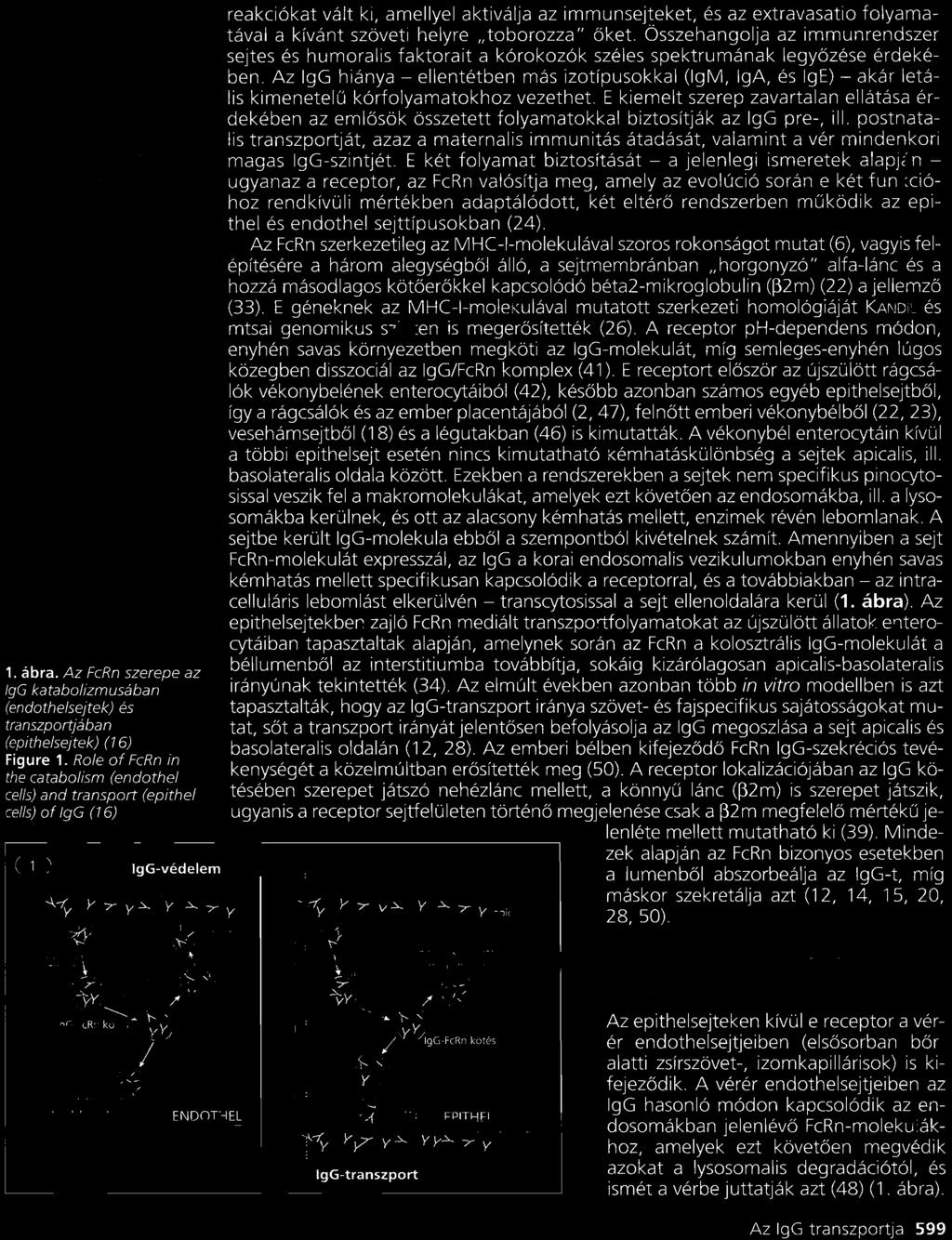
143


144

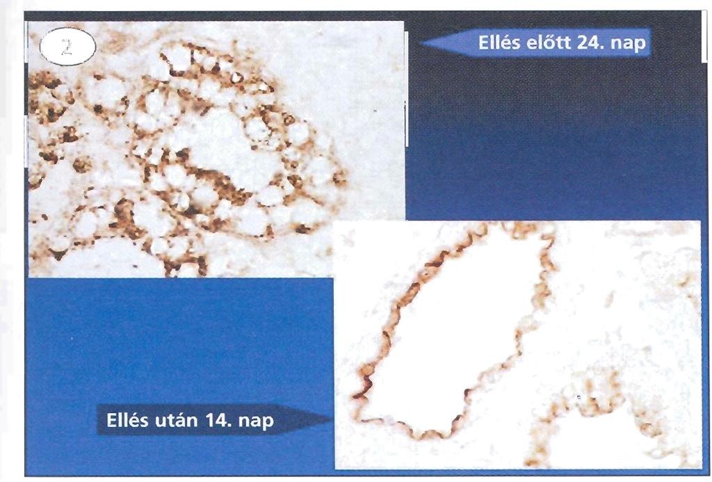
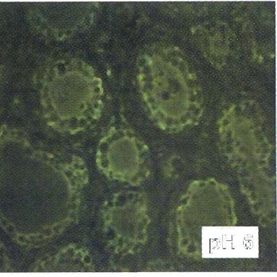

145


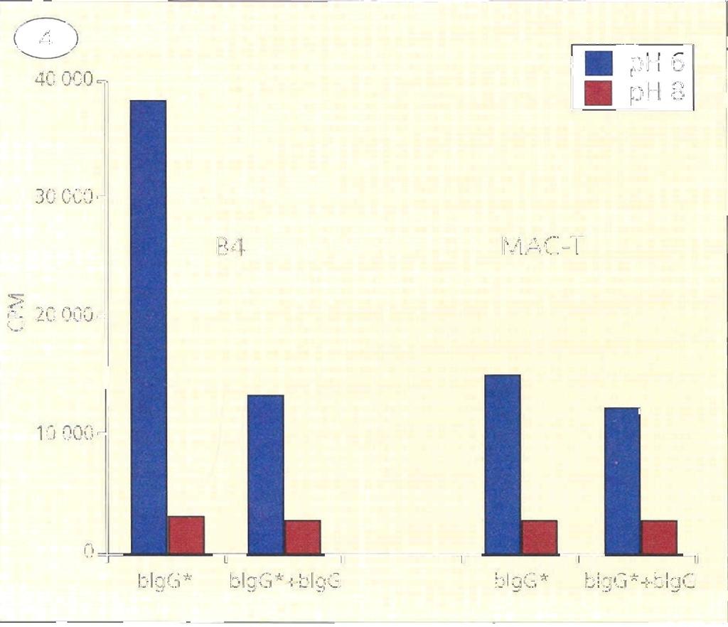
146
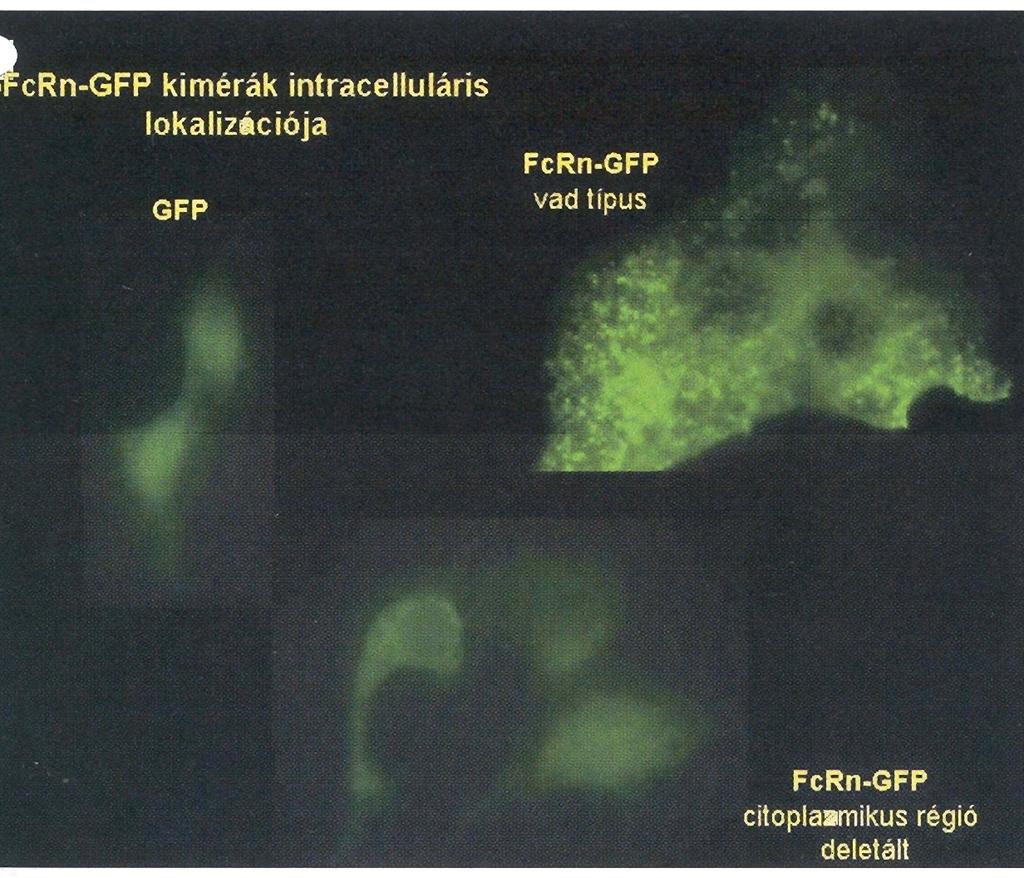
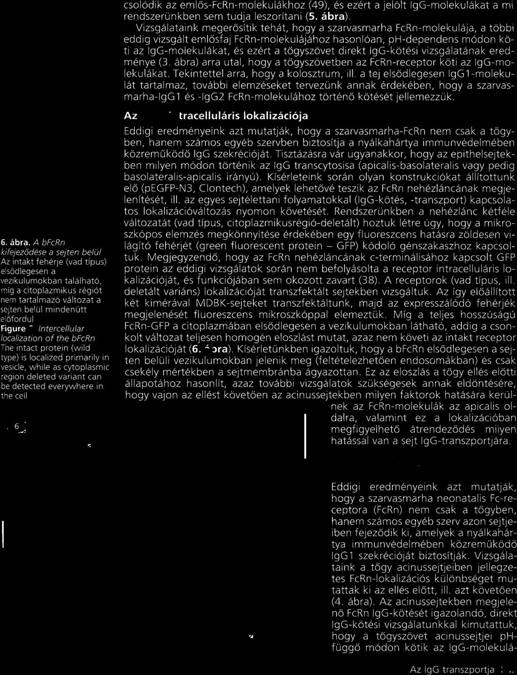
147
148
149
150 Veterinary Immunology and Immunopathology 98 (2004) The neonatal Fc receptor (FcRn) is expressed in the bovine lung Balázs Mayer a, Zsuzsanna Kis a, Győző Kaján a,lászló V. Frenyó a, Lennart Hammarström b, Imre Kacskovics a,* a Department of Physiology and Biochemistry, Faculty of Veterinary Science, Szent István University, István u.2, H 1078 Budapest, Hungary b Center for Biotechnology, Karolinska Institute, Novum, SE Huddinge, Sweden Received 10 July 2003; accepted 22 October 2003 Abstract In neonatal calves, maternal immunoglobulin (Ig) is transferred into respiratory secretion which contributes to protection against pathogens. The early predominance of IgG1 in respiratory tract secretions is progressively reduced in favor of IgA by age but in the lower, bronchoalveolar system secreted IgG remains the dominant secreted Ig even in adulthood. The trans-epithelial transport of secretory IgA into mucosal secretions is carried out by the polymeric Ig receptor. However, the mechanism by which IgG crosses epithelial cells to provide defense on mucosal surfaces is still unknown. In order to investigate the possibility that the neonatal Fc receptor, FcRn is involved in this transport we have first analyzed the localization of this receptor in the upper and lower respiratory tracts. Consistent with the in situ hybridization data, immunohistochemistry showed undetectable expression in the tracheal epithelial cells, relatively weak expression in epithelial cells of the bronchi, apparent staining those lining the bronchioli and randomly scattered signal over the alveolar tissue. The bovine FcRn may thus play a role in IgG transport across mucosal epithelial barriers as a trafficking receptor and ensure IgG predominance in the lower respiratory tract. # 2003 Elsevier B.V. All rights reserved. Keywords: Neonatal Fc receptor; Lung; Respiratory tract; IgG transport; Cow 1. Introduction Mucosal immune system responds to and protects against microbes that enter body through mucosal surfaces of the gastrointestinal, genitourinary and respiratory tracts. On these surfaces, IgA and IgG Abbreviations: FcRn, neonatal Fc receptor; DEPC, dietyl pyrocarbonate; DIG, digoxigenin * Corresponding author. Present address: PO Box 2, H-1400 Budapest, Hungary. Tel.: þ ; fax: þ address: ikacsko@univet.hu (I. Kacskovics). in secretions contribute to humoral immunity (Mestecky and Russell, 1998; Russell and Mestecky, 2002; Brandtzaeg, 2003). In cattle, IgA is the major Ig in saliva, tears and nasal secretions while IgG predominates in colostrum, milk, gastrointestinal (Newby and Bourne, 1976a; Butler, 1983) and genitourinary tract secretions (Duncan et al., 1972; Corbeil et al., 1976). The Ig composition of the respiratory secretions is similar to other species in that there is a predominance of IgA in the upper respiratory tract with an increase of IgG in the secretions from the lower respiratory tract (Wilkie, /$ see front matter # 2003 Elsevier B.V. All rights reserved. doi: /j.vetimm
151 86 B. Mayer et al. / Veterinary Immunology and Immunopathology 98 (2004) ). In calves, IgG1 is the major Ig in tracheal washings while IgA and IgM are present in smaller quantities up to 6 weeks of age (Morgan et al., 1981). The predominance of IgG1 over IgG2 in mucosal fluids supports the concept of a special role for IgG1 in mucosal immunity in ruminants, which is supported by the fact that bovine IgG1 but not IgG2 is as resistant to proteolysis as IgA (Newby and Bourne, 1976b). The trans-epithelial transport of secretary IgA into mucosal secretions is carried out by the polymeric Ig receptor (Mostov and Deitcher, 1986), that has also been identified in cattle (Kulseth et al., 1995; Verbeet et al., 1995). However, the mechanism by which IgG crosses epithelial cells to provide defense on mucosal surfaces is still unknown. The MHC-I related, heterodimeric neonatal Fc receptor (FcRn) (Simister and Mostov, 1989) binds IgG in a ph dependent manner and was first described as an IgG transporter in the neonatal gut of rodents (Rodewald, 1976; Ahouse et al., 1993) and in the human placenta (Leach et al., 1996). Following its first characterization, FcRn expression has been widely detected in epithelial cells of different tissues (e.g. adult human intestine and kidney; rodent liver and mammary gland) (Ghetie and Ward, 2000; Haymann et al., 2000). Other investigations have revealed that this receptor, expressed in capillary endothelial cells, plays a role in IgG homeostasis (reviewed in Ghetie and Ward, 2000). Besides rodents and human, the FcRn encoding gene has been cloned and characterized in cow, sheep, pig and possum (Adamski et al., 2000; Kacskovics et al., 2000; Mayer et al., 2002; Schnulle and Hurley, 2003). In our previous study the obvious change in the subcellular localization of the receptor in the mammary epithelial cells around the time of parturition in ewes and its presence in the crypt epithelial cells of the neonatal lamb led to the hypothesis, that this receptor was involved in IgG1 secretion across epithelial cells in ruminants (Mayer et al., 2002). A more recent study indicated bronchial FcRn expression in human, macaque and mouse and found that this receptor absorbed IgG from the lumen (Spiekermann et al., 2002). We have therefore analyzed the FcRn expression in the bovine respiratory system. The presence of this receptor in the lower respiratory tract suggests a role in mediating IgG transport in the bovine lung. 2. Materials and methods 2.1. Samples for histology Lung tissue specimens and trachea were harvested from a 1-year-old Holstein Friesian bull and two 4- year-old cows at a local slaughterhouse and placed in 4% paraformaldehyde in PBS treated with diethyl pyrocarbonate (DEPC; Sigma Aldrich Co., St. Louis, MO). After overnight fixation the tissues were embedded in paraffin. Paraffin blocks of lung tissue from two 10-week-old Holstein Friesian calves were kindly provided by László Stipkovits (Veterinary Medical Research Institute, Hungarian Academy of Sciences, Budapest, Hungary) In situ hybridization A segment, showing the lowest homology to MHC- I, from a bovine FcRn cdna clone prepared in our laboratory (Kacskovics et al., 2000) was amplified by PCR (primers: B7: 5 0 -GGCGACGAGCACCACT- CAC-3 0, B8: 5 0 -GATTCCCGGAGGTCWCACA-3 0 ). There is a sequence identity between bovine and sheep FcRn corresponding the aforementioned primer sequences. Therefore, the same procedure was done to generate a bovine FcRn specific probe incorporated digoxigenin-11-dutp (Boehringer Mannheim, Mannheim, Germany) as described in a previous study on sheep FcRn (Mayer et al., 2002) with the only modification using the QIAquick Gel Extraction kit (QIA- GEN GmbH, Hilden, Germany) to purify the template segment for the labeling PCR. This DIG-labelled probe was applied to in situ hybridization. Five micrometer thick sections were cut from the paraffin blocks and attached to silanized slides. After deparaffination an in situ hybridization protocol was performed as described previously (Mayer et al., 2002) with some modifications. Briefly, the bovine FcRn specific DIG-labelled probe was used at final concentration about 0.5 ng/ml, the hybridization temperature was adjusted to 45 8C and the dilution of the anti-dig antibody (Boehringer Mannheim) was 500-fold Immunohistochemistry For immunohistochemistry, an affinity purified antiserum (raised against the peptide CLEW-
 85 89 87 KEPPSMRLKAR representing the highly conserved 173 186 aminoacids of FcRn a-chain plus an N terminal Cys for conjugation) was used at")
152 B. Mayer et al. / Veterinary Immunology and Immunopathology 98 (2004) KEPPSMRLKAR representing the highly conserved aminoacids of FcRn a-chain plus an N terminal Cys for conjugation) was used at final concentration of 120 mg/ml. Sections were incubated with affinity-purified anti-fcrn at 4 8C overnight and for 1 h at room temperature and then with biotinylated goat anti-rabbit IgG for 30 min at room temperature. The second antibody was detected with the aid of peroxidase labeled avidin (Vectastain ABC kit, Vector Laboratories, Burlingame, CA). The production of antisera and the immunohistochemical method used have been published previously (Mayer et al., 2002). 3. Results and discussion In situ hybridization with digoxigenin-labeled DNA probe complementary to an FcRn a-chain mrna segment (B7 B8) detected strong FcRn expression in bronchiolar epithelial cells of the lung sections (Fig. 1a). Alveolar tissue also revealed positive staining but it could not be assigned to epithelial or endothelial cells specifically. A randomly scattered signal was observed over the tissue that could originate from the staining of macrophages which have been reported to express FcRn in humans (Zhu et al., 2001). Control sections hybridized with a sense DNA probe derived from the same segment (B7 B8) exhibited weak, nonspecific background signal (Fig. 1b). Consistent with the in situ hybridization data, the immunohistochemical method using the affinity purified anti-fcrn antibody, resulted in randomly scattered signal over the alveolar tissue (Fig. 2a) and apparent staining of the epithelial cells lining the bronchioli and somewhat weaker signal in the bronchus epithelium (Fig. 2b and c). We could not detect FcRn expression in the epithelial layer of the trachea either by immunohistochemistry (Fig. 2d) or by in situ hybridization. In neonatal calves, maternal Ig is transferred into respiratory tract secretions and contributes to local protection (Belknap et al., 1991). The early predominance of IgG1 in respiratory tract secretion is progressively reduced in favor of IgA but in the lower, bronchoalveolar system the secreted IgG predominates in adult animals (Wilkie, 1982). In this study, we have found that the bovine FcRn, in accordance with the results obtained by other species (Spiekermann et al., 2002), is expressed mainly in epithelial cells of the lower airways and in the alveoli of the lung, where IgG is the major Ig in the secretion. In the upper respiratory tract, IgA is the dominant isotype (Wilkie, 1982) and consistent with this observation, we could not detect FcRn expression in the epithelial layer of the trachea. Pertaining to the function of the FcRn in the bronchoalveolar system, Spiekermann and colleagues Fig. 1. In situ hybridization of bull lung sections with a digoxigenin-labeled DNA probe derived from the B7 B8 region of bfcrn a-chain cdna: (a) bronchiolus in a lung section hybridized with an antisense probe; (b) section from the same area hybridized with a sense probe as a negative control. Sections from cows and calves exhibited similar results (data not shown).
 immunostained alveolar area, A indicates the lumen of an alveolus, arrows indicate scattered staining of")
153 88 B. Mayer et al. / Veterinary Immunology and Immunopathology 98 (2004) Fig. 2. The localization of FcRn expression using immunohistochemistry on lung sections of a cow: (a) immunostained alveolar area, A indicates the lumen of an alveolus, arrows indicate scattered staining of the alveoli, magnification: 20; (b) bronchiolus, magnification: 40; (c), bronchus, magnification: 20; (d) trachea (section from a bull), E stands for the epithelial layer, L means the lumen of the trachea, magnification: 20. have recently demonstrated FcRn-dependent absorption of a bioactive Fc-fusion protein in a mouse model. Although, they have not investigated whether FcRn is involved in secretion of IgG into the lumen, they have proposed that FcRn mediates a steady-state and dynamic distribution of IgG across the mucosal barriers (Spiekermann et al., 2002). More recently, Kim and Malik (2003) have confirmed FcRn expression in rat alveolar epithelial cell monolayers and reported bidirectional fluxes of biotinylated rat IgG across these cells which could be saturated and inhibited in the presence of excess unlabelled rat Fc. They concluded that alveolar epithelial IgG transport occurs via FcRnmediated transcytosis. In addition, they proposed that translocation of IgG via paracellular routes by restricted passive diffusion does not appear to be the primary route under physiological conditions (Kim and Malik, 2003). 4. Conclusions We speculate that the bovine FcRn may have a role to ensure the IgG predominance at the lower bronchoalveolar system and function at this site as a trafficking receptor to mediate host defence or immunsurveillance or both. Further in vivo investigations are needed to test this hypothesis. Acknowledgements The authors are grateful to Ágnes Mészáros for preparation of the paraffin sections and to Ágnes Méhes and Ilona Horn performing some of the immunohistochemistry experiments. This work was supported by the National Research Foundation of Hungary (OTKA T035209, T030304).
154 B. Mayer et al. / Veterinary Immunology and Immunopathology 98 (2004) References Adamski, F.M., King, A.T., Demmer, J., Expression of the Fc receptor in the mammary gland during lactation in the marsupial Trichosurus vulpecula (brushtail possum). Mol. Immunol. 37, Ahouse, J.J., Hagerman, C.L., Mittal, P., Gilbert, D.J., Copeland, N.G., Jenkins, N.A., Simister, N.E., Mouse MHC class I- like Fc receptor encoded outside the MHC. J. Immunol. 151, Belknap, E.B., Baker, J.C., Patterson, J.S., Walker, R.D., Haines, D.M., Clark, E.G., The role of passive immunity in bovine respiratory syncytial virus-infected calves. J. Infect. Dis. 163, Brandtzaeg, P., Role of secretory antibodies in the defence against infections. Int. J. Med. Microbiol. 293, Butler, J.E., Bovine immunoglobulins: an augmented review. Vet. Immunol. Immunopathol. 4, Corbeil, L.B., Hall, C.E., Lein, D., Corbeil, R.R., Duncan, J.R., Immunoglobulin classes in genital secretions of mycoplasmainfected and normal heifers. Infect. Immun. 13, Duncan, J.R., Wilkie, B.N., Hiestand, F., Winter, A.J., The serum and secretory immunoglobulins of cattle: characterization and quantitation. J. Immunol. 108, Ghetie, V., Ward, E.S., Multiple roles for the major histocompatibility complex class I- related receptor FcRn. Annu. Rev. Immunol. 18, Haymann, J.P., Levraud, J.P., Bouet, S., Kappes, V., Hagege, J., Nguyen, G., Xu, Y., Rondeau, E., Sraer, J.D., Characterization and localization of the neonatal Fc receptor in adult human kidney. J. Am. Soc. Nephrol. 11, Kacskovics, I., Wu, Z., Simister, N.E., Frenyo, L.V., Hammarstrom, L., Cloning and characterization of the bovine MHC class I-like Fc receptor. J. Immunol. 164, Kim, K.J., Malik, A.B., Protein transport across the lung epithelial barrier. Am. J. Physiol. Lung Cell Mol. Physiol. 284, L247 L259. Kulseth, M.A., Krajci, P., Myklebost, O., Rogne, S., Cloning and characterization of two forms of bovine polymeric immunoglobulin receptor cdna. DNA Cell Biol. 14, Leach, J.L., Sedmak, D.D., Osborne, J.M., Rahill, B., Lairmore, M.D., Anderson, C.L., Isolation from human placenta of the IgG transporter, FcRn, and localization to the syncytiotrophoblast: implications for maternal fetal antibody transport. J. Immunol. 157, Mayer, B., Zolnai, A., Frenyo, L.V., Jancsik, V., Szentirmay, Z., Hammarstrom, L., Kacskovics, I., Redistribution of the sheep neonatal Fc receptor in the mammary gland around the time of parturition in ewes and its localization in the small intestine of neonatal lambs. Immunology 107, Mestecky, J., Russell, M.W., Passive and active protection against disorders of the gut. Vet. Q. 20 (Suppl. 3), Morgan, K.L., Bourne, F.J., Newby, T.J., Bradley, P.A., Humoral factors in the secretory immune system of ruminants. Adv. Exp. Med. Biol. 137, Mostov, K.E., Deitcher, D.L., Polymeric immunoglobulin receptor expressed in MDCK cells transcytoses IgA. Cell 46, Newby, T.J., Bourne, F.J., 1976a. The nature of the local immune system of the bovine small intestine. Immunology 31, Newby, T.J., Bourne, F.J., 1976b. Relative resistance of bovine and porcine immunoglobulins to proteolysis. Immunol. Commun. 5, Rodewald, R., ph-dependent binding of immunoglobulins to intestinal cells of the neonatal rat. J. Cell Biol. 71, Russell, M.W., Mestecky, J., Humoral immune responses to microbial infections in the genital tract. Microbes Infect. 4, Schnulle, P.M., Hurley, W.L., Sequence and expression of the FcRn in the porcine mammary gland. Vet. Immunol. Immunopathol. 91, Simister, N.E., Mostov, K.E., An Fc receptor structurally related to MHC class I antigens. Nature 337, Spiekermann, G.M., Finn, P.W., Ward, E.S., Dumont, J., Dickinson, B.L., Blumberg, R.S., Lencer, W.I., Receptor-mediated immunoglobulin G transport across mucosal barriers in adult life: functional expression of FcRn in the mammalian lung. J. Exp. Med. 196, Verbeet, M.P., Vermeer, H., Warmerdam, G.C., de Boer, H.A., Lee, S.H., Cloning and characterization of the bovine polymeric immunoglobulin receptor-encoding cdna. Gene 164, Wilkie, B.N., Respiratory tract immune response to microbial pathogens. J. Am. Vet. Med. Assoc. 181, Zhu, X., Meng, G., Dickinson, B.L., Li, X., Mizoguchi, E., Miao, L., Wang, Y., Robert, C., Wu, B., Smith, P.D., Lencer, W.I., Blumberg, R.S., MHC class I-related neonatal Fc receptor for IgG is functionally expressed in monocytes, intestinal macrophages, and dendritic cells. J. Immunol. 166,
155 Veterinary Immunology and Immunopathology 108 (2005) Isolation of the gene encoding the bovine neonatal Fc receptor Márton Doleschall a, Yaofeng Zhao b, Balázs Mayer a, Lennart Hammarström b, Imre Kacskovics a, * a Department of Physiology and Biochemistry, Faculty of Veterinary Science, Szent Istvan University, P.O. Box 2, H-1400 Budapest, Hungary b Division of Clinical Immunology, Department of Laboratory Medicine, F79 Karolinska University Hospital Huddinge, Karolinska Institute, SE Stockholm, Sweden Abstract The role of an IgG-Fc receptor (FcRn) that resembles a class I MHC Ag in transporting IgGs through epithelial cells was recently shown in selected species. Here we report our preliminary characterization of a clone encoding the alpha chain of the bovine FcRn from a BAC library. The recombinant BAC DNAwas digested, analyzed by Southern blot hybridization and a bovine FcRn positive 9 kb long fragment was subcloned and partially sequenced. The exon/intron organization of the bovine FcRn alpha chain gene was deduced by comparison with its cdna sequence. The sequence revealed a similar organization to the human and mouse FcRn alpha chain genes. The bovine FcRn alpha chain gene has acquired several repetitive sequences in its 5 0 -flanking region, including multiple SINE and LINE elements. Potential binding sites for transcription factors within the 5 0 -flanking sequence were identified using TESS and TFSEARCH programs. The 5 0 -flanking region of the bfcrn alpha chain gene was analyzed for its ability to directly express the luciferase reporter gene in bovine mammary gland epithelial cells. Transient transfection of a luciferase construct revealed that there was promoter activity in the region 1787 to +92 of the 5 0 -flanking sequence. # 2005 Elsevier B.V. All rights reserved. Keywords: Immunoglobulin; Maternal antibodies; Mucosal immunity; Bovine Fc receptor; Gene regulation 1. Introduction The mucosal immune system responds to and protects against microbes that enter the body through mucosal surfaces of the gastrointestinal, genitourinary and respiratory tracts. On these surfaces, IgA and IgG in * Corresponding author. Tel.: ; fax: address: kacskovics.imre@aotk.szie.hu (I. Kacskovics). secretions contribute to humoral immunity. The transepithelial transport of secretory IgA into mucosal secretions is carried out by the polymeric Ig receptor (Mostov and Deitcher, 1986), that has also been identified in cattle (Kulseth et al., 1995; Verbeet et al., 1995). The mechanism by which IgG reaches luminal secretions is unknown. Recent evidence has pointed toward a role for the neonatal Fc receptor (FcRn) in these processes. The neonatal Fc receptor (FcRn) is composed of two subunits, b2-microglobulin and an /$ see front matter # 2005 Elsevier B.V. All rights reserved. doi: /j.vetimm
156 146 M. Doleschall et al. / Veterinary Immunology and Immunopathology 108 (2005) integral membrane polypeptide homologous the MHC class I proteins (Simister and Mostov, 1989). It binds IgG in a ph-dependent manner and was first described as an IgG transporter in the neonatal gut of rodents (Rodewald, 1976). Following its first characterization, FcRn expression has been widely detected in epithelial cells of different tissues suggesting that it may modulate IgG transport at these sites, as well as in capillary endothelial cells, where it plays a critical role in IgG homeostasis (reviewed in Ghetie and Ward, 2000). The bovine neonatal Fc receptor (bfcrn) has been characterized and its expression has been found in multiple tissues, among the mammary gland, small intestine, kidney and liver (Kacskovics et al., 2000). In our previous studies, the obvious change in the subcellular localization of the receptor in the mammary epithelial cells around the time of parturition in ewes and its presence in the crypt epithelial cells of the neonatal lamb (Mayer et al., 2002) as well as in the lower respiratory tract (Mayer et al., 2004) led to the hypothesis that this receptor is involved in IgG1 transport across these barriers and by analogy with the human and mouse FcRn, it is expected to protect circulating IgG from catabolism. This hypothesis is further supported by the fact that allotypic variants of both the heavy and the light chains of the bfcrn influence serum IgG concentration in newborn calves (Clawson et al., 2004; Laegreid et al., 2002). Insufficient information is currently available regarding the regulation of the FcRn expression. Whereas the sequences of the human and murine FcRn genes, and the promoter sequence of the possum, have been reported (Kandil et al., 1995; Mikulska et al., 2000; Western et al., 2003), the elements that are critical in directing FcRn promoter activity in these species have been only partially identified. Recent reports on the transcriptional regulation of the rat FcRn heavy chain has revealed that the Sp family of transcription factors control its basal expression (Jiang et al., 2004), while in the case of mouse, NF1, SP1 and Ets were identified as possible transcriptional regulators (Tiwari and Junghans, 2005). We report here the exon/intron organization of the gene encoding bovine alpha chain (FCGRT). The structure reveals a similar organization to the human and mouse FCGRTs. We also report the generation of a luciferase construct containing the 5 0 -flanking region of this gene which shows promoter activity in a bovine mammary epithelial cell line. 2. Materials and methods 2.1. Cloning the gene of the bfcrn heavy chain To isolate the bfcrn gene a BAC library was used (The Resource Center of the German Human Genome Project at the Max-Planck-Institute for Molecular Genetics). The library was made from DNA from lymphocytes of an adult male (2 years) Bos taurus Jersey, then partially digested with EcoRI and subsequently ligated into the pbace3.6 BAC vector. Screening of the bfcrn heavy chain positive BAC clone was performed by two primers, which were designed based on the bfcrn cdna sequence. Three rounds of screening of bovine super-pool DNA (BAC library DNA) gave one FcRn positive BAC clone with about 110 kb insert containing the whole bfcrn gene. As a following step the bfcrn heavy chain DNA was sequenced with primers that anneal to the exons of the gene Cloning the upstream flanking region The recombinant BAC DNA was digested with BamHI, and the digested DNA was separated on an agarose gel. Southern blot detected a 9 kb long positive band using a DNA fragment from alpha1 domain, as a probe. The 9 kb long BamHI fragment was then subcloned into the pgem-11zf(+) vector. An additional subcloning process resulted a 2 kb of the promoter segment with exon 1 until exon 3 in the same vector. The insert was then completely sequenced by ABI Prism BigDye Terminator Cycle sequencing Ready Reaction Kit (AB1, 373A-Stretch, Perkin- Elmer) in the Cybergene Company (Huddinge Sweden) Analysis of the 5 0 -flanking sequence Approximately 1800 bp of the region upstream of the translation start of the bovine FcRn alpha chain was screened for interspersed repeats and low complexity DNA sequences using the RepeatMasker program (Smit et al., ). Potential binding sites for
157 M. Doleschall et al. / Veterinary Immunology and Immunopathology 108 (2005) Fig. 1. The exon/intron organization of the bovine FcRn alpha chain. The bovine FcRn alpha chain gene contains seven exons (filled rectangles) and six introns (empty rectangles) spanning, where the relative lengths of them are proportional (total length is about 6.5 kb). transcription factors within the 5 0 -flanking sequence were identified using TESS (Schug and Overton, 1997) in the combined string and weight matrix search mode and TFSEARCH (Heinemeyer et al., 1998) matrix analyses. Comparisons among the bovine, human and mouse Fcgrt promoter sequences (1.8 kb upstream regions) were performed using the conserved regulatory elements anchored alignment analysis (CON- REAL; Berezikov et al., 2004), that allows identification of transcription factor binding sites (TFBS) that are conserved between two orthologous promoter sequences Transient transfection Bovine mammary epithelial cells (MAC-T (Huynh et al., 1991), a kind gift from Dr. A.J. Guidry, USDA) were grown in modified DMEM supplemented with 10% FCS, 2 mm glutamine, insulin (5 mg/ml), hydrocortisone (5 mg/ml), penicillin (100 IU/ml) and streptomycin (100 mg/ml) (Sigma Aldrich). A luciferase construct containing the 5 0 -flanking region (bases 1787 to +92) of the bovine FcRn gene was generated by PCR amplification and the fragment was subcloned into the promoterless luciferase expression vector pgl3 (Promega). Cells were transfected by the calcium phosphate co-precipitation method (Ausubel et al., 1989). Luciferase activity was determined by using the Bright-Glo Luciferase Assay System (Promega) in a luminometer (Luminoskan Ascent, Thermo Labsystems, Vantaa, Finland). The hcmv-bgal plasmid carrying the human CMV promoter linked to the b-gal was used to normalize the cell transfection efficiency. The b-gal activity was measured as described previously (Ausubel et al., 1989). 3. Results and discussion 3.1. Analysis of the gene encoding the bovine FcRn heavy chain To obtain genomic DNA encoding the bfcrn, a bovine BAC library was screened using a PCR method. After three rounds of screening, one positive clone (BBI_B750B15194) was identified. The clone was amplified and partially sequenced using primers which were designed based on the bovine FcRn cdna sequence (Kacskovics et al., 2000), which was also used to analyze the exon/intron organization of the bovine FcRn alpha chain. The bovine FcRn alpha chain gene contains seven exons and six introns spanning (total length is about 6.5 kb) and therefore the exon/intron organization is the same as that of the mouse and human FcRn alpha chain genes (Kandil et al., 1995; Mikulska et al., 2000) (Fig. 1) Analyzing the 5 0 -flanking sequence of the bovine Fcgrt The recombinant BAC DNA was then partially mapped and an approximately 2 kb of the promoter Fig. 2. Schematic diagram of the bovine specific repetitive sequences in the 5 0 -flanking region of the bovine Fcgrt gene.
158 148 M. Doleschall et al. / Veterinary Immunology and Immunopathology 108 (2005) Fig. 3. Predicted transcription factor binding sites within the 5 0 -flanking sequence of the bfcrn alpha chain analyzed by TESS and TFSEARCH programs (Heinemeyer et al., 1998; Schug and Overton, 1997). TSS: transcription start site. segment with exon 1 until exon 3 was completely sequenced and analyzed. We have first examined the 5 0 -flanking sequence by RepeatMasker (Smit et al., ), which revealed several bovine specific interspersed repeats, except a 500 bp upstream region of the transcription initiation site (Fig. 2). Potential binding sites for transcription factors within the 5 0 -flanking sequence were identified using TESS (Schug and Overton, 1997) in the combined string and weight matrix search mode and TFSEARCH (Heinemeyer et al., 1998) matrix analyses (Fig. 3). Like its mouse and human homologues, the 5 0 -flanking region of the bfcrn alpha gene does not harbour an obvious TATA box, but contains a potential Sp1 site close to the transcription start site that may stimulate constitutive promoter activity as it has been recently demonstrated for the rat FcRn promoter (Jiang et al., 2004). Similar to its human counterpart, the bovine promoter has multiple C/EPB b transcription binding sites between 600 and 500 bp, indicating potential regulation of the gene by interleukin-6 as previously suggested (Mikulska et al., 2000). Another potential candidate for regulating the human FcRn transcription is AP-1 (Mikulska and Simister, 2000), which was not detected in the bovine promoter. In the more distant segment upstream of the transcriptional start site (between 1800 and 800) multiple potential binding sites were localized, such as the prolactin response factor MGF/STAT5 binding site which has also been found in the possum promoter sequence based on a similar database search (Western et al., 2003). Possible binding sites for interferon response elements IRF1, IRF2 and members of the nuclear factor kb family were also proposed in the bovine promoter which may indicate responsiveness of the bfcrn in inflammatory reactions (Fig. 3). A previous comparison of about 2 kb upstream region of the mouse and human Fcgrt genes emphasized considerable divergence in the promoter sequence (Tiwari and Junghans, 2005). We have analyzed the similarity between the bovine and human, and also the bovine and mouse 1.8 kb upstream regions of the Fcgrt genes. CONREAL analysis (Berezikov et al., 2004) has revealed a few conserved TFBS regions between the bovine and human genes (Fig. 4), however a similar study has found no such similarity between the bovine and mouse genes (data not shown). These data further highlight considerable divergence in the Fcgrt promoter sequences, which reflect differences in their developmental and tissue regulation. To test for promoter activity, fragments of the flanking sequence of the bfcrn gene were PCR Fig. 4. Comparison of the bovine and human 1.8 kb upstream regions of the Fcgrt genes using the CONREAL program, which allows identification of transcription factor binding sites (TFBS) using the TRANSFAC database (v.8.4) that are conserved between two orthologous promoter sequences (Berezikov et al., 2004).

159 M. Doleschall et al. / Veterinary Immunology and Immunopathology 108 (2005) Fig. 5. Relative luciferase activity obtained from MAC-T cells transiently transfected with promoterless pgl3 and bfp2-pgl3 which encodes the 5 0 -flanking region of the bovine FcRn a-chain gene. Transfection efficiency was normalized by b-gal activity. Results represent the mean S.E.M. of six independent transfection experiments. subcloned into the promoterless luciferase expression vector pgl3 (Promega). We found that the construct containing the cloned 5 0 -flanking region (bfp2) supported transcription of the luciferase reporter in MAC-T cells (Fig. 5). These data indicate that there is a promoter activity in the region 1802 to +92, and suggest that there are multiple positive regulatory elements throughout this region. Although much more needs to be done before we understand how transcription of the bovine FcRn alpha chain gene is regulated, the sequences we obtained should provide a basis for future studies. Acknowledgement This project was supported by the Ministry of Education, Republic of Hungary (NKFP 4/040/2001) and the Swedish Research Council. References Ausubel, F.M., Brent, R., Kingston, R.E., Moore, D.D., Seidman, J.G., Smith, J.A., Struhl, K., Current Protocols in Molecular Biology. Greene Publishing Associates, Wiley/Interscience, John Wiley & Sons. Berezikov, E., Guryev, V., Plasterk, R.H., Cuppen, E., CON- REAL: conserved regulatory elements anchored alignment algorithm for identification of transcription factor binding sites by phylogenetic footprinting. Genome Res. 14, Clawson, M.L., Heaton, M.P., Chitko-McKown, C.G., Fox, J.M., Smith, T.P., Snelling, W.M., Keele, J.W., Laegreid, W.W., Beta-2-microglobulin haplotypes in U.S. beef cattle and association with failure of passive transfer in newborn calves. Mamm. Genome 15, Ghetie, V., Ward, E.S., Multiple roles for the major histocompatibility complex class I-related receptor FcRn. Annu. Rev. Immunol. 18, Heinemeyer, T., Wingender, E., Reuter, I., Hermjakob, H., Kel, A.E., Kel, O.V., Ignatieva, E.V., Ananko, E.A., Podkolodnaya, O.A., Kolpakov, F.A., Podkolodny, N.L., Kolchanov, N.A., Databases on transcriptional regulation: TRANSFAC, TRRD and COMPEL. Nucleic Acids Res. 26, Huynh, H.T., Robitaille, G., Turner, J.D., Establishment of bovine mammary epithelial cells (MAC-T): an in vitro model for bovine lactation. Exp. Cell Res. 197, Jiang, L., Wang, J., Solorzano-Vargas, R.S., Tsai, H.V., Gutierrez, E.M., Ontiveros, L.O., Kiela, P.R., Wu, S.V., Martin, M.G., Characterization of the rat intestinal Fc receptor (FcRn) promoter: transcriptional regulation of FcRn gene by the Sp family of transcription factors. Am. J. Physiol. Gastrointest. Liver Physiol. 286, G922 G931. Kacskovics, I., Wu, Z., Simister, N.E., Frenyo, L.V., Hammarstrom, L., Cloning and characterization of the bovine MHC class I-like Fc receptor. J. Immunol. 164, Kandil, E., Noguchi, M., Ishibashi, T., Kasahara, M., Structural and phylogenetic analysis of the MHC class I-like Fc receptor gene. J. Immunol. 154, Kulseth, M.A., Krajci, P., Myklebost, O., Rogne, S., Cloning and characterization of two forms of bovine polymeric immunoglobulin receptor cdna. DNA Cell Biol. 14, Laegreid, W.W., Heaton, M.P., Keen, J.E., Grosse, W.M., Chitko- McKown, C.G., Smith, T.P., Keele, J.W., Bennett, G.L., Besser, T.E., Association of bovine neonatal Fc receptor alphachain gene (FCGRT) haplotypes with serum IgG concentration in newborn calves. Mamm. Genome 13, Mayer, B., Kis, Z., Kajan, G., Frenyo, L.V., Hammarstrom, L., Kacskovics, I., The neonatal Fc receptor (FcRn) is expressed in the bovine lung. Vet. Immunol. Immunopathol. 98, Mayer, B., Zolnai, A., Frenyo, L.V., Jancsik, V., Szentirmay, Z., Hammarstrom, L., Kacskovics, I., Redistribution of the sheep neonatal Fc receptor in the mammary gland around the time of parturition in ewes and its localization in the small intestine of neonatal lambs. Immunology 107, Mikulska, J.E., Pablo, L., Canel, J., Simister, N.E., Cloning and analysis of the gene encoding the human neonatal Fc receptor. Eur. J. Immunogenet. 27, Mikulska, J.E., Simister, N.E., Analysis of the promoter region of the human FcRn gene. Biochim. Biophys. Acta 1492, Mostov, K.E., Deitcher, D.L., Polymeric immunoglobulin receptor expressed in MDCK cells transcytoses IgA. Cell 46, Rodewald, R., ph-dependent binding of immunoglobulins to intestinal cells of the neonatal rat. J. Cell Biol. 71, Schug, J., Overton, G.C., TESS: transcription element search system on the WWW. Computational Biology and Informatics Laboratory, School of Medicine, University of Pennsylvania.
160 150 M. Doleschall et al. / Veterinary Immunology and Immunopathology 108 (2005) Simister, N.E., Mostov, K.E., An Fc receptor structurally related to MHC class I antigens. Nature 337, Smit, A., Hubley, R., Green, P., RepeatMasker Open Tiwari, B., Junghans, R.P., Functional analysis of the mouse Fcgrt 5 0 proximal promoter. Biochim. Biophys. Acta 1681, Verbeet, M.P., Vermeer, H., Warmerdam, G.C., de Boer, H.A., Lee, S.H., Cloning and characterization of the bovine polymeric immunoglobulin receptor-encoding cdna. Gene 164, Western, A.H., Eckery, D.C., Demmer, J., Juengel, J.L., McNatty, K.P., Fidler, A.E., Expression of the FcRn receptor (alpha and beta) gene homologues in the intestine of suckling brushtail possum (Trichosurus vulpecula) pouch young. Mol. Immunol. 39,
161 Journal of Dairy Research (2005) f Proprietors of Journal of Dairy Research doi: /s Printed in the United Kingdom Expression of the neonatal Fc receptor (FcRn) in the bovine mammary gland Balázs Mayer 1,Márton Doleschall 1, Balázs Bender 2,János Bartyik 3, Zsuzsanna Bősze 2, László V. Frenyó 1 and Imre Kacskovics 1 * 1 Department of Physiology and Biochemistry, Faculty of Veterinary Science, Szent István University, PO Box 2, H-1400 Budapest, Hungary 2 Department of Animal Biology, Agricultural Biotechnology Center, H-2100 Gödöllő, Hungary 3 Agricultural Company of Enying, H-8155 Kiscséripuszta, Hungary In ruminants, protective immunoglobulins are transferred to the newborn via colostrum to mediate maternal immunity. There is a high selectivity in the transport of immunoglobulins from the maternal plasma across the mammary barrier into the colostrum, and only IgG1 is transferred in large amounts. We have recently analysed the expression of the neonatal Fc receptor (FcRn) in sheep mammary gland around parturition. Re-analysing this issue in bovine confirmed our previous data indicating that FcRn is homogeneously localized in the mammary gland acinar cells before parturition, however a remarkable difference was observed in the pattern after calving, where only the apical side of the cells was strongly stained. The presence of the FcRn in the acinar epithelial cells of the mammary gland and the obvious change in distribution before and after parturition indicate that FcRn plays an important role in the IgG transport during colostrum formation in ruminants. Keywords: FcRn, mammary gland, non-lactating, parturition, bovine, IgG. Maternal IgG endows the fetus with protection against congenital infection and also provides adequate immunity for the first weeks of independent life, since at birth the offspring is exposed to a similar antigenic environment as its mother. In mammals, the offspring acquire maternal IgGs either before or after birth. In ruminants, protective immunoglobulins are transferred to the newborn via colostrum to mediate passive immunity. There is a high selectivity in the transport of immunoglobulins from the maternal plasma across the mammary barrier into the colostrum, and only IgG1 is transferred in large amounts (reviewed in Butler 1999). Upon ingestion of the colostrum, the immunoglobulins are transported across the intestinal barrier of the neonate into its blood. This intestinal passage appears to be nonspecific, and later, a large proportion of the absorbed IgG1 has been suggested to be recycled back into several mucosal surfaces, like the intestinal lumen (Newby & Bourne 1976; Besser et al. 1988) and respiratory tract (Wilkie 1982). *For correspondence; Kacskovics.Imre@aotk.szie.hu Preferential binding of IgG1 to the mammary epithelial cells was previously shown near parturition (Kemler et al. 1975; Sasaki et al. 1977; Barrington et al. 1997) and these cells were reported to stain prepartum with anti-igg1 serum (Leary et al. 1982). There is a rapid drop in the concentration of all lacteal immunoglobulins immediately postpartum (Butler 1983) and the selectivity of this transfer has led to the speculation that FcRn might be involved in this process. The neonatal Fc receptor (FcRn) was first identified in rodents as the receptor that transfers maternal immunoglobulins (IgGs) from mother to newborn via the neonatal intestine (Rodewald 1976). Since then, this receptor has been detected in different epithelial cells which delivers IgG across these barriers, as well as in endothelial cells which are responsible for the maintenance of serum IgG levels (reviewed in Ghetie & Ward 2000). FcRn binds IgG in a strictly ph dependent manner (binding occurs at ph 6, but not at ph 7. 4) and consists of a heterodimer of an integral membrane glycoprotein, similar to MHC class I a-chains, and b2-microglobulin (Simister & Mostov 1989). One of several functions described for FcRn is the regulation of IgG isotype transport into milk as it was
162 2 B Mayer and others localized to the epithelial cells of the mammary gland in lactating mice. Analysis of the transfer of Fc fragments and IgG which have different affinities for FcRn indicated that it prevents IgG from being secreted into milk (Cianga et al. 1999). In addition to this finding, the expression of FcRn in the mammary gland of other species like possum (Trichosurus vulpecula) (Adamski et al. 2000), swine (Schnulle & Hurley 2003), ruminants (Kacskovics et al. 2000; Mayer et al. 2002) and human (Cianga et al. 2003) were shown, however, how FcRn is involved in the mammary IgG transport in these species has not been directly assessed. We have previously shown that the sheep FcRn is expressed exclusively in the epithelial cells of the acini in the mammary gland, and there was a remarkable cellular redistribution of this receptor around parturition with a downward expressional trend postpartum. We also found its presence in the crypt epithelial cells (that secrete IgG1 into the lumen) of the neonatal lamb (Mayer et al. 2002). Others have found that there is a correlation between allotypes of the cattle FcRn heavy and light chains and the IgG1 content in their calves (Laegreid et al. 2002; Clawson et al. 2004). In a more recent study we demonstrated bovine FcRn expression in epithelial cells of the lower airways and in the alveoli of the lung, where IgG is the major immunoglobulin in secretion (Mayer et al. 2004). These findings led to the hypothesis, that this receptor was indeed involved in IgG1 secretion in these cells in ruminants. Although, the function of the sheep mammary gland is considered to be highly similar to its cattle s counterpart, and FcRn transcripts have already been detected in the bovine mammary gland from non-lactating animals (Kacskovics et al. 2000), we decided to analyse the localization of FcRn expression in the bovine mammary gland around the time of parturition. Based on this survey cow FcRn shows similar expression and localization pattern as was observed in ewes. Materials and Methods Samples for histology Mammary gland tissue from a non-lactating 4-year-old Holstein-Friesian cow was sampled at a local slaughterhouse and the specimens were fixed in 4% paraformaldehyde (PFA) in PBS treated with dietyl-pyrocarbonate (Sigma-Aldrich Co., St. Louis, MO). Biopsies (16 gauger10-cm length biopsy needle; Magnum, Bard, Covington, GA) were collected from the mammary gland (length of sample notch: 1, 9 cm) of 3-year-old Holstein-Friesian cows 14, 7 and 1 days prepartum, on the day of calving, and 7, 14 days postpartum, under local anaesthesia, as reported previously (Colitti et al. 2000). To prevent local infection, Aureomycin Violet Spray (Fort Dodge Animal Health, Overland Park, KS) was applied. Samples were harvested for reverse transcriptase-pcr into liquid nitrogen or for immunohistochemistry into freshly made 4% PFA. Biopsy experiments were approved by the Animal Care and Ethics Committee of the Faculty of Veterinary Science, Szent István University (Ref: 23/B/2000) and complied with the Hungarian Code of Practice for the Care and Use of Animals for Scientific Purposes. Reverse transcriptase-pcr Total RNA was extracted (by using TRIzol Reagent, Gibco BRL-Life Technologies Inc., Gaithersburg, MD) from frozen mammary gland biopsy samples. Two micrograms RNA were reverse transcribed by using Moloney-murine leukemia virus (M-MLV) reverse transcriptase enzyme (Promega, Madison, WI) and the (dt)17-adapter primer as recommended by the manufacturer. PCR was performed to obtain a 134 bp long bovine FcRn alpha-chain specific amplicon of the cytoplasmic and 3 -untranslated region ( bp, NM_176657; Kacskovics et al. 2000). The amplified segment was separated by electrophoresis on 1% agarose gel and stained with ethidium bromide. In situ hybridization After overnight fixation, non-lactating mammary tissue samples were embedded in paraffin. Subsequently, 5 mm thick sections were cut and placed onto Superfrost slides. In situ hybridization was performed as described previously (Mayer et al. 2004). Briefly, an antisense fragment of the cytoplasmic and 3 -untranslated region (914 to 1280 bp; NM_176657; Kacskovics et al. 2000) from a bovine cdna clone was DIG-dUTP labelled by a linear PCR method. After deparaffination, the sections were digested by proteinase K (Boehringer Mannheim, Mannheim, Germany), and postfixed with 4% PFA in order to stop the digestion. Subsequently, the specimens were washed and then the DIG-labelled probe was added (final concentration: ng/ml). After the initial 3 5 min denaturation step at 94 8C, the ISH was carried out overnight at 45 8C on an in situ block. Detection was performed with DIG Nucleic Acid Detection kit (Boehringer Mannheim) according to the instructions of the manufacturer. Immunohistochemical staining Sections from biopsies were prepared similarly as completed on non-lactating mammary gland tissue samples by in situ hybridization. For immunohistochemistry, an affinity purified antiserum (raised against the peptide CLEWKEPPSMRLKAR representing the highly conserved aminoacid residues of bovine FcRn alpha-chain plus an N terminal Cys for conjugation) was used at final concentration of 120 mg/ml. Non-lactating mammary gland and mammary biopsy sections were incubated with affinity-purified anti-fcrn at 4 8C overnight and for 1 h at
 Mammary tissue was hybridized with an anti-sense bovine FcRn specific, digoxigenin-labelled probe, scale bar indicates 50 mm; (b) negative control hybridized with a sense probe derived from the")
163 Bovine FcRn in the mammary gland 3 Fig. 1. In situ hybridization and immunohistochemistry on a non-lactating mammary gland section. (a) Mammary tissue was hybridized with an anti-sense bovine FcRn specific, digoxigenin-labelled probe, scale bar indicates 50 mm; (b) negative control hybridized with a sense probe derived from the same fragment; (c) immunostaining of non-lactating mammary gland with affinity purified anti-fcrn rabbit serum, scale bar indicates 20 mm. L, lumen of the acini. room temperature and then with biotinylated goat antirabbit IgG for 30 min at room temperature. The secondary antibody was detected using peroxidase-labelled avidin (Vectastain ABC kit, Vector Laboratories, Burlingame, CA). The production of antisera and the immunohistochemical protocol have been published previously (Mayer et al. 2002). Results and Discussion In the frame of this study, we intended to analyse the expression and localization of the FcRn in the bovine mammary gland in non-lactating animals and also around parturition. First we analysed FcRn heavy chain expression in non-lactating mammary gland sections by in situ hybridization and immunohistochemistry. In order to detect the FcRn heavy chain transcripts, a digoxigeninlabeled probe complementary to a segment of the transmembrane, cytoplasmic and 3 -untranslated regions of the bovine FcRn alpha-chain cdna was used, as it shows low degree of similarity to the bovine MHC class I molecule (Kacskovics et al. 2000). The anti-sense probe detected FcRn mrna in the acinar and ductal epithelial cells. We noticed scattered staining in the interstitium (which could possibly be due to staining of macrophages), but could not detect FcRn expression in vascular endothelium (Fig. 1a). Sections hybridized with a sense probe derived from FcRn alpha-chain cdna exhibited weak non-specific background signal (Fig. 1b). Immunohistochemistry using affinity purified anti-fcrn rabbit sera confirmed our in situ hybridization data as we could observe diffuse signal over acinar epithelial cells (Fig. 1c). Consistent with the in situ hybridization data there was no signal in endothelial cells (Fig. 1c). This finding is in good agreement with our previous result which indicated the presence of the bfcrn heavy chain transcripts in bovine non-lactating mammary gland (Kacskovics et al. 2000), and presents similar result to a more recent publication which demonstrated FcRn expression in the epithelial but not in the endothelial of the human mammary gland. Noteworthy, that the localization of the human FcRn was primarily intracellular, too (Cianga et al. 2003). Detecting FcRn in bovine mammary gland confirms earlier data, which demonstrated FcRn expression in other species (Cianga et al. 1999; Adamski et al. 2000; Kacskovics et al. 2000; Schnulle & Hurley 2003). After having established that our immunohistological method is reliable for detection of FcRn in bovine mammary tissue sections, a series of mammary gland biopsy sections collected around parturition were incubated with the anti-fcrn sera. We found the most significant change of the FcRn localization in the acini.
, whereas apical signal appeared 1 (d), 7 (e) and 14 (f) days postpartum. L, lumen of the acini.")
164 4 B Mayer and others Fig. 2. Immunohistochemical analyses of bovine mammary gland biopsies around parturition. Diffuse FcRn expression was detected 14 (a) and 7 (b) days prepartum in the acinar cells. Transition from diffuse to apical staining was observed in these cells on the day of calving (c), whereas apical signal appeared 1 (d), 7 (e) and 14 (f) days postpartum. L, lumen of the acini. Scale bar indicates 20 mm. Biopsies 14 and 7 days prepartum showed diffuse signal in the acinar cells (Fig. 2a b), however on the day of calving (after parturition) and 7 and 14 days postpartum the distribution of FcRn alpha-chain dramatically changed and only the apical/luminal sides of the epithelial cells were stained (Fig. 2d f). Noteworthy, we found a transient state situation one day before parturition, where a continuous shift of FcRn from the intracellular to the apical side was visible (Fig. 2c). The presence of bovine FcRn alpha-chain transcripts was detected throughout the whole examined period (data not shown), which supports our immunohistological data and exclude the possibility of some staining artifact after parturition. As concerning the endothelial cells, we could find weak FcRn expression before parturition which may increase the efficacy of the transport of IgG from circulation toward the epithelial cells. However, the lack of FcRn expression in endothelial cells can not completely inhibit IgG accumulation in the interstitium, since a previous study has indicated the presence of both IgG1 and IgG2 in the stroma of the mammary gland during lactation (Leary et al. 1982), when we could not detect FcRn in endothelial cells. Immunohistochemical data around parturition in the cows underlines our previous results in ewes (Mayer et al. 2002). Based on these findings, and given that we have
165 Bovine FcRn in the mammary gland 5 also found FcRn expression in other mucosal epithelial cells which have been previously shown to secrete IgG1 (Mayer et al. 2002; Mayer et al. 2004), along with the fact that FcRn is considered as the only receptor able to transport monomeric IgG through polarized epithelial cells (reviewed in Rojas & Apodaca 2002), it is tempting to speculate that FcRn secrete IgG1 at these sites in ruminants. However, it should be mentioned, that FcRn was originally considered as a receptor transporting IgG from the apical to basolateral direction in rodents and human (Simister & Mostov 1989; Story et al. 1994). Furthermore, previous analysis suggested that in the lactating mice FcRn appears to play a role in recycling rather than secreting IgG in the mammary gland (Cianga et al. 1999). Since then much evidence has been accumulated indicating significant basolateral-apical transport even in these species (Dickinson et al. 1999; Ramalingam et al. 2002; Schlachetzki et al. 2002; Yoshida et al. 2004). These findings point to the fact that the FcRn mediated IgG transcytosis is a cell type and possibly a species specific process, hence it can not be excluded that FcRn in the bovine mammary gland secretes IgG into the colostrum/ milk rather than recycles it to the circulation. This question emphasizes the need of an established polarized monolayer system that allows studies on FcRn mediated IgG transcytosis in cattle. Our ongoing experiments, in collaboration with others, are focused on the binding affinity of the bovine IgG1, IgG2 and IgG3 to the bovine FcRn. We also study the role of the FcRn in endothelial cells both in the mammary gland which may contribute to the IgG secretion into colostrum and also systematically, where it may regulate IgG homeostasis, as is the case in rodents and human (reviewed in Lobo et al. 2004). The receptor which is involved in IgG1 secretion in the bovine mammary gland is under strict hormonal regulation (Barrington et al. 1999, 2001). In order to better understand how the expression of the FcRn genes is regulated around calving we analysed its promoter in reporter gene assays. Furthermore, we created and started to characterize three transgenic mouse lines, which carry the bovine FcRn alpha chain on a BAC clone (Bender et al. 2004). The BAC construct was selected for this purpose because it is expected to ensure high level and position-independent expression in transgenic animals. Preliminary RT-PCR and Northern analyses revealed bovine FcRn alpha chain expression in the intestine and liver of newborn transgenic animals. In summary, our data revealed that FcRn is expressed in the bovine acinar epithelial cells of the mammary gland and shows different intracellular localization during colostrogenesis, lactogenesis, lactation and in non-lactation, where it is hypothesized to mediate IgG transfer into colostrum and later into milk. Since we do not have information if the altered localization of the FcRn receptor does influence IgG transport during these phases of lactation, further studies are awaited to clarify this. We are indebted to Ágnes Mészáros who prepared the tissue sections for histology and we also thank Ágnes Méhes for technical assistance in immunohistochemistry. This work was supported by the National Research Fund of Hungary (OTKA ) and the Ministry of Education, Republic of Hungary (OMFB /2002). References Adamski FM, King AT & Demmer J 2000 Expression of the Fc receptor in the mammary gland during lactation in the marsupial Trichosurus vulpecula (brushtail possum). Molecular Immunology 37(8) Barrington GM, Besser TE, Davis WC, Gay CC, Reeves JJ & McFadden TB 1997 Expression of immunoglobulin G1 receptors by bovine mammary epithelial cells and mammary leukocytes. Journal of Dairy Science 80(1) Barrington GM, Besser TE, Gay CC, Davis WC, Reeves JJ, McFadden TB & Akers RM 1999 Regulation of the immunoglobulin G1 receptor: effect of prolactin on in vivo expression of the bovine mammary immunoglobulin G1 receptor. Journal of Endocrinology 163(1) Barrington GM, McFadden TB, Huyler MT & Besser TE 2001 Regulation of colostrogenesis in cattle. Livestock Production Science 70(1 2) Bender B, Bodrogi L, Yaofeng Z, Kacskovics I, Hammarstrom L & Bosze Z 2004 Generation of bovine FcRn alpha chain BAC transgenic mice. Tissue Antigens 64(4) Besser TE, McGuire TC, Gay CC & Pritchett LC 1988 Transfer of functional immunoglobulin G (IgG) antibody into the gastrointestinal tract accounts for IgG clearance in calves. Journal of Virology 62(7) Butler JE 1983 Bovine immunoglobulins an augmented review. Veterinary Immunology and Immunopathology 4(1 2) Butler JE 1999 Immunoglobulins and immunocytes in animal milks. In Mucosal Immunology, pp (Ed. PL Ogra). New York: Academic Press Cianga P, Cianga C, Cozma L, Ward ES & Carasevici E 2003 The MHC class I related Fc receptor, FcRn, is expressed in the epithelial cells of the human mammary gland. Human Immunology 64(12) Cianga P, Medesan C, Richardson CA, Ghetie V & Ward ES 1999 Identification and function of neonatal Fc receptor in mammary gland of lactating mice. European Journal of Immunology 29(8) Clawson ML, Heaton MP, Chitko-McKown CG, Fox JM, Smith TP, Snelling WM, Keele JW & Laegreid WW 2004 Beta-2-microglobulin haplotypes in U.S. beef cattle and association with failure of passive transfer in newborn calves. Mammalian Genome 15(3) Colitti M, Stradaioli G & Stefanon B 2000 Effect of alpha-tocopherol deprivation on the involution of mammary gland in sheep. Journal of Dairy Science 83(2) Dickinson BL, Badizadegan K, Wu Z, Ahouse JC, Zhu X, Simister NE, Blumberg RS & Lencer WI 1999 Bidirectional FcRn-dependent IgG transport in a polarized human intestinal epithelial cell line. Journal of Clinical Investigation 104(7) Ghetie V & Ward ES 2000 Multiple roles for the major histocompatibility complex class I-related receptor FcRn. Annual Review of Immunology Kacskovics I, Wu Z, Simister NE, Frenyo LV & Hammarstrom L 2000 Cloning and characterization of the bovine MHC class I-like Fc receptor. Journal of Immunology 164(4) Kemler R, Mossmann H, Strohmaier U, Kickhofen B & Hammer DK 1975 In vitro studies on the selective binding of IgG from different species to tissue sections of the bovine mammary gland. European Journal of Immunology 5(9) Laegreid WW, Heaton MP, Keen JE, Grosse WM, Chitko-McKown CG, Smith TP, Keele JW, Bennett GL & Besser TE 2002 Association of bovine neonatal Fc receptor alpha-chain gene (FCGRT) haplotypes with serum IgG concentration in newborn calves. Mammalian Genome 13(12)
166 6 B Mayer and others Leary HL Jr, Larson BL & Nelson DR 1982 Immunohistochemical localization of IgG1 and IgG2 in prepartum and lactating bovine mammary tissue. Veterinary Immunology and Immunopathology 3(5) Lobo ED, Hansen RJ & Balthasar JP 2004 Antibody pharmacokinetics and pharmacodynamics. Journal of Pharmacological Sciences 93(11) Mayer B, Kis Z, Kajan G, Frenyo LV, Hammarstrom L & Kacskovics I 2004 The neonatal Fc receptor (FcRn) is expressed in the bovine lung. Veterinary Immunology and Immunopathology 98(1 2) Mayer B, Zolnai A, Frenyo LV, Jancsik V, Szentirmay Z, Hammarstrom L & Kacskovics I 2002 Redistribution of the sheep neonatal Fc receptor in the mammary gland around the time of parturition in ewes and its localization in the small intestine of neonatal lambs. Immunology 107(3) Newby TJ & Bourne FJ 1976 The nature of the local immune system of the bovine small intestine. Immunology 31(3) Ramalingam TS, Detmer SA, Martin WL & Bjorkman PJ 2002 IgG transcytosis and recycling by FcRn expressed in MDCK cells reveals ligand-induced redistribution. EMBO Journal 21(4) Rodewald R 1976 ph-dependent binding of immunoglobulins to intestinal cells of the neonatal rat. Journal of Cell Biology 71(2) Rojas R & Apodaca G 2002 Immunoglobulin transport across polarized epithelial cells. Nature Reviews Molecular Cell Biology 3(12) Sasaki M, Larson BL & Nelson DR 1977 Kinetic analysis of the binding of immunoglobulins IgG1 and IgG2 to bovine mammary cells. Biochimica et Biophysica Acta 497(1) Schlachetzki F, Zhu C & Pardridge WM 2002 Expression of the neonatal Fc receptor (FcRn) at the blood-brain barrier. Journal of Neurochemistry 81(1) Schnulle PM & Hurley WL 2003 Sequence and expression of the FcRn in the porcine mammary gland. Veterinary Immunology and Immunopathology 91(3 4) Simister NE & Mostov KE 1989 An Fc receptor structurally related to MHC class I antigens. Nature 337(6203) Story CM, Mikulska JE & Simister NE 1994 A major histocompatibility complex class I-like Fc receptor cloned from human placenta: possible role in transfer of immunoglobulin G from mother to fetus. Journal of Experimental Medicine 180(6) Wilkie BN 1982 Respiratory tract immune response to microbial pathogens. Journal of American Veterinarian Medical Association 181(10) Yoshida M, Claypool SM, Wagner JS, Mizoguchi E, Mizoguchi A, Roopenian AC, Lencer WL & Blumberg RS 2004 Human neonatal fc receptor mediates transport of IgG into luminal secretions for delivery of antigens to mucosal dendritic cells. Immunity 20(6)
167 International Immunology, Vol. 18, No. 4, pp doi: /intimm/dxh393 ª The Japanese Society for Immunology All rights reserved. For permissions, please FcRn mediates elongated serum half-life of human IgG in cattle Imre Kacskovics 1, Zsuzsanna Kis 1, Balázs Mayer 1, Anthony P. West Jr 2, Noreen E. Tiangco 2, Mulualem Tilahun 3,László Cervenak 4, Pamela J. Bjorkman 2, Richard A. Goldsby 3, Ottó Szenci 5 and Lennart Hammarström 6 1 Department of Physiology and Biochemistry, Faculty of Veterinary Science, Szent István University, Budapest, Hungary 2 Division of Biology and Howard Hughes Medical Institute, California Institute of Technology, Pasadena, CA, USA 3 Department of Biology, Amherst College, Amherst, MA, USA 4 Research Group of Metabolism and Atherosclerosis, Hungarian Academy of Sciences, Semmelweis University, Budapest, Hungary 5 Clinics for Large Animals, Faculty of Veterinary Science, Szent István University, Üll}o, Hungary 6 Division of Clinical Immunology, Karolinska Hospital, Huddinge, Sweden Keywords: antibody, clearance, interaction, therapeutic antibodies, transchromosomic cattle Abstract IgG has the longest survival time in the circulation of the Ig classes and the lowest fractional catabolic rate. The neonatal Fc receptor (FcRn) plays an important role in regulating these processes. Recently, we have cloned the bovine neonatal Fc receptor (bfcrn) alpha chain and detected its expression in various epithelial cells which are mediating IgG secretion. However, its function in IgG homeostasis has not been investigated. In the current study, we analyzed the binding affinity of bovine and human IgGs to bfcrn using surface plasmon resonance and by in vitro radioreceptor binding assays. As human IgG binds stronger to the bfcrn, than bovine IgG at ph 6, we subsequently analyzed its catabolism in normal and transchromosomic calves that produce human Igs. Pharmacokinetic studies showed that human IgG had ~33 days serum half-life both in normal and transchromosomic calves, which is more than two times longer than its bovine counterpart. We also demonstrate FcRn expression in endothelial cells and in the kidney which are supposed to be involved in IgG metabolism. These data suggest that bfcrn is involved in IgG homeostasis in cattle and furthermore, that the transchromosomic calves producing human Igs can effectively protect their human IgGs which have implications for successful large-scale production of therapeutic antibodies. Introduction Human polyclonal antibodies can be used for a wide variety of therapeutic applications, including treatment of antibioticresistant infections, immune deficiencies and various autoimmune diseases. However, as they are available only from human donors who cannot be readily hyperimmunized, supply is limited. Transgenic mice carrying human Ig loci have previously been created (1 3) and are useful for the derivation of human mabs antibodies. However, large-scale production of human antibodies cannot be gained from these animals. To address this need, transchromosomic calves carrying the human Ig heavy-chain and light-chain loci have recently been produced. These animals express diversified transcripts and human Igs have been detected in the blood (4). Since the majority of human therapeutic antibodies are of the IgG class, it is of importance to understand human IgG homeostasis in cattle. The metabolism of IgG differs from those of the other classes of Igs, in that IgG has the longest survival time in the circulation and the lowest fractional catabolic rate (5). IgG elimination is likely dominated by affinity for the neonatal Fc receptor (FcRn), and the nature of and affinity for the specific target of the antibody (6, 7). However, other factors may contribute to the rate of antibody elimination, including the immunogenicity of the antibody (8), the degree and nature of the glycosylation of the antibody (9 11) and the susceptibility of the antibody to proteolysis (12). The FcRn is a heterodimer that comprises a transmembrane a chain with structural homology to the extracellular domains Correspondence to: I. Kacskovics; kacskovics.imre@aotk.szie.hu Received 19 October 2005, accepted 3 January 2006 Transmitting editor: A. Falus Advance Access publication 15 February 2006
168 526 FcRn protects human IgG in the cattle of the a chain of MHC class I molecules, and a light chain consisting of beta 2-microglobulin (b2m) (13). FcRn mediates both transcytosis of maternal IgG to the fetus or neonate and IgG homeostasis in adults (14). Evidence for the latter role initially derived from studies indicates an unusually short serum half-life of IgG in b2m-deficient mice (15 17). This observation led to the generation of mutant mouse Fc fragments with enhanced binding to FcRn and increased serum persistence in mice (18), and in a more recent study a similar observation has been published with mutant human IgGs in rhesus monkeys (19). Earlier pharmacokinetic studies indicated that most plasma proteins, including IgG, were catabolized in close contact with the vascular space, which led to the hypothesis that the catabolic site for IgG and other proteins was most likely the vascular endothelium (5). Analyses in mice have proven that IgG homeostasis is indeed maintained by vascular endothelial cells (20). Interestingly, FcRn also binds albumin and prolongs its halflife in a concentration-dependent manner like is seen for IgG (21, 22). In cattle, the serum IgG level is ~20 mg ml ÿ1, and the two major IgG isotypes (IgG1, IgG2) are present in nearly equal amounts. The half-lives of IgG1 and IgG2 have been reported in several studies but the values are divergent between publications [reviewed in (23)]. However, the data indicate that they both fall in the range of days (24, 25) with a slightly longer half-life for IgG2 (26, 27). It is worth mentioning, that in addition to vascular catabolism, a significant amount of IgG1 is secreted onto mucosal surfaces that may influence its apparent half-life in serum [reviewed in (28)]. The clearance rates of other bovine Ig classes, similar to other mammalian species, are much shorter, with half-lives of 4.8, 3.4 and 1.9 days for IgM, IgA and IgE, respectively (25). In ruminants, the FcRn a chain has been cloned, characterized and its deduced amino acid sequence shows the highest similarity to human neonatal Fc receptor (hfcrn) among the non-primate species (29, 30). Although the presence of FcRn transcripts in multiple mucosal epithelial cells, which are considered to secrete IgG, has been confirmed (30, 31), its exact role in IgG transport has not been elucidated and even less is known about its function in IgG homeostasis. In this study, we have analyzed the interaction of human and bovine IgG with bovine neonatal Fc receptor (bfcrn) by surface plasmon resonance (SPR), radioreceptor assays and the half-life of human IgG in normal and transchromosomic calves producing human Igs. Methods Expression and purification of the soluble bovine FcRn The soluble bovine FcRn (sbfcrn) heavy-chain cdna ( bp; GenBank AF139106) was PCR amplified with Deep Vent polymerase (New England Biolab, Beverly, MA, USA) from a eucaryotic expression construct described before (29). The forward primer contains an EcoRI site, a Kozak sequence (italic) and the coding region (underlined) (bfcrnbacu_for: 59-ATC AGA ATT CCC TATAAATAT GCG GCT TCC CCG GCC T- 39); the reverse primer contains the gene-specific coding regions (underlined), a factor Xa site (italic), a 63His tag coding sequence, a stop codon and an EcoRI site (bfcrnbacu_rev: 59-ATC AGA ATT CCT AAT GAT GAT GAT GAT GAT GAC GAC CTT CGA TCA GCT CCA CCG TGA GGG GCT-39). The lightchain (bovine beta 2-microglobulin; bb2m) cdna was PCR amplified ( bp; GenBank X69084) with Deep Vent polymerase from a construct which was a kind gift from Shirley Ellis (Compton, UK) (32). The forward primer contains a BamHI site, a Kozak sequence (italic) and the coding region (underlined) (BB2MBACU_for: 59-ATC AGG ATC CCC TAT AAA TAT GGC TCG CTT CGT GGC CTT-39), the reverse primers contain a BamHI site with the coding region (BB2MBACU_rev: 59-ATC AGG ATC CTG CTG CTT ACA GGT CTC GAT-39). The sbfcrn and bb2m fragments were digested with EcoRI and BamHI, respectively, and ligated into pacuw51 baculovirus transfer vector (BD Biosciences, San Diego, CA, USA). Clones harboring plasmid constructs with the desired orientation of the gene fragments were fully sequenced. Recombinant bfcrn was purified from supernatants of baculovirus-infected insect cells (High-5; Invitrogen, Carlsbad, CA, USA), buffer exchanged to 50 mm phosphate at ph 8.0, followed by Ni-NTA chromatography (Ni-NTA superflow, Qiagen, Chatsworth, CA, USA). Protein from an imidazole elution was further purified by gel filtration chromatography and then analyzed on a 15% SDS PAGE gel, followed by Coomassie blue staining. SPR experiments A Biacore 2000 biosensor system was used to assay the interaction of human and bovine IgGs with bfcrn and hfcrn (33). The biosensor system includes a chip with a dextrancoated gold surface to which one protein (the ligand ) is covalently immobilized. Binding of an injected protein (the analyte ) to the immobilized protein results in changes in the SPR that are proportional to the amount of bound protein and read out in real time as resonance units (RU). hfcrn and bfcrn were immobilized by amine coupling chemistry on seperate flowcells at similar coupling densities (hfcrn, 1700 RU; bfcrn, 1800 RU), and a third flowcell was mock coupled using buffer to serve as a blank. Serial dilutions of bovine and human IgGs (Jackson ImmunoResearch Laboratories, West Grove, PA, USA) were injected at room temperature in 50 mm sodium phosphate, ph 6.0, and 150 mm NaCl. Binding reactions were allowed to closely approach or to reach equilibrium and K D s were derived by non-linear regression analysis of plots of R eq (the equilibrium binding response) versus the analyte concentration. Binding data were fit to a model with two classes of non-interacting binding sites as described for the analysis of FcRn with IgG (33, 34). This model assumes that there are two classes of IgG binding sites on the chip and derives macroscopic equilibrium dissociation constants (K D1 and K D2 ) and the precentage of the total response due to each class of binding sites (f 1 and f 2 ). The higher affinity binding constant (K D1 ), which reflects IgG being bound by two immobilized FcRn molecules (one by each heavy-chain Fc domain), is comparable to the affinities measured in cell binding assays involving membrane-bound FcRn (35, 36).
169 bfcrn stable-transfected cells bfcrn heavy-chain stable-transfected bovine mammary epithelial cell line (MAC-T; a kind gift from A. Guidry, USDA, Beltsville, MD, USA) was generated as described previously (29). Briefly, a construct of the full length of bfcrn heavy-chain cdna cloned into the pci-neo eucaryotic expression vector (Promega Corp., Madison, WI, USA) was transfected into MAC-T cells using Ca-phosphate (37, 38). Cells were then selected with G418 (600 lg ml ÿ1 ) and individual G418- resistant colonies were tested for the presence of the bfcrn by western blot using an affinity-purified specific antibody against an oligopeptide (CLEWKEPPSMRLKAR, amino acids ) of bfcrn (30). ph-dependent IgG binding and uptake of the transfected and untransfected cells were analyzed by single-point competitive binding assay according to the protocol of Story et al. (39). Briefly, bovine and human IgGs (Sigma Aldrich, St Louis, MO, USA) were labeled with Na 125 I to a specific activity of ~10 Ci lmol ÿ1 using Chloramine-T (Sigma Aldrich). The cells expressing the bfcrn were first washed with DMEM, ph 6 or 7.5. Then, bovine- 125 I-IgG or human- 125 I-IgG in DMEM, ph 6.0 or 7.5 with or without unlabeled bovine, human IgG and chicken IgY (Sigma Aldrich) in 1000 molar excess was added. The cells were allowed to bind and internalize IgG for 2 h at 37 C and then unbound ligand was removed with washes of chilled PBS, ph 6.0 or 7.5. Bound radioligand was measured in a gamma counter. Viability of the cells before and after the assay was checked by trypan blue exclusion assay. Competitive binding assays For the competitive binding assay, we measured the binding of a single concentration of radiolabeled human IgG in the presence of various concentrations of unlabeled human and bovine IgGs on transfected cells. The binding assay was performed as it was indicated above, except we performed the assay only at ph 6 with increasing amount (2-fold serial dilutions from to 0.06-fold molar excess) of unlabeled bovine and human IgG, as competitors. The cells were allowed to bind and take up IgG for 2 h at 37 C; then unbound ligand was removed with washes of chilled PBS (ph 6.0). Inhibitory concentration 50% (IC 50 ) values were calculated using Prism for Windows, version 4.0 (GraphPad Software, San Diego, CA, USA). FcRn detection in an endothelial cell line (bovine aortic endothelial cells), in tissue capillary endothelial cells and in the kidney Reverse transcription PCR. RNA was isolated from primary bovine aortic endothelial cells (baecs, Cambrex Bio Science, Walkersville, MD, USA) by TRIzolÒ reagent (Invitrogen). cdna was synthesized using standard methods and was subsequently used in PCR with a primer pair specific for bfcrn (matching or complementary to bases or ). PCR products were analyzed by agarose gel electrophoresis. Immunohistochemistry. Intestinal, skin and kidney samples were collected at the slaughterhouse from healthy Holstein cattle. Samples were analyzed by immunohistochemistry as described previously (30). Briefly, the samples were treated FcRn protects human IgG in the cattle 527 with 1% H 2 O 2 to inactivate endogenous peroxidases, and then blocked by Tris-buffered saline containing 5% BSA. Sections were subsequently incubated with affinity-purified anti-fcrn in 1% BSA and then with biotinylated goat anti-rabbit IgG. The secondary antibody was detected with Vectastain ABC kit (Vector Laboratories, Burlingame, CA, USA) and 3,39- diaminobenzidine (Sigma Chemical Co., St Louis, MO, USA) was used as a color substrate. In vivo studies of human IgG and chicken IgY half-lives in calves Four Holstein calves (heifers), ~200 kg, were used to analyze the human IgG clearance. Following a pre-bleed, each calf was injected intravenously (i.v.) with 2 g (10 mg per body weight kilogram; BW kg ) of human IgG (Gammonativ intended for intravenous use was a kind gift from Octapharma, Stockholm, Sweden) in 50 mg ml ÿ1 saline solution and during the next 35 days, periodic blood samples were collected and evaluated for their content of human IgG. A quantitative ELISA employing an unlabeled affinitypurified goat anti-human polyclonal antibody (goat anti-human IgG (H + L); Pierce Biotechnology Inc., Rockford, IL, USA) as capture reagent and an HRP-conjugated affinity-purified polyclonal goat anti-human IgG (Southern Biotechnology Associates, Inc., Birmingham, AL, USA) as detecting reagent was used to evaluate plasma concentrations of human IgG during the course of the experiment. The peroxidase-conjugated antibody was detected using o-phenylenediamine (Fluka Chemie GmbH, Buchs, Switzerland) as the substrate. Samples were assayed in triplicate. Analysis of the mean human IgG concentration of the animals in the first 6.8 days was done by fitting the data to the two-compartmental model using WinNonLin Professional, version 4.1 (Pharsight, Mountain View, CA, USA). The individual elimination curves were used to obtain the pharmacokinetic parameters. The individual values were averaged, and the standard deviation was calculated. Bovine IgG specific to the human IgG was detected by an ELISA assay employing the human IgG as capture reagent and after blocking (in 0.02 M Tris buffer containing 0.1% Tween 20 and 1mg ml ÿ1 ovalbumin for 1 h at room temperature), incubating with a peroxidase-conjugated affinity-purified goat anti-bovine IgG (H + L) (Jackson ImmunoResearch Laboratories, Inc.) as a detecting reagent. Samples were assayed in triplicate. In a control experiment, we injected 1 g (5 mg per BW kg )of chicken IgY which was purified from egg yolk (a kind gift from Vilmos Palya, CEVA Phylaxia, Budapest, Hungary) i.v. into two Holstein calves weighing ~200 kg and during the next 21 days periodic blood samples were collected. A quantitative ELISA employing a monoclonal anti-chicken IgY (Biodesign International, Saco, ME, USA) as capture reagent and an HRP-conjugated affinity-purified polyclonal rabbit anti-chicken IgY (Fc) (Biodesign International) as detecting reagent was used to evaluate plasma concentrations of chicken IgY during the course of the experiment. Samples were assayed in triplicate. Pharmacokinetic parameters were calculated by non-compartmental method, using the computer program WinNonLin Professional, version 4.1 (Pharsight). Serum concentration time curve was calculated using the linear
170 528 FcRn protects human IgG in the cattle trapezoidal rule; however this was estimated by extrapolation from the apparent linear portion of the semi-logarithmic plot after the predicted steady-state situation (day 2) and before the change in the kinetics due to the onset of the immune response against the chicken IgY. The individual elimination curves were used to obtain the pharmacokinetic parameters. The individual values were averaged. All experimental procedures were approved by the Animal Care and Ethics Committee of the Faculty of Veterinary Science, Szent István University and complied with the Hungarian Code of Practice for the Care and Use of Animals for Scientific Purposes. In vivo studies of human IgG half-life in human artificial chromosome-transgenic cattle Two calves, weighing ~300 kg, 104 (#) and 1153 ($) were derived by cloning from a line of primary bovine fibroblasts that had been derived from an Angus Simmental cross and transfected with a human artificial chromosome that bore the complete unrearranged human lambda (Igk) and heavy-chain (IgH) loci. Following a pre-bleed, each calf was injected i.v. with 6 g (20 mg per BW kg ) of human IgG (Panglobulin intended for intravenous use, Blood Transfusion Service, Swiss Red Cross, Berne, Switzerland) in 60 ml of saline and during the next 42 days, periodic blood samples were collected in EDTA tubes and evaluated for their content of human IgG. A quantitative ELISA employing an unlabeled affinity-purified bovine anti-human polyclonal antibody as a capture reagent and a biotinylated affinity-purified bovine anti-human polyclonal antibody as a detecting reagent was used to evaluate plasma concentrations of human IgG during the course of the experiment. Samples were assayed in triplicate. The mean human IgG concentration data of the two animals were fitted with a two-compartmental model using WinNonLin Professional, version 4.1 (Pharsight). The individual elimination curves were used to obtain the pharmacokinetic parameters. The individual values were averaged. All experimental procedures were approved by the Animal 325 Care and Ethics Committee of Hematech, LLC (limited liability company) where all of the animal experiments were conducted using procedures that were deemed in full compliance with the rules of the institutional IACUC (Institutional Animal Care and Use Committee) which was chartered by the federal government of the United States. Results SPR experiments Recombinant sbfcrn was generated, purified and then analyzed by Coomassie blue staining which detected two bands at molecular weights of 31 and 12 kda, representing the sbfcrn and bb2m, respectively (data not shown). The interaction of bovine and human IgG with bfcrn and hfcrn at ph 6.0 was investigated using a SPR assay. bfcrn and hfcrn (33) were covalently immobilized onto a biosensor chip, and a series of injections containing 2 nm 10 lm human or bovine IgG were allowed to reach or nearly reach equilibrium (Fig. 1A and B). These data were fit to a model with two classes of non-interacting binding sites, yielding two equilibrium dissociation constants, K D1 and K D2, along with their fraction occupancies, f 1 and f 2 (Table 1). For the high-affinity population (K D1 ), which generally accounted for the majority of the binding response, the dissociation constants were 0.45 nm for human IgG over bfcrn, 2.0 nm for bovine IgG over bfcrn, 4.9 nm for human IgG over hfcrn and 430 nm for bovine IgG over hfcrn (Fig. 1 and Table 1). Generating a stable transfectant bovine cell line (B4) and testing its binding ability to bovine and human IgG G418-selected bfcrn-transfected cell clones were assessed by western blot using rabbit anti-peptide antisera raised against an epitope of the bfcrn heavy chain (amino acids ) (30). From among several resistant bfcrn/mac-t clones, we selected one (B4) which strongly expressed a ~40-kDa band which is consistent with the known molecular weight of the bfcrn a chain (29, 30). There was no hybridization in the untransfected MAC-T cells (Fig. 2A). Fig. 1. Biosensor analyses of bfcrn and hfcrn interacting with bovine and human IgG. (A) Equilibrium binding of bovine and human IgG to bfcrn (1800 RU) immobilized on a biosensor chip is plotted as a function of the log of the IgG concentration. Best-fit binding curves, modeled as two classes of non-interacting binding sites, are superimposed on the binding data. (B) Equilibrium binding of bovine and human IgG to hfcrn (1700 RU).
171 To determine whether the bfcrn/mac-t clone (B4) expressed a functional FcR, we first measured its binding to radiolabeled bovine IgG. We could detect specific binding at ph 6 in the case of B4 cells, but not in the untransfected control. There was no specific binding at ph 7.5 (Fig. 2B). Next, we analyzed their binding to bovine IgG, human IgG and Table 1. Binding affinities derived from the data in panels A and B of Fig. 1 Immobilized bfcrn K D1 (nm) f 1 (%) K D2 (nm) f 2 (%) Immobilized hfcrn K D1 (nm) f 1 (%) K D2 (nm) f 2 (%) Bovine IgG Human IgG FcRn protects human IgG in the cattle 529 chicken IgY. We found that B4 cells bound bovine and human IgG specifically at ph 6.0 but not at ph 7.5, and there was no binding of the chicken IgY. Although, the amount and the specific activity of the labeled bovine and human IgGs were similar, human IgG binding was much greater (>10 times) than bovine IgG uptake. The binding of bovine IgG was completely displaced by non-labeled bovine and human IgG, but not by chicken IgY, added in 1000-fold molar excess. This single-point competitive binding assay also revealed that radiolabeled human IgG was completely displaced by excess of nonlabeled human IgGs, but only partially by adding 1000-fold molar excess of non-labeled bovine IgGs (Fig. 2C). We have also checked viability of cells before and after binding and found no differences (viability exceeded 98% in both cases). Although, the single-point competitive binding assay suggested that bfcrn preferably binds human IgG, we performed a more comprehensive analysis to accurately evaluate the Fig. 2. In vitro radioreceptor assays of bfcrn interacting with bovine IgG, human IgG and chicken IgY. (A) Functional expression of bfcrn in transfected (B4) and untransfected (MAC-T) bovine mammary epithelial cell lines. Western blot of total cellular protein (10 lg per lane) by using affinity-purified rabbit antisera raised against amino acids of the a2 a3 domains (30). (B) Bovine- 125 I-IgG binding by bfcrn transfected (B4) and untransfected MAC-T cells. Assays were done at 37 C without (filled columns) and with (open columns) competing-unlabeled bovine IgG, at ph 6.0 or 7.5. Each column represents the mean cell-associated radioactivity in three replicates; bars show the standard error of the mean. (C) Binding ability for bovine IgG, human IgG and chicken IgY to transfected MAC-Tcells (B4). B4 cells bound bovine and human IgG specifically at ph 6.0 but not at ph 7.5. Human IgG binding was much greater as compared with the bovine IgG uptake. The binding of bovine IgG was completely displaced by non-labeled bovine and human IgG, but not with chicken IgY, added in 1000-fold molar excess. Radiolabeled human IgG was completely displaced by excess of non-labeled human IgGs, but only partially by adding 1000-fold molar excess of non-labeled bovine IgGs.
172 530 FcRn protects human IgG in the cattle binding affinity of these two IgG molecules to the bfcrn. In this assay, the binding of a single concentration of radiolabeled human IgG in the presence of various concentrations of unlabeled human and bovine IgGs was measured by bfcrntransfected (B4) cells. Strong human and weaker bovine IgG binding was observed, since unlabeled human IgG effectively displaced its radiolabeled form even by ~30-fold molar excess, while bovine IgG did not completely displace the radiolabeled human IgG even at a 1000-fold molar excess (Fig. 3A). Comparison of the IC 50 values indicated that human IgG showed an increased in binding to bfcrn at ph 6 of ~43- fold better than the bovine IgG, whereas the IC 50 values were ÿ9 and ÿ8 M for the human and bovine IgG, respectively (Fig. 3B). FcRn detection in an endothelial cell line (baec), in tissue capillary endothelial cells and in the kidney First, reverse transcription PCR was used to detect bfcrn heavy-chain transcripts from primary bovine arterial endothelial cells that resulted a bfcrn-specific fragment (data not shown). We then analyzed small intestinal tissue and skin sections to reveal their bfcrn expression. FcRn expression was detected in the endothelial cells of the intestinal (duodenum) sub-mucosal connective tissue and skin using affinity-purified anti-fcrn rabbit sera (Fig. 4A and B). We subsequently investigated whether, under physiologic conditions, FcRn was expressed in adult kidneys. Normal bovine kidney tissues were stained with an antiserum to bfcrn using immunohistochemistry, and demonstrated weak, nonspecific staining in the glomeruli and granular staining on proximal tubular cells, associated with the basal side. No staining was detected in the interstitium, medulla or distal tubular cells (Fig. 4C). Pharmacokinetics of human IgG and chicken IgY in normal calves The pharmacokinetic behavior of the human IgG was first examined in four normal calves. Ten milligrams per BW kg Fig. 3. Competitive binding assay of bfcrn interaction with bovine and human IgG by bfcrn-transfected (B4) cells. (A) Human and bovine IgGs were tested in a competitive binding assay to analyze their binding affinity to bfcrn. The assay was performed at ph 6, with increasing amount (2-fold serial dilutions from to 0.06-fold molar excess) of unlabeled bovine and human IgG, as competitors. (B) The mean radioactivity was plotted against the competitor concentration (M) for each Ig. The data are representative of three independent experiments. The top of the curve is the plateau at a value equal to radiolabeled human IgG binding in the absence of the competing-unlabeled human or bovine IgGs. The bottom of the curve is a plateau equal to non-specific binding. The concentration of unlabeled IgG that produces radioligand binding half-way between the upper and lower plateaus is called the IC 50.IC 50 values were calculated using Prism for Windows, version 4.0 (GraphPad Software).

173 FcRn protects human IgG in the cattle 531 human IgG were injected and its levels in serum were measured with a sandwich ELISA. The ELISA was human IgG specific and did not show cross reactivity with bovine Igs. The clearance curves was triphasic, with phase 1 (alpha phase) representing equilibration between the intravascular and extravascular compartments, phase 2 (beta phase) representing a slow elimination, while phase 3 showed an increased rate of removal of human IgG from the intravascular space. Mathematical modeling of phases 1 and 2 until days 6.8 have shown good correlation to the general scheme of FcRnmediated IgG pharmacokinetics (6), hence we calculated the alpha- and beta-phase half-lives of human IgG in this time frame. The estimated alpha-phase half-lives were (mean 6 SD), while the beta-phase half-life was days, based on the two-compartmental modeling analysis. However, the latter data showed a high uncertainty due to the short period of time that could be analyzed (Fig. 5A and Table 2). We also analyzed the anti-human IgG antibody level in the sera of the calves with an ELISA which detected bovine IgG specific to human IgG. We found that anti-human IgG antibody first appeared ~7 days after intravenous administration of human IgG and then its level continuously increased. Following the appearance of endogenous anti-human IgG antibodies, human IgG concentrations in plasma rapidly decreased and therefore presence of these antibodies coincided with phase 3, representing an increased clearance of the human IgG (Fig. 5A). The pharmacokinetic behavior of the chicken IgY was also analyzed in two normal calves. Chicken IgY level was measured in sera and found a marked acceleration of clearance compared with the human IgG. Due to the fast catabolic rate, alpha and beta phases were not distinguisable. Furthermore, from day 8, there was a sudden drop of the IgY level probably due to antibody response against it. Levels of non-neutralizing antibodies were not assessed in this experiment. The half-life for chicken IgY in these animals was only 2.39 days, analyzed between days 2 (from the presumed steady-state situation after injection) and 6 (Table 2). Fig. 4. Immunohistochemical analysis of the cattle small intestine (duodenum), skin and kidney was performed using affinity-purified anti-fcrn rabbit sera. FcRn expression was detected in the endothelial cells of the sub-mucosal connective tissue in the duodenum (A) and subcutaneous connective tissue (B); asterisks indicate blood vessels). In a normal bovine kidney tissue (g, glomerulus; pt, proximal tubule; dt, distal tubule) the assay demonstrated weak, nonspecific staining in the glomeruli and granular staining was observed on proximal tubular cells, associated with the basal side. No staining was detected in the interstitium, medulla or distal tubular cells (C). Sections were counterstained using Mayer s hematoxylin. Bars ¼ 50 lm. Pharmacokinetics of human IgG in transgenic calves We substantially performed a similar experiment in cloned transchromosomic calves producing human Igs (4), where immunogenicity of human IgG does not pose a problem. Additionally, these animals had relatively low level of endogenous human Igs (human Ig levels prior to injection of human IgG was ~13 lg ml ÿ1 ) and injecting 20 mg per BW kg human IgG i.v. resulted a concentration of ~200 lg ml ÿ1. We could therefore effectively use this model to accurately analyze the human IgG catabolism in cattle. The human IgG concentration in these animals was ~200 lg ml ÿ1 after phase 1 of the clearance curve (alpha phase) which correlated well with the human IgG concentration in our first calves in the same phase (~90 lg ml ÿ1 ), since the latter animals received twice as much human IgG per BW kg.as expected, we did not observe a change in the kinetics of phase 2 (beta phase) due to the onset of the immune response and hence we could analyze the half-life of human IgG in cattle for 42 days. Based on the two-compartmental modeling

174 532 FcRn protects human IgG in the cattle Fig. 5. Pharmacokinetic parameters. (A) Pharmacokinetic behavior of the human IgG was first examined in normal Holstein calves. A total of 10 mg per BW kg human IgG was injected in the animals and their sera levels were measured with a sandwich ELISA during the course of the experiment. Samples were assayed in triplicate and error bars represent the standard deviation. Black line indicates the first 6.8 days of the mean human IgG concentration of a representative animal which were fitted with a two-compartmental model using WinNonLin (Pharsight). This period is further emphasized by a gray rectangle. Based on this analysis, the estimated human IgG half-life of alpha phase was 0.35 day, while the betaphase half-life was 32.9 days, although it has a high uncertainty due to the short period that could be analyzed. In the following days, the mean human IgG data were plotted without analysis. Calves generated immune response to the human IgG as it can be seen by the dotted line and open circles. (B) Pharmacokinetics of human IgG in transchromosomic calves. The modeled data (simulated based on the geometric mean of the primary pharmacokinetic parameters) as well as the observed mean serum antibody concentration (lg ml ÿ1 ) for the two animals were plotted as a function of time (days after injection) of human IgG. Samples were assayed in triplicate and error bars represent the standard deviation. Based on the two-compartmental modeling analysis, the half-life of alpha phase was 0.32 day, while the beta-phase half-life was days. Table 2. Pharmacokinetic parameters of human IgG in normal (WT-hIgG) and transchromosomic calves (TC-hIgG) as well as chicken IgY in normal calves (WT-cIgY) Dose (mg/bw kg ) Analyzed time frame (days) C max (lg/ml) CL (ml/day) AUC (day 3 lg/ml) t 1/2 (days) n WT-hIgG WT-cIgY (28 49) 6892 ( ) ( ) 2.39 ( ) 2 TC-hIgG ( ) ( ) ( ) ( ) 2 Values represent either the mean and the range of two calves or mean 6 SD where four calves were used. C max, maximum serum antibody concentration; CL, clearence; AUC, area under the curve; t 1/2, elimination (beta phase) half-life in serum. analysis the half-life of phase 1 was 0.32 day, while the betaphase half-life was days (Fig. 5B and Table 2). Discussion In the current study, we have analyzed the role of the bfcrn in IgG catabolism in normal and transchromosomic cattle that produce human Igs (4). We first analyzed the binding affinity of the bovine and human IgGs to the bfcrn on SPR and found that in both cases specific interaction occurred at ph 6, however there was no interaction at ph 7.5. Surprisingly, human IgG bound much stronger to bfcrn than bovine IgG did (Fig. 1 and Table 1). In a control experiment, we found very weak, basically insignificant binding of bovine IgG to hfcrn, reaffirming the previous data (40). In our SPR experiments, FcRn was immobilized rather than IgG to mimic the physiological situation in which membrane-bound FcRn interacts with soluble IgG and to facilitate comparisons with previous biosensor-based assays. The data do not fit well to a one-site model, and were therefore fit to a two-site model, as described in our previous publication (33). Consistent with the results of the SPR studies, we detected specific binding of both the bovine and human IgG to the bfcrn stably transfected epithelial cells at ph 6, but not at ph 7.5, in a single-point competitive binding assay (Fig. 2). Noteworthy, the increased binding of human IgG to bfcrn at ph 6 did not exhibit a parallel increase in binding at ph 7.5 (Figs 1 and 2), hence human IgG should not be trapped in cells that express FcRn, as occurs with some mutant human IgG1 molecules in mice (41). We could not observe specific interaction between chicken IgY and bfcrn. It has been long known, that mammalian FcRn does not bind to IgY (42, 43) and mammalian IgGs do not transfer into the chick embryo (44), suggesting that a distinct receptor might be involved in IgY transport. This receptor, a phospholipase A2R homolog, has been recently isolated and characterized (45). In a competitive binding assay, comparison of the IC 50 values indicated that human IgG showed an increase in binding to bfcrn at ph 6 of ~40-fold better than the bovine IgG (Fig. 3). This result correlates well with our SPR results (Fig. 1 and Table 1), which indicated that the affinity of bfcrn for human IgG (K D1 = 0.45 nm) was many times higher than for bovine IgG (K D1 = 2 nm). The difference in binding affinities
175 between bovine and human IgG to bfcrn, analyzed by the in vitro receptor assay is greater than that we found by, possibly due to the condition we used. FcRn is localized primarily intracellularly in epithelial and endothelial cells and only a small fraction of it participates in IgG binding on the cell surface (43, 46). It is also directly involved in exocytic events involving transported IgG at the plasma membrane (47). In our case, the receptor assays occurred at ph 6 which allowed more efficient IgG binding to the surface bfcrn in the transfected MAC-T cells and hence, more effective uptake relative to the untransfected MAC-T cells where only fluid phase processes (Fig. 2B). Furthermore, the condition of the assay (ph 6) results poor release of the IgG during exocytic events involving FcRn at the plasma membrane. Thus, the IgG that has higher affinity to the receptor accumulates at substantially higher level in the cells, as we observed in the case of human IgG comparing it to the bovine IgG (Fig. 2C). The situation described here reflects a recent finding, in which the binding characteristics of a mutated human IgG that has higher affinity and reduced ph dependence to FcRn were analyzed (48). Although, the binding experiments in that case were performed at ph 7.2, the reduced ph dependence of the mutated IgG allowed effective binding to the cell surface and poor release during exocytosis, resulting in similarly high accumulation of the mutated molecule (48). As our competitive binding assay was also performed at ph 6, the competition between human and bovine IgGs occurred primarily at the cell surface due to the FcRn molecules that locate originally, albeit at low concentrations, in the membrane and to those receptors that are involved in the exocytosis and thus the data we got reflects to the affinity difference between human and bovine IgGs (Fig. 3). Biochemical studies of membrane-bound and soluble forms of FcRn demonstrate that IgG binding is ph dependent: there is strong binding (nanomolar K D ) under the acidic conditions (ph ~6.0) found in endosomal compartments and in rodent intestines, while at the slightly basic ph of blood (ph 7.4), there is no detectable binding (14). From mutagenesis and crystallographic studies, the mechanism of this ph dependence has been shown to involve chemical rather than conformational changes (49 51). Specifically, there are attractive interactions at acidic ph between protonated histidines on Fc and negatively charged side chains on FcRn, which are lost upon deprotonation of the Fc histidines at basic ph (50). The sequences of FcRn genes isolated from other organisms, including pig (52), sheep (30), macaque (GenBank accession number AAL92101), brushtail possum (53) and cow (29), suggest that the features described above for rodent and hfcrn proteins are shared in the other mammalian orthologs. A comparison between rat, mouse, hfcrn and bfcrn alpha chain and b2m residues which are supposed to be involved in binding to IgG molecules have been extensively analyzed based on a crystallography analysis of a rat FcRn-heterodimeric Fc complex (50). This study compared important residues in the interaction and found that amino acid residues of rat Glu117 and Glu132 are conserved (although Glu132 is replaced by Asp132 in humans and cows), indicating that the two ph-dependent salt bridges involving amino acid residues His310 and His435 of the IgG/Fc would still form at ph 6. Asp137 is not conserved, as the hfcrn sequence has Leu (neutral) and the bfcrn has Arg at this position. In the rat, FcRn protects human IgG in the cattle 533 the FcRn Asp137 interacts with His436 of IgG, however, His436 is not conserved in all Fcc chains and neither the human nor the bovine has His at that position (Tyr or Phe in human Fcc chains and Tyr in the bovine Fcc chains). It is interesting that the counterpart of Asp137 is a positively charged residue (Arg) in the cattle [and also in the sheep and pig sequences (30, 52)], which would imply that it cannot be interacting with a positively charged His on IgG Fc, so the prediction would be that bfcrn should not bind any rat IgG subclass as they all have His at position 436. Another residue Trp133, which is a solvent-exposed tryptophan in FcRn that is required for binding between rat FcRn and Fc, is conserved in all sequences analyzed so far. Residues 250 and 251, both are histidines in rat FcRn and interact with a negatively charged residue, Glu89, on rat b2m in the FcRn dimer, which have been detected in some crystal forms (54). Positions 250 and 251 are conserved as His in all FcRn sequences, however, b2m position 89 is not conserved as human and bovine have Gln at this position, and hence this interaction does not take place in all species. Based on the similarities between hfcrn and bfcrn heavy chain and b2m sequences, listed above, one would not predict an increased affinity of the bfcrn to human IgG. Earlier pharmacokinetic studies have indicated that the catabolic site for IgG and other proteins was most likely the vascular endothelium (5). Distribution studies of murine IgG1, Fc fragments and anti-fcrn antibodies have demonstrated that the major sites of FcRn function in adult, non-pregnant mice are skin and muscle, with a lesser involvement of liver and adipose tissue (20). Supportive of this hypothesis are the findings that FcRn alpha chain is expressed in many tissues, and it has been detected in both mouse and human endothelial cells (20, 55). In accordance with these results, we could amplify FcRn alpha-chain cdna from primary baec and also show its expression in capillary endothelial cells of the skin and small intestine (Fig. 4). FcRn is also expressed in human renal glomerular epithelial cells and in the brush border of the proximal tubular cells (56). More recently, it has been demonstrated that monolayers of a cell line derived from proximal tubular cells are able to transcytose intact IgG (57). These data led to the suggestion that FcRn may play a role in the reabsorption of filtered IgG, and hence the minimization of the role of urinary excretion as a route of IgG elimination (6). We demonstrated the expression of FcRn alpha-chain mrna in a bovine kidney epithelial cell line (MDBK) in our earlier study (29). Here we have shown the presence of the FcRn alpha chain in the basal side of the proximal tubular cells of the kidney, but not in the glomerular epithelial cells (Fig. 4). Although, the cellular localization of bfcrn is different in the proximal tubular epithelial cells compared with its human counterpart (56), we suggest that the function should be similar, as urine in healthy cattle also contains only trace amounts of IgG (28). The elimination of IgG is known to be concentration dependent, where half-life decreases as a function of increasing serum IgG concentrations (5). Therefore, we used a single intravenous injection of human IgG dose of 10 and 20 mg per BW kg in our in vivo models, since this amount is considered to lead to insignificant changes in the apparent elimination rate constant of IgG (6). As with other drugs, the overall extent of distribution of the injected Ig is typically quantified by inferring the ratio of the mass of the Ig in the body at the steady state
176 534 FcRn protects human IgG in the cattle and the concentration of Ig in plasma at steady state (58). For most antibodies, tissue : blood concentration ratios are found to be in the range of (59, 60). Given that the plasma volume is 3 5% of the total body volume, Ig in plasma are expected to comprise ~20 50% of all antibodies in the body (6). In our study, the plasma steady-state concentration (2 3 days after injection) of the human IgG was ~90 lg ml ÿ1 and ~200 lg ml ÿ1 in normal and transchromosomic calves, respectively (Fig. 5). These values indicate that the plasma of these calves contained 30 50% of the injected human IgG, which fits well to the model in which antibody does not bind with high affinity to extravascular sites (6) and also to an earlier study suggesting that bovine IgG1 and IgG2 were divided almost equally between the intra- and extravascular pools before parturition, in cows (61). In normal calves, the injected human IgG led to an immune response which we could detect at ~7 days following administration, with a peak antibody concentration observed at ~16 days. We also found that the presence of anti-human IgG antibodies led to an increased elimination rate of the human IgG (Fig. 5). These results are in good agreement with previous studies, regarding the time course (62) and pharmacokinetic effect of induced antibody responses (63). Determination of the human IgG beta-phase half-life resulted in a value of ~33 days (Fig. 5A and Table 2), although with high uncertainty, due to the relatively short period. We also found that the chicken IgY, which does not bind to bfcrn (Fig. 2), is cleared rapidly (Table 2), as it has been indicated in previous studies in cattle (64) and mouse (17). To accurately analyze the catabolism of human IgG in cattle, we used transchromosomic calves, in which human IgG administration does not elicit an antibody response. Therefore, we did not observe a change in the kinetics of the beta phase and hence, we could accurately analyze the mean elimination (beta phase) half-life of human IgG in a relatively long time scale, in these animals. Based on these results, the betaphase half-life of the human IgG was ~33 days (Fig. 5B and Table 2), which is times longer compared with its bovine counterpart, supposing that the beta-phase half-life of bovine IgG is in the region of days (24, 25). This result agrees well with data of a most recent study in which mutated human IgG2 molecules, which bound 28-fold stronger to hfcrn as compared with wild-type antibody at ph 6, but did not interact at ph 7.5, showed a 2-fold longer serum half-life in rhesus monkeys (19). Worth mentioning, that human IgG binds much stronger to the bfcrn as compared with the hfcrn (>10 order magnitude, Fig. 1 and Table 1) which is also in good agreement to the longer half-life of human IgG in cattle in contrast to its half-life in humans (21 days). In summary, we have demonstrated that bfcrn binds both bovine and human IgGs in a ph-dependent manner. Furthermore, bfcrn is expressed, among other tissues, in capillary endothelial cells and in the kidney which are involved in IgG metabolism. We could also show a longer half-life for the human IgG in calves, indicating that there is correlation between ph-dependent binding affinity of IgG antibodies to FcRn and their serum half-lives, as has been previously demonstrated in mice and rhesus monkeys. These data suggest that FcRn is involved in IgG homeostasis in cattle and furthermore, that transchromosomic calves producing human Igs can effectively protect their human IgG. The latter has implications for the successful large-scale production of therapeutic antibodies in these animals. Future studies should be aimed at testing the different human and bovine IgG isotypes interaction with the bfcrn, the role of bfcrn in secreting IgG onto the mucosal surfaces and into colostrum/ milk and how this process influences human or bovine IgG metabolism in these animals. Acknowledgements We thank Viktória Juhász for exellent technical assistance in the competitive binding assays on stably transfected cells. We also thank for Drs. Csaba Bajcsy and András Horváth for their help with the normal calf experiments. This study was supported by grants from the Ministry of Education, Republic of Hungary Grant (OMFB 01605/2002) and National Research Fundation of Hungary (OTKA ) to I.K., National Institutes of Health (R37 AI ) to P.J.B. and NSF- MCB to R.A.G. Abbreviations baec bovine aortic endothelial cell bfcrn bovine neonatal Fc receptor bb2m bovine beta 2-microglobulin BW kg body weight kilogram b 2 m beta 2-microglobulin FcRn neonatal Fc receptor hfcrn human neonatal Fc receptor IC 50 inhibitory concentration 50% i.v. intravenously RU resonance unit sbfcrn soluble bovine FcRn SPR surface plasmon resonance References 1 Bruggemann, M. and Neuberger, M. S Strategies for expressing human antibody repertoires in transgenic mice. Immunol. Today 17: Tomizuka, K., Shinohara, T., Yoshida, H. et al Double transchromosomic mice: maintenance of two individual human chromosome fragments containing Ig heavy and kappa loci and expression of fully human antibodies. Proc. Natl Acad. Sci. USA 97: Magadan, S., Valladares, M., Suarez, E. et al Production of antigen-specific human monoclonal antibodies: comparison of mice carrying IgH/kappa or IgH/kappa/lambda transloci. Biotechniques 33: Kuroiwa, Y., Kasinathan, P., Choi, Y. J. et al Cloned transchromosomic calves producing human immunoglobulin. Nat. Biotechnol. 20: Waldmann, T. A. and Strober, W Metabolism of immunoglobulins. Prog. Allergy 13:1. 6 Lobo, E. D., Hansen, R. J. and Balthasar, J. P Antibody pharmacokinetics and pharmacodynamics. J. Pharm. Sci. 93: Ghetie, V. and Ward, E. S Transcytosis and catabolism of antibody. Immunol. Res. 25:97. 8 Kuus-Reichel, K., Grauer, L. S., Karavodin, L. M., Knott, C., Krusemeier, M. and Kay, N. E Will immunogenicity limit the use, efficacy, and future development of therapeutic monoclonal antibodies? Clin. Diagn. Lab. Immunol. 1: Wawrzynczak, E. J., Cumber, A. J., Parnell, G. D., Jones, P. T. and Winter, G Blood clearance in the rat of a recombinant mouse monoclonal antibody lacking the N-linked oligosaccharide side chains of the CH2 domains. Mol. Immunol. 29: Meier, W., Gill, A., Rogge, M. et al Immunomodulation by LFA3TIP, an LFA-3/IgG1 fusion protein: cell line dependent
177 glycosylation effects on pharmacokinetics and pharmacodynamic markers. Ther. Immunol. 2: Newkirk, M. M., Novick, J., Stevenson, M. M., Fournier, M. J. and Apostolakos, P Differential clearance of glycoforms of IgG in normal and autoimmune-prone mice. Clin. Exp. Immunol. 106: Gillies, S. D., Lo, K. M., Burger, C., Lan, Y., Dahl, T. and Wong, W. K Improved circulating half-life and efficacy of an antibodyinterleukin 2 immunocytokine based on reduced intracellular proteolysis. Clin. Cancer Res. 8: Simister, N. E. and Mostov, K. E An Fc receptor structurally related to MHC class I antigens. Nature 337: Ghetie, V. and Ward, E. S Multiple roles for the major histocompatibility complex class I- related receptor FcRn. Annu. Rev. Immunol. 18: Ghetie, V., Hubbard, J. G., Kim, J. K., Tsen, M. F., Lee, Y. and Ward, E. S Abnormally short serum half-lives of IgG in beta 2-microglobulin-deficient mice. Eur. J. Immunol. 26: Junghans, R. P. and Anderson, C. L The protection receptor for IgG catabolism is the beta2-microglobulin-containing neonatal intestinal transport receptor. Proc. Natl Acad. Sci. USA 93: Israel, E. J., Wilsker, D. F., Hayes, K. C., Schoenfeld, D. and Simister, N. E Increased clearance of IgG in mice that lack beta 2-microglobulin: possible protective role of FcRn. Immunology 89: Ghetie, V., Popov, S., Borvak, J. et al Increasing the serum persistence of an IgG fragment by random mutagenesis. Nat. Biotechnol. 15: Hinton, P. R., Johlfs, M. G., Xiong, J. M. et al Engineered human IgG antibodies with longer serum half-lives in primates. J. Biol. Chem. 279: Borvak, J., Richardson, J., Medesan, C. et al Functional expression of the MHC class I-related receptor, FcRn, in endothelial cells of mice. Int. Immunol. 10: Chaudhury, C., Mehnaz, S., Robinson, J. M. et al The major histocompatibility complex related Fc receptor for IgG (FcRn) binds albumin and prolongs its lifespan. J. Exp. Med. 197: Kim, J., Bronson, C. L., Hayton, W. L. et al Albumin turnover: FcRn-mediated recycling saves as much albumin from degradation as the liver produces. Am. J. Physiol. Gastrointest. Liver Physiol 290:G Butler, J. E Biochemistry and biology of ruminant immunoglobulins. Prog. Vet. Microbiol. Immunol. 2:1. 24 Brandon, M. R. and Lascelles, A. K Relative efficiency of absorption of IgG1, IgG2, IgA and IgM in the newborn calf. Aust. J. Exp. Biol. Med. Sci. 49: Nielsen, K., Sheppard, J., Holmes, W. and Tizard, I Experimental bovine trypanosomiasis. Changes in the catabolism of serum immunoglobulins and complement components in infected cattle. Immunology 35: Nansen, P Metabolism of bovine immunoglobulin-g. Munksgaard, Copenhagen. 27 Husband, A. J., Brandon, M. R. and Lascelles, A. K Absorption and endogenous production of immunoglobulins in calves. Aust. J. Exp. Biol. Med. Sci. 50: Butler, J. E Bovine immunoglobulins: an augmented review. Vet. Immunol. Immunopathol. 4: Kacskovics, I., Wu, Z., Simister, N. E., Frenyo, L. V. and Hammarstrom, L Cloning and characterization of the bovine MHC class I-like Fc receptor. J. Immunol. 164: Mayer, B., Zolnai, A., Frenyo, L. V. et al Redistribution of the sheep neonatal Fc receptor in the mammary gland around the time of parturition in ewes and its localization in the small intestine of neonatal lambs. Immunology 107: Mayer, B., Kis, Z., Kajan, G., Frenyo, L. V., Hammarstrom, L. and Kacskovics, I The neonatal Fc receptor (FcRn) is expressed in the bovine lung. Vet. Immunol. Immunopathol. 98: Ellis, S. A., Braem, K. A. and Payne, L. K Nucleotide sequence of cattle beta 2-microglobulin cdna. Immunogenetics 38: West, A. P., Jr and Bjorkman, P. J Crystal structure and immunoglobulin G binding properties of the human major histocompatibility complex-related Fc receptor. Biochemistry 39:9698. FcRn protects human IgG in the cattle Vaughn, D. E. and Bjorkman, P. J High-affinity binding of the neonatal Fc receptor to its IgG ligand requires receptor immobilization. Biochemistry 36: Mackenzie, N Fc receptor-mediated transport of immunoglobulin across the intestinal epithelium of the neonatal rodent. Immunol. Today 5: Israel, E. J., Simister, N., Freiberg, E., Caplan, A. and Walker, W. A Immunoglobulin G binding sites on the human foetal intestine: a possible mechanism for the passive transfer of immunity from mother to infant. Immunology 79: Ausubel, F. M., Brent, R., Kingston, R. E. et al Current Protocols in Molecular Biology. Greene Publishing Associates and Wiley-Interscience, John Wiley & Sons., Hoboken, NJ, USA. 38 Gorman, C. M., Gies, D. R. and McCray, G Transient production of proteins using an adenovirus transformed cell line. DNA Protein Eng. Tech. 2:3. 39 Story, C. M., Mikulska, J. E. and Simister, N. E A major histocompatibility complex class I-like Fc receptor cloned from human placenta: possible role in transfer of immunoglobulin G from mother to fetus. J. Exp. Med. 180: Ober, R. J., Radu, C. G., Ghetie, V. and Ward, E. S Differences in promiscuity for antibody-fcrn interactions across species: implications for therapeutic antibodies. Int. Immunol. 13: Dall Acqua, W. F., Woods, R. M., Ward, E. S. et al Increasing the affinity of a human IgG1 for the neonatal Fc receptor: biological consequences. J. Immunol. 169: Halliday, R The absorption of antibodies from immune sera by the gut of the young rat. Proc. R. Soc. Lond. B 143: McCarthy, K. M., Yoong, Y. and Simister, N. E Bidirectional transcytosis of IgG by the rat neonatal Fc receptor expressed in a rat kidney cell line: a system to study protein transport across epithelia. J. Cell Sci. 113: Brambell, F. W. R The Transmission of Passive Immunity from Mother to Young. North-Holland Publishing, Amsterdam. 45 West, A. P., Jr, Herr, A. B. and Bjorkman, P. J The chicken yolk sac IgY receptor, a functional equivalent of the mammalian MHC-related Fc receptor, is a phospholipase A(2) receptor homolog. Immunity 20: Ober, R. J., Martinez, C., Vaccaro, C., Zhou, J. and Ward, E. S Visualizing the site and dynamics of IgG salvage by the MHC class I-related receptor, FcRn. J. Immunol. 172: Ober, R. J., Martinez, C., Lai, X., Zhou, J. and Ward, E. S Exocytosis of IgG as mediated by the receptor, FcRn: an analysis at the single-molecule level. Proc. Natl Acad. Sci. USA 101: Vaccaro, C., Zhou, J., Ober, R. J. and Ward, E. S Engineering the Fc region of immunoglobulin G to modulate in vivo antibody levels. Nat. Biotechnol. 23: Kim, J. K., Tsen, M. F., Ghetie, V. and Ward, E. S Localization of the site of the murine IgG1 molecule that is involved in binding to the murine intestinal Fc receptor. Eur. J. Immunol. 24: Martin, W. L., West, A. P., Jr, Gan, L. and Bjorkman, P. J Crystal structure at 2.8 A of an FcRn/heterodimeric Fc complex: mechanism of ph-dependent binding. Mol. Cell 7: Vaughn, D. E. and Bjorkman, P. J Structural basis of phdependent antibody binding by the neonatal Fc receptor. Structure 6: Schnulle, P. M. and Hurley, W. L Sequence and expression of the FcRn in the porcine mammary gland. Vet. Immunol. Immunopathol. 91: Adamski, F. M., King, A. T. and Demmer, J Expression of the Fc receptor in the mammary gland during lactation in the marsupial Trichosurus vulpecula (brushtail possum). Mol. Immunol. 37: Burmeister, W. P., Gastinel, L. N., Simister, N. E., Blum, M. L. and Bjorkman, P. J Crystal structure at 2.2 A resolution of the MHC-related neonatal Fc receptor. Nature 372: Ward, E. S., Zhou, J., Ghetie, V. and Ober, R. J Evidence to support the cellular mechanism involved in serum IgG homeostasis in humans. Int. Immunol. 15: Haymann, J. P., Levraud, J. P., Bouet, S. et al Characterization and localization of the neonatal Fc receptor in adult human kidney. J. Am. Soc. Nephrol. 11:632.
178 536 FcRn protects human IgG in the cattle 57 Kobayashi, N., Suzuki, Y., Tsuge, T., Okumura, K., Ra, C. and Tomino, Y FcRn-mediated transcytosis of immunoglobulin G in human renal proximal tubular epithelial cells. Am. J. Physiol. Renal Physiol. 282:F Gibaldi, M. and Perrier, D Pharmacokinetics, 2nd Rev Ex edn. Marcel Dekker, New York. 59 Zhu, H., Melder, R. J., Baxter, L. T. and Jain, R. K Physiologically based kinetic model of effector cell biodistribution in mammals: implications for adoptive immunotherapy. Cancer Res. 56: Molthoff, C. F., Pinedo, H. M., Schluper, H. M., Nijman, H. W. and Boven, E Comparison of the pharmacokinetics, biodistribution and dosimetry of monoclonal antibodies OC125, OV-TL 3, and 139H2 as IgG and F(ab9)2 fragments in experimental ovarian cancer. Br. J. Cancer 65: Sasaki, M., Davis, C. L. and Larson, B. L Production and turnover of IgG1 and IgG2 immunoglobulins in the bovine around parturition. J. Dairy Sci. 59: Gobburu, J. V., Tenhoor, C., Rogge, M. C. et al Pharmacokinetics/dynamics of 5c8, a monoclonal antibody to CD154 (CD40 ligand) suppression of an immune response in monkeys. J. Pharmacol. Exp. Ther. 286: Richter, W. F., Gallati, H. and Schiller, C. D Animal pharmacokinetics of the tumor necrosis factor receptor-immunoglobulin fusion protein lenercept and their extrapolation to humans. Drug Metab. Dispos. 27: Erhard, M. H., Gobel, E., Lewan, B., Losch, U. and Stangassinger, M Systemic availability of bovine immunoglobulin G and chicken immunoglobulin Y after feeding colostrum and whole egg powder to newborn calves. Arch. Tierernahr. 50:369.
179 ARTICLE IN PRESS Developmental & Comparative Immunology Developmental and Comparative Immunology 30 (2006) Cloning and characterization of the dromedary (Camelus dromedarius) neonatal Fc receptor (drfcrn) Imre Kacskovics a,, Bala zs Mayer a, Zsuzsanna Kis a,lászlo V. Frenyo a, Yaofeng Zhao b, Serge Muyldermans c, Lennart Hammarstro m b a Department of Physiology and Biochemistry, Faculty of Veterinary Science, Szent István University, Istvan utca 2. H-1078, Budapest, Hungary b Clinical Immunology (F79), Karolinska University Hospital, Huddinge, SE Stockholm, Sweden c Laboratory of Cellular & Molecular Immunology, Department of Molecular & Cellular Interactions, Vlaams Interuniversitair Instituut voor Biotechnologie, Vrije Universiteit Brussel, Pleinlaan 2, 1050 Brussel, Belgium Received 10 January 2006; received in revised form 23 February 2006; accepted 25 February 2006 Available online 24 March 2006 Abstract The full length cdna of the dromedary neonatal Fc receptor (drfcrn) a chain was isolated and found that it is similar to the neonatal Fc receptor (FcRn) of other species with a high overall similarity to ruminant FcRn a chains. The drfcrn/ Fc contact residues are highly conserved and predicted to bind both conventional (IgG1) and heavy chain (IgG2a, IgG3) antibodies. Using immunohistochemistry, we detected its expression in the hepatocytes and in epithelial cells of portal bile ductuli and also in the mammary gland acini and ducti. Remarkably, Ser313, that was identified to be crucial for apical to basolateral transcytosis, is substituted in the drfcrn a chain. The full length of the dog and orangutan FcRn a chains was also identified from databases. Analyzing the phylogenetic relatedness of this gene we found that dromedary clustered together with artiodactyls, dog is located between artiodactyls and primates, where the orangutan was branched, reflecting the accepted evolutionary relationships. r 2006 Elsevier Ltd. All rights reserved. Keywords: Antibodies; Fc receptors; Dromedary; Mammary gland; Liver 1. Introduction Although the camel is a ruminant (or a pseudoruminant), its immunoglobulin G (IgG) isotypes are distinct from those of other ruminants. The emergence of the Camelidae (camels and llamas) Corresponding author. Tel.: ; fax: address: kacskovics.imre@aotk.szie.hu (I. Kacskovics). within the Artiodactyls was accompanied by the appearance of additional classes of functional antibodies, composed solely of heavy chains [1]. Unlike their conventional IgG1 counterpart, camelid IgG2 and IgG3 do not associate with light chains and are referred to as heavy-chain antibodies (HCAbs). These HCAbs lack the first domain of the constant region (C H 1), which is encoded in the genome but is spliced out during mrna processing. Thus, the antigen-binding site of these HCAbs is composed of a single variable domain (reviewed in X/$ - see front matter r 2006 Elsevier Ltd. All rights reserved. doi: /j.dci
180 ARTICLE IN PRESS 1204 I. Kacskovics et al. / Developmental and Comparative Immunology 30 (2006) [2]). Investigations of camelid IgGs have focused largely on manipulating their variable domains for medical and biotechnological applications [3]. Conversely, little is known of the functional contributions of camelid IgGs to immune defense though camelid HCAbs may have distinct roles in combating infections. Their discrete antigen-binding domains, extended CDRs, and increased tissue accessibility enable HCAbs to bind epitopes otherwise inaccessible to conventional antibodies. The significance of these properties in the passive transfer of immunity to neonates, in immune defense and their catabolism are however largely unknown. A major problem in camel productivity is the high mortality rate of the calves in the first 3 months of life [4]. In ruminant species maternal antibodies are acquired from the colostrum during the first day after birth rather than transplacentally [5,6] and, like in other ruminants, IgG accounts for about 90% of camel colostral immunoglobulins [7]. The neonatal Fc receptor (FcRn) was first identified in rodents as the receptor that transfers maternal immunoglobulins (IgGs) from mother to newborn via the neonatal intestine [8]. Since then, this receptor has been detected in different epithelial cells which deliver IgG across these barriers, as well as in endothelial cells which are responsible for the maintenance of serum IgG levels (reviewed in [9]). FcRn binds IgG in a strictly ph-dependent manner (binding occurs at ph 6.0, but not at ph 7.4) and consists of a heterodimer of an integral membrane glycoprotein, similar to MHC class I a-chains, and b2-microglobulin [10]. Interestingly, FcRn also binds albumin and prolongs its half-life in a concentration-dependent manner similar to IgG [11,12]. One of several functions described for FcRn is the regulation of IgG transport into milk as it is localized in the epithelial cells of the mammary gland in lactating mice. Analysis of the transfer of Fc fragments and IgG which have different affinities for FcRn, indicated that it prevents IgG from being secreted into milk in mice [13]. Expression of FcRn in the mammary gland of other species like possum (Trichosurus vulpecula) [14], swine [15], cow and sheep [16 18] and human [19] has also been demonstrated. However, its role in mammary IgG transport in these species has not been directly assessed. As FcRn plays a central role in regulating the metabolism of IgG and albumin in mammals, we cloned and characterized this receptor in dromedary. 2. Materials and methods 2.1. Cloning of the drfcrn a chain cdna A dromedary neonatal Fc receptor (drfcrn) cdna fragment was first cloned using reverse transcription-polymerase chain reaction (RT- PCR). Total RNA isolated from liver of an adult dromedary by TRIzol s Reagent (Invitrogen, Carlsbad, CA) was reverse transcribed using Superscript II (Invitrogen, Carlsbad, CA) with the (dt)17- adapter primer (5 0 -GACTCGAGTCGACATC- GA(T) ). A segment spanning the a1 and a2 domains was amplified by PCR using the following primers: BORE3; 5 0 -CGC AGC ART AYC TGA SCT ACA A-3 0 and BORE4; GGC TCC TTC CAC TCC AGG TT-3 0, which were designed based on the sequence homology of the published bovine and sheep FcRn sequences [16,20]. Primer degeneracy is indicated by the IUPAC (International Union of Pure and Applied Chemistry) nucleotide ambiguity codes that we have used (R ¼ A=G; Y ¼ C=T; W ¼ A=T; S ¼ C=G). To obtain the full length of drfcrn cdna, we used rapid amplification of the cdna ends (RACE) technique to isolate and clone the unknown and 3 0 -ends. The cdna, described above, was then subjected to 3 0 RACE-PCR amplification using the adapter primer (5 0 -GAC TCG AGT CGA CAT CG-3 0 ) and a drfcrn specific primer (CARE1; 5- AGA CGA CCG ATC TGA GGG ACA A-3 0 ). The remaining 5 0 -end portion of the drfcrn was isolated using the 5 0 RACE System for Rapid Amplification of cdna Ends, Version 2.0 (Invitrogen, Carlsbad, CA). Briefly, total RNA was reverse transcribed using an FcRn-specific, degenerated oligonucleotide (BORE8: 5 0 -GAT TCC CGG AGG TCW CAC-3 0 ). After first strand synthesis, the original mrna template was removed by treatment with RNase. Unincorporated dntps, primer and proteins were separated from cdna using a GlassMaxs Spin Cartridge. A homopolymeric tail was then added to the 3 0 -end of the cdna using TdT and dctp. PCR amplification was accomplished using Taq polymerase, a nested FcRn-specific primer (BORE5; 5 0 -CTGCTGCG- TCCACTTGATA-3 0 ) and a deoxyinosine-containing anchor primer. Finally, the whole coding region was amplified by DRO5 (5 0 -CCA GGT CTC CCG
181 ARTICLE IN PRESS I. Kacskovics et al. / Developmental and Comparative Immunology 30 (2006) CTG AGG A-3 0 ) and DRO3 (5 0 -GGC AGT TGC TGG GAA CGC AT-3 0 ) which bind to the and 3 0 -untranslated regions that were designed based on sequences of 5 0 and 3 0 RACE PCR products. The amplified cdna segments were analyzed by gel-electrophoresis, cloned into the pgem-t vector (Promega Corp., Madison, WI) and sequenced. In general, sequencing was performed using fluorescent dye-labeled (BigDye) terminators by an automated fluorescent sequencer (ABI Prism 3100 Genetic Analyzer capillary sequencer) in the Biological Research Center (Szeged, Hungary) Immunohistochemistry In order to analyze drfcrn expression we used liver and mammary gland samples from a nonlactating animal, embedded in paraffin, which were kind gifts from Dr. Ahmed Sayed-Ahmed (Mansoura, Egypt). Tissue samples were sectioned (5 mm), placed on silanized slides and placed in 1% H 2 O 2 for 15 min to inactivate endogenous peroxidases. After washing in distilled water (2 10 min) and Tris-buffered saline (TBS) for 10 min, blocking was performed in TBS containing 5% bovine serum albumin (BSA) for an hour. Sections were incubated with an affinity purified antiserum (raised against the peptide CLEWKEPPSMRLKAR (Agricultural Biotechnology Center, Hungary) representing the highly conserved aminoacid residues of bovine FcRn a chain plus an N terminal Cys for conjugation) that was used at final concentration of 120 mg/ml, at 4 1C overnight and for 1 h at room temperature and then with biotinylated goat antirabbit IgG (Vectastain ABC kit, Vector Laboratories, Burlingame, CA) for 30 min at room temperature. The secondary antibody was detected using peroxidase-labeled avidin (Vectastain ABC kit, Vector Laboratories, Burlingame, CA) and color was developed using 0.25 mg/ml 3,3 0 -diaminobenzidine (Sigma Chemical Co., St. Louis, MO) in Tris buffer. The specimens were then rinsed in distilled water, air dried and mounted with Entellan (Merck, Germany). The production of antiserum and the immunohistochemical protocol have been published previously [20]. A negative control was provided by incubating slides with rabbit affinity purified anti-igy antibody (Pierce Biotechnology, Inc., Rockford, IL) at the same concentration (120 mg/ml) instead of the anti-fcrn antibody before the other exposures DNA computations Sequence editing, comparisons were accomplished using GeneDoc version [21]. Distances were calculated with MegAlign 4.0 (DNASTAR, Inc., Madison, WI, USA). FcRn sequences were searched in DDBJ/EMBL/GenBank databases using the BLAST program. The amino acid sequences were aligned using the ClustalX program v1.83 [22]. The resultant alignment was used in the construction of the phylogenetic tree by the neighbor-joining (NJ) method [23], the minimum evolution (ME) method [24] with the use of MEGA v3.1 [25], based on the amino-p distance model. The reliability of interior branches was tested using the bootstrap test, in which the amino acid data are randomly sampled (with replacement) and resultant tree topologies compared with the original neighbor-joining tree. To assess the reliability of the clusters, 1000 bootstrap replications were performed [26]. 3. Results 3.1. Isolation of the dromedary FcRn a chain cdna To isolate a fragment of the drfcrn, we first synthesized cdna from the RNA isolated from dromedary liver, as it expresses FcRn in other species [16,27,28]. PCR amplification using a degenerate and a conserved primer, BORE3 and BORE4, respectively, yielded a DNA fragment of about 420 bp. The degenerate primer was designed based on two conserved segments of rat [29], mouse [30] and human FcRn [31] sequences, and has been successfully used to amplify bovine and sheep FcRn sequences [16,20]. The amplified DNA was ligated into a pgem-t vector and one of the clones was completely sequenced. The data were compared to other DDBJ/EMBL/GenBank sequences using the BLAST programs, and showed high homology to all FcRn a chain cdna sequences analyzed to date. The amplified segment spans the a1 and a2 domains. We then performed 3 0 -RACE, using dromedary specific CARE1 and the adapter primer which generated a DNA fragment of 1.2 kb. Several of the clones obtained were completely sequenced. The insert contained a segment of the a1, the full length of the a2, a3 domains, the transmembrane (TM) domain, the cytoplasmic (CYT) domain and ended with the 3 0 -untranslated (3 0 -UT) region. The total length of the insert was 1206 bp excluding the
182 ARTICLE IN PRESS 1206 I. Kacskovics et al. / Developmental and Comparative Immunology 30 (2006) poly(a)-tail. The 5 0 portion of the drfcrn was obtained by applying a 5 0 -RACE technique. The amplification, in which we used BORE5 and the adapter primers, produced a 500 bp DNA fragment, which then was ligated into the pgem-t vector and several clones were sequenced. These contained 534 bp, which included the missing untranslated (5 0 -UT) region, signal and a1 domains. Clones derived from 5 0 and 3 0 -RACE amplification had an overlap of 201 bp and therefore, it was possible to obtain a composite DNA sequence of 1522 bp, encompassing the entire drfcrn a chain cdna. This was confirmed by sequencing the whole coding region re-amplified by DRO5 and DRO3 primers. The sequence data derived from the clones resulted in a composite cdna sequence of 1499 bp, encompassing part of the 5 0 -untranslated region, the entire coding segment and the 3 0 -untranslated region of the drfcrn heavy chain cdna. (The sequence data have been submitted to the NCBI Nucleotide Sequence Databases under the accession number: AY894681) (Fig. 1). The data were compared to other published FcRn a chain sequences, and showed a high homology to the coding region of the bovine, ovine, swine and human FcRn (86.4%, 86.1%, 84.5%, 81.5%, respectively), a moderate homology to rat and mouse (68.6% and 69.2%, respectively) and a low homology when compared to possum sequences (55.4%) Analysis of drfcrn expression in dromedary tissues We subsequently investigated the drfcrn expression and localization on liver and mammary gland samples derived from a non-lactating animal by using immunohistochemistry. Based on the deduced amino acid sequence data we could find that the epitope which is detected by our affinity purified FcRn specific antiserum [20] is completely conserved and identical in the drfcrn a chain (aa: ). Thus, it could be used to detect the drfcrn a chain protein by immunohistochemistry. Analyzing the liver, we detected diffuse staining in hepatocytes (Fig. 2a) and in epithelial cells of portal bile ductuli. However, staining was not observed in the branches of portal veins and hepatic arteries (Fig. 2b). We detected a marked staining of the epithelial cells of the acini and ducti in the mammary gland. The staining was diffuse in the epithelial cells, but emphasized at the apical side. Interestingly, we found differences in staining intensity among the acinar cells. We could not detect FcRn expression in the mammary gland blood vessels (Fig. 2c). Affinity purified IgY specific rabbit serum used as a negative control yielded a faint, non-specific signal (Fig. 2d) Identification of FcRn a chain genes in orangutan and dog During screening of databases, we identified the full length of the FcRn a chain sequences from orangutan and dog. The orangutan (Pongo pygmaeus) sequence was derived from a cdna clone isolated and sequenced by The German cdna Consortium (Genbank accession no. CR859421), while the dog (Canis familiaris) sequence (Genbank accession no. XP_533618) was predicted by automated computational analysis and derived from annotated genomic sequence (NW_876270) using gene prediction method: GNOMON, supported by EST evidence. These sequences, when translated, showed regions of high homology with previously identified FcRn a chain protein sequences. These sequences have not been confirmed at the cdna level and are thus available in the Third Party Annotation Section of the DDBJ/EMBL/GenBank databases Alignment of the deduced FcRn a chain protein sequences Fig. 3 shows the deduced amino acid sequence of the drfcrn as compared to those of the human [31], orangutan (CR859421), macaque [32], ovine [20], bovine [16], swine [33], canine (XP_533618), rat [29], mouse [30] and possum [14] that can be retrieved from published sequences publicly available in DDBJ/EMBL/GenBank databases. The full-length transcript of the drfcrn a chain we isolated is composed of three extracellular domains (a1 a2 a3), a TM region and a CYT tail. Domain-by-domain sequence distance analysis was performed of the FcRn a1, a2 anda3 amino acid sequences, based upon sequence alignment employing default parameters of the ClustalX method, MegAlign program, Lasergene Navigator (DNAstar) (Table 1). The data showed a high homology to a1, a2 and a3 domains, where the average distances were 21.9 (19.1), 27.7 (22.1) and 21.3 (14.6) in case of a1, a2 and a3 domains, respectively. Numbers in parenthesis mean average
183 ARTICLE IN PRESS I. Kacskovics et al. / Developmental and Comparative Immunology 30 (2006) Fig. 1. The nucleotide sequence and deduced amino acid sequence of the dromedary FcRn a chain. The potential ATG start codon is marked by bold characters, while the consensus initiation site and also the polyadenylation signal AATAAA in the 3 0 -UT is underlined. Exon-intron organization is made after the human FcRn a chain gene (FCGRT) [56]. distances calculated without the possum sequence (the most diverse ortholog). Thus, the most conserved domain was a3 of the FcRn a chain. Similarly to the cdna sequence comparison, the bovine and ovine sequences showed the highest homology to the drfcrn, while the possum sequence showed the greatest difference in all three extracellular domains. A comparison between rat, mouse, human and bovine FcRn a chain and b2m residues which are supposed to be involved in binding to IgG molecules have been extensively analyzed based on

184 ARTICLE IN PRESS 1208 I. Kacskovics et al. / Developmental and Comparative Immunology 30 (2006) Fig. 2. Immunohistochemical analyses of dromedary mammary gland and liver sections in a non-lactating animal using affinity purified anti-fcrn serum. In liver, hepatocytes have been diffusely stained. Tissue section is shown at low magnification in panel 2a, whereas the portal area of the liver (indicated in the rectangle) is demonstrated at higher magnification in panel 2b. Endothelial cells from branches of the portal vein, pv and portal hepatic artery, pha have not been stained. However, epithelial cells of the portal bile ductule, bd and hepatocytes, h have revealed FcRn a chain expression. Strong FcRn expression can be detected in the acinar and ductal cells in the mammary gland (2c). FcRn a chain is localized diffusely in the cell with a more intense apical staining, where l indicates lumen of the acini. Noteworthy are the differences in staining intensity among acinar cells. Affinity purified IgY specific rabbit serum used as a negative control yielded a faint, non-specific signal (Fig. 2d). Scale bars 50 mm. a crystallography analysis of a rat FcRn heterodimeric Fc complex [34]. This study compared important residues in the interaction and found that the rat Glu117, Glu118, Glu132, Trp133, Glu137 (aa numbering follows the rat sequence) are highly conserved in mouse, human and bovine and mutation of them result significant loss in binding affinity. By comparing all aligned FcRn a chain sequences in Fig. 3, we found that among the indicated ones, Glu117 is replaced by Leu117 (neutral, radical substitution) only in possum, Glu118 is replaced by Asp118 (non-radical substitution) in canine and possum, while Glu132 is replaced by Asp132 (non-radical substitution) in all other sequences except the mouse and swine. This indicates that the two ph-dependent salt bridges involving amino acid residues His310 and His435 of the IgG/Fc would still form at ph 6.0 (except maybe the possum since Glu117 to Leu117 is a radical change). Another residue, a solvent exposed tryptophan (Trp133) in FcRn that is required for binding between rat FcRn and Fc, is conserved in all sequences, except in possum (where there is a Thr which is a radical change). Although, Asp137 (acidic) was proven as important in rat FcRn/IgG interaction, it is not conserved, as the human, orangutan, macaque and possum FcRn sequences have Leu (neutral) and the bovine, ovine, swine and drfcrn sequences have Arg (basic) at this position. In the rat, the FcRn Asp137 interacts with His436 of IgG, however, His436 is not conserved in all Fcg chains and hence it is probably not significant in the binding in other species. From among the dromedary IgG isotypes, IgG1
 1203 1215 1209 Fig. 3.")
185 ARTICLE IN PRESS I. Kacskovics et al. / Developmental and Comparative Immunology 30 (2006) Fig. 3. Domain-by-domain alignment of the predicted amino acid sequences for human [31], orangutan (CAH91593), macaque (AAL92101), ovine [20], bovine [16] dromedary (AY894681), swine [33], canine (XP ), rat [29], mouse [30], possum [14] FcRn a chain sequences. Structural and functional features are highlighted, potential N-linked glycosylation sites (N X S or N X T, where X is any amino acid except proline) at positions 87, 128, 225 (present in mouse and rat FcRn), 145 (present in possum) and 104 (present in all FcRn species) are indicated by empty and filled triangles to denote non-conserved and conserved sites, respectively. Numbering is based on the rat FcRn sequence. Residues at the interface between rfcrn and Fc based on a crystallography analysis of a rat FcRn heterodimeric Fc complex [34] are labeled with asterisks. Conserved His at position 166 is considered to bind to albumin and indicated by plus sign (Dr. Clark Anderson, personal communication). FcRn has been shown to have two endocytosis signals, di-leucine motif (DDxxxLL indicated by # characters) and tryptophan-based motifs (W indicated by character) [52]. Consensus residues are assigned based on the number of occurrences of the character in the column, emphasizing the degree of conservation. The higher the conservation in a column the darker the background of the character [21]. (CAD13185) and both of the heavy chain antibodies, IgG2a [35] and IgG3 [36] have been characterized, while in the dog four IgG isotypes (IgGA, IgGB, IgGC and IgGD) have been identified [37]. Analyzing their FcRn contact residues, we found that these residues are highly conserved in
186 ARTICLE IN PRESS 1210 I. Kacskovics et al. / Developmental and Comparative Immunology 30 (2006) Table 1 Domain-by-domain sequence analysis of FcRn a chain genes was performed, based upon sequence alignment employing default parameters of the ClustalX method and Lasergene Navigator (DNAstar) Orangutan Macaque Ovine Bovine Dromedary Swine Dog Rat Mouse Possum Human Orangutan Macaque Ovine Bovine a Dromedary Swine Dog Rat 57.8 Mouse Human Orangutan Macaque Ovine a Bovine Dromedary Swine Dog Rat 42.4 Mouse Human Orangutan Macaque Ovine a Bovine Dromedary Swine Dog Rat 41.8 Mouse Human Orangutan Macaque a1 a2 a Ovine Bovine Dromedary Swine Dog Rat 43 Mouse these two species. The only exception is the dog IgGC where the hydrophobic Ile253 is substituted by the hydrophilic Thr253 (Table 2, Fig. 4). Potential N-linked glycosylation sites (N X S or N X T; where X is any amino acid except proline) at positions 87 (present in mouse and rat sequences), 104 (present in all FcRn species), 128 (present in mouse and rat sequences), 145 (present in possum FcRn) 225 (present in rat and mouse FcRn sequences). His at position 168, considered to bind to albumin (Dr. Clark L. Anderson, The Ohio State University, Columbus, Ohio, USA, personal communication), is conserved and present in all FcRn a chain sequences thus far characterized.
187 ARTICLE IN PRESS I. Kacskovics et al. / Developmental and Comparative Immunology 30 (2006) Table 2 Residues known [34] or predicted to be involved in FcRn IgG interaction IgG subclasses FcRn residue R H O B D C Fc residue R H B D C ABCD abc 117 E E E E E E 310 HHHH HHHH HHH HHH HHHH 118 E E E E E D 311 QRQQ QQQQ QQQ QQQ QQQQ 132 E D D D D D 435 HHHH HHRH HHH HHH HHHH 133 W W W W WW 253 I I I I I I I I I I I I I I I I T I 137 D L L R R E 436 HHHH YYFY YYY S SY YYYY b2 m residue Fc residue 1 I I O I # V 307 PPPP TTTT RPR TTT PPPP 2 Q Q Q Q # Q 288 QQQQ KKKK TRR NNN KKNK The following species are indicated: rat (R), human (H), orangutan (O), bovine (B), dromedary (D), canine (C). Hash mark (#) indicates that dromedary b2 m has not been characterized. Orangutan IgG subclasses have not characterized and hence their Fc residues that are interacting with FcRn are missing. Conserved amino acid residues of the cytoplasmic tail of FcRn are Arg302 and Arg304, Pro308 and Ala309, Trp311 and the subsequent amino acid residue, which is Ile/Leu/Met312. Other conserved residues are Gly316, Asp318, Gly320, and the Leu322/Leu323 pair. A conserved Trp311 starts the tryptophan motif and the following residue is conserved, too (Ile/Leu/Met312). The 313 residue is usually a serine, but there are exceptions, as it is replaced by alanine in the dromedary, and by phenylalanine in the possum. The 314 residue is a leucin (dog, rodents, primates) or a phenylalanine (ungulates) thus the tryptophan motif corresponds to the WxxL or WxxF in case of the FcRn sequences; the only exception is the possum (Arg314). Ser319 which can be phosphorylated in rat [38] shows identity only in the mouse but is radically changed in all other species to Arg319, or His319 in swine. The well-characterized DDxxxLL signal is conserved, and although in bovine and ovine the first aspartic acid is replaced by glutamic acid, it is not a radical change. Compared to rodents and primates FcRn sequences, the cytoplasmic tail of FcRn in other species is 10 amino acids shorter (possum has some insertions Arg316 and Ala317), although no functional features have been allocated to this region Phylogenetic relationships of FcRn sequences We subsequently constructed a phylogenetic tree containing the a1-a2-a3 domains of the possum, mouse, rat, dog, pig, dromedary, sheep, cattle, macaque, orangutan and human FcRn amino acid sequences. As shown in Fig. 5, the dromedary sequence clusters together with those of other ruminants (sheep and cattle), whereas that of dog is located between the artiodactyls and primates, and orangutan clusters to the primates. 4. Discussion Of the three IgG subclasses described in camelids, IgG2 and IgG3 are distinct in that they do not incorporate light chains [1]. These HCAbs constitute 50% of the IgG in llama serum and as much as 75% of the IgG in camel serum [39]. The half-life of these isotypes has not been selectively analyzed and the literature contains only a single report indicating that the mean half-life of IgG in the related neonatal llamas and alpacas is 15.7 days [40], similar to that found in cattle where both IgG1 and IgG2 subclasses fall in the range of days [41]. It is well documented that the high mortality rate of camel calves in the first 3 months after birth constitutes a major problem in camel productivity [4]. In ruminant species, like in most livestock mammals, maternal antibodies are acquired from the colostrum during the first day after birth rather than transplacentally [5,6]. In camels, like in other ruminants, IgG accounts for about 90% of colostral immunoglobulins, and the average IgG concentration in the camel colostrum is higher than in that of cattle [42]. The colostrum in camels contains
 1203 1215 100 65 64 98 100 68 100 100 humanfcrn orangutanfcrn macaquefcrn ovinefcrn bovinefcrn dromedaryfcrn swinefcrn dogfcrn ratfcrn mousefcrn")
188 ARTICLE IN PRESS 1212 I. Kacskovics et al. / Developmental and Comparative Immunology 30 (2006) humanfcrn orangutanfcrn macaquefcrn ovinefcrn bovinefcrn dromedaryfcrn swinefcrn dogfcrn ratfcrn mousefcrn possumfcrn 0.05 Fig. 5. Phylogeny of FcRn amino acid sequences. Rooted neighbor-joining tree of the a1 a2 a3 domains of the FcRn molecules. The possum FcRn gene was used as outgroup. The bootstrap percentage from 1000 replicates is indicated at each node. The dromedary FcRn clusters together with other ruminants and formed an isolated clade with the pig, as another artiodactyls, compared in this study. The dog FcRn is located between artiodactyls and primates, reflecting the accepted evolutionary relationships among eutherian (placental) mammals. Scale bar indicates genetic distance. Fig. 4. Rat FcRn receptor model in complex with Fc. The FcRn (blue), b2 m (gold) of the receptor and CH2 CH3 dimer (cyan and green) of the Fc are shown. The side-chains of interacting amino-acid pairs are shown in stick representation and coloured in CPK. The FcRn and CH2 CH3 interacting amino acids are labelled (blue and black, respectively) and underlined if notable mutation occurred between rat and dromedary molecules. The top gives an overall view of the receptor Ig complex, and at the bottom is a cross-eye stereo presentation of the enlarged area within the square (dotted lines) of the upper part. X-ray data were from the crystal structure of the complex of rat neonatal Fc receptor Fc (PDB entries: 1I1A) [34]. conventional IgG1 and heavy chain antibodies (IgG2, IgG3) at levels that are proportional to their serum concentration based on our preliminary analysis (unpublished data). A previous study has also indicated that camel colostrum contains all three IgG isotypes and hence differs from other ruminants where colostrum contains primarily IgG1 [7]. Our previous data indicate that FcRn is localized in the mammary gland acinar cells of sheep and cattle, and that there is a remarkable change in its localization pattern around calving, suggesting that FcRn plays an important role in the IgG transport during colostrum formation in ruminants [18,20]. Moreover, FcRn is also involved in the regulation of the IgG serum half-life (reviewed in [43]), which we have recently confirmed by analyzing the human IgG (that binds strongly to bovine FcRn) catabolism in cattle [44]. The purpose of our investigation was to characterize the drfcrn, in order to have a better insight of the IgG metabolism in this species. First, we have isolated the full length cdna of the drfcrn a chain (Fig. 1) and found that it is similar to the FcRn of other species described so far and shows the highest overall similarity to the other ruminant FcRn a chains. We investigated the drfcrn expression and localization on liver and mammary gland samples derived from a non-lactating animal by using immunohistochemistry. Analyzing the liver, we detected diffuse staining in the hepatocytes (Fig. 2a) and in epithelial cells of portal bile ductuli (Fig. 2b), however staining was not observed in the branches of portal veins and hepatic arteries. In a previous study, FcRn detected in hepatocytes was hypothesized to mediate transport of IgG from serum to bile [27]. Yet another investigation suggested that it recycles IgG into circulation rather than secretes it into bile [28]. The latter role is supported by the fact that bile contains only trace amount of IgG in ruminants [41]. However, the role
189 ARTICLE IN PRESS I. Kacskovics et al. / Developmental and Comparative Immunology 30 (2006) of FcRn expressed in hepatocytes has been recently studied with regard to albumin homeostasis and the data suggested that presecretory degradation and recycling is unlikely to occur in the liver [12] and thus the role of the FcRn in this organ remains to be elucidated. We also detected diffuse staining in the epithelial cells of the mammary gland acini and ducti, but not in the blood vessels. Interestingly, we found differences in the staining intensity among the acinar cells (Fig. 2c) which has not been observed in other ruminants. The general image resembled our previous findings in the ewes and cows during involution [18,20] suggesting that FcRn is involved in the colostral IgG transport in dromedaries. Comparison of the deduced amino acid sequences of the FcRn a chains indicates high homology among the species, and exon4 encoding the a3 domain shows the greatest conservation (Fig. 3. and Table 1) as has been observed in other MHC class I genes [45,46]. However, the a1 and a2 domains of FcRn a chains are also well conserved as FcRn is a non-polymorphic member of its related classical MHC class Ia molecules which are extremely polymorphic in these domains, a feature that is related to their role in antigen presentation. The FcRn/Fc binding interface (Fig. 4) spans a large surface area and is highly complementary. On FcRn, the Fc binding site encompasses the C- terminal portion of the a2 domain a helix and the first residues of b2m. On Fc, the FcRn binding site encompasses the residues identified by DeLano et al. [47] as common to all interactions involving the Fc Cg2 Cg3 interface (residues in the Cg2 A B loop and residues in strand G of the Cg3 domain). In addition, the FcRn footprint on Fc includes residues in the Cg2 E F loop ( ). The center of the FcRn/Fc interface includes a core of hydrophobic residues in which FcRn Trp133 contacts Fc Ile253 and b2m Ile1 from FcRn contacts Fc Pro307. Surrounding the hydrophobic core containing Fc Ile253 is a network of salt bridges involving FcRn residues Glu117, Glu118, Glu132, and Asp137 and Fc residues His310, Arg311, His435 and, in rodents, His436 [34]. These contact residues in the dromedary and dog FcRn and IgG molecules are well conserved, except the dog IgGC that has a radical substitution at position 253 (Table 2, Fig. 4). As Ile253 is involved in the hydrophobic pocket formed by the FcRn Fc interaction, the hydrophobic Ile substitution by the hydrophilic Thr may disrupt the FcRn/Fc interface. Thus, we propose that both dromedary IgGs and the dog IgGA, IgGB and IgGD are able to bind to the FcRn, although future studies should confirm this prediction. FcRn transports IgG bi-directionally through epithelial cells or recycles it into circulation in vascular endothelial cells (reviewed in [48]). Phosphorylation of FcRn regulates transcytosis of this receptor. Previous experiment suggests that Ser313 and Ser319, in the cytoplasmic domains of the rat FcRn, are the main sites for receptor phosphorylation, and when Ser313 is mutated to an Ala residue, apical-to-basolateral transcytosis, but not basolateral-to-apical transcytosis, is significantly impaired. The S319A mutation slows transcytosis, but to a lesser degree than the S313A mutant [38]. Comparing the aligned FcRn a chain cytoplasmic regions, we found that Ser313 is highly conserved among the species. However, dromedary has an Ala and possum has a Phe substitution which cannot be phosphorylated, and that may hypothetically lead to reduced apical to basolateral transcytosis or the complete lack of it. Therefore, the mutation of Ser313 in these species may potentially result in an FcRn that is involved in secretion but not in IgG absorption. In addition, bidirectional IgG transport that has been described in human and rat models [49,50] and also in human intestine [51] is questioned to occur in these species. Alternatively, the FcRn mediated IgG transcytosis is differently regulated in dromedary and possum. This challenging hypothesis should be validated by in vitro models. The less important Ser319 [38] is not conserved as only the rodents have Ser and primates have the related Thr, while all other species have Val or Ile at this position. FcRn has two endocytosis signals, one based on Trp311 and Leu314 [52] and the other on Asp317/ Asp318 and Leu322/Leu323 [52,53]. More recent studies have revealed the basolateral sorting signals in FcRn overlap the endocytosis signals. One of these signals WLSL (aa ), resembles a YXXF motif, but with a tryptophan in place of the critical tyrosine residue; the other is a DDXXXLL signal (aa: ) [54]. Although, the second signal is highly conserved as only the cattle and sheep have a non-radical Asp317Glu substitution, the first shows a radical change as Leu314 has been replaced in all artiodactyls (bovine, ovine, swine and dromedary) by Phe314 and in possum by Arg314. As suggested, these species might not use the tryptophan-based sequence for
190 ARTICLE IN PRESS 1214 I. Kacskovics et al. / Developmental and Comparative Immunology 30 (2006) basolateral targeting, relying instead on their conserved DDXXXLL sequences. Alternatively, their basolateral-targeting machinery might differ from that in rat cells and recognize WXXF as is the case in artiodactyls [54]. Analyzing the phylogenetic relatedness of the extracellular domains (a1 a2 a3) we found that dromedary clustered together with other ruminants and formed an isolated clade with the pig, another artiodactyl. The dog FcRn is located between artiodactyls and primates, reflecting the accepted evolutionary relationships among eutherian (placental) mammals [55]. This indicates that the dromedary, canine and orangutan FcRn genes identified in this study represent bona fide FcRn homologues (Fig. 5). The present study confirms that FcRn is expressed in the dromedary liver and mammary gland. Further study is required to confirm that the FcRn expressed in the dromedary mammary gland is responsible for the extensive IgG transport occurring during colostrogenesis. Based on our analysis the dromedary FcRn/Fc contact residues are highly conserved and thus both regular and heavy chain antibodies are predicted to bind to this receptor and hence, FcRn mediates their transepithelial transport and controls their homeostasis. Acknowledgements The authors are grateful to A gnes Méhes performing some of the immunohistochemistry experiments. This work was supported by the National Research Foundation of Hungary (OTKA T049015) and the Swedish Research Council. References [1] Hamers-Casterman C, Atarhouch T, Muyldermans S, Robinson G, Hamers C, Songa EB, et al. Naturally occurring antibodies devoid of light chains. Nature 1993; 363: [2] Conrath KE, Wernery U, Muyldermans S, Nguyen VK. Emergence and evolution of functional heavy-chain antibodies in Camelidae. Dev Comp Immunol 2003;27: [3] Revets H, De Baetselier P, Muyldermans S. Nanobodies as novel agents for cancer therapy. Expert Opin Biol Ther 2005;5: [4] Wernery U. Camelid immunoglobulins and their importance for the new-born a review. J Vet Med B Infect Dis Vet Public Health 2001;48: [5] Goldman AS. Evolution of the mammary gland defense system and the ontogeny of the immune system. J Mammary Gland Biol Neoplasia 2002;7: [6] Butler JE. Immunoglobulins and immunocytes in animal milks. In: Ogra PL, editor. Mucosal immunology. second ed. New York: Academic Press; p [7] Azwai SM, Carter SD, Woldehiwet Z. Immunoglobulins of camel (Camelus dromedarius) colostrum. J Comp Pathol 1996;114: [8] Rodewald R. ph-dependent binding of immunoglobulins to intestinal cells of the neonatal rat. J Cell Biol 1976;71: [9] Ghetie V, Ward ES. Multiple roles for the major histocompatibility complex class I- related receptor FcRn. Annu Rev Immunol 2000;18: [10] Simister NE, Mostov KE. An Fc receptor structurally related to MHC class I antigens. Nature 1989;337: [11] Chaudhury C, Mehnaz S, Robinson JM, Hayton WL, Pearl DK, Roopenian DC, et al. The major histocompatibility complex-related Fc receptor for IgG (FcRn) binds albumin and prolongs its lifespan. The Journal of Experimental Medicine 2003;197: [12] Kim J, Bronson CL, Hayton WL, Radmacher MD, Roopenian DC, Robinson JM, et al. Albumin turnover: FcRn-mediated recycling saves as much albumin from degradation as the liver produces. Am J Physiol Gastrointest Liver Physiol [13] Cianga P, Medesan C, Richardson JA, Ghetie V, Ward ES. Identification and function of neonatal Fc receptor in mammary gland of lactating mice. Eur J Immunol 1999;29: [14] Adamski FM, King AT, Demmer J. Expression of the Fc receptor in the mammary gland during lactation in the marsupial Trichosurus vulpecula (brushtail possum). Mol Immunol 2000;37: [15] Schnulle PM, Hurley WL. Sequence and expression of the FcRn in the porcine mammary gland. Vet Immunol Immunopathol 2003;91: [16] Kacskovics I, Wu Z, Simister NE, Frenyo LV, Hammarstrom L. Cloning and characterization of the bovine MHC class I-like Fc receptor. J Immunol 2000;164: [17] Mayer B, Zolnai A, Frenyo LV, Jancsik V, Szentirmay Z, Hammarstrom L, et al. Localization of the sheep FcRn in the mammary gland. Vet Immunol Immunopathol 2002;87: [18] Mayer B, Doleschall M, Bender B, Bartyik J, Bosze Z, Frenyo LV, et al. Expression of the neonatal Fc receptor (FcRn) in the bovine mammary gland. J Dairy Res 2005;72(Spec No: ). [19] Cianga P, Cianga C, Cozma L, Ward ES, Carasevici E. The MHC class I related Fc receptor, FcRn, is expressed in the epithelial cells of the human mammary gland. Hum Immunol 2003;64: [20] Mayer B, Zolnai A, Frenyo LV, Jancsik V, Szentirmay Z, Hammarstrom L, et al. Redistribution of the sheep neonatal Fc receptor in the mammary gland around the time of parturition in ewes and its localization in the small intestine of neonatal lambs. Immunology 2002;107: [21] Nicholas KB, Nicholas HBJ, GeneDoc: a tool for editing and annotating multiple sequence alignments, [22] Thompson JD, Gibson TJ, Plewniak F, Jeanmougin F, Higgins DG. The CLUSTAL_X windows interface: flexible strategies for multiple sequence alignment aided by quality analysis tools. Nucleic Acids Res 1997;25: [23] Saitou N, Nei M. The neighbor-joining method: a new method for reconstructing phylogenetic trees. Mol Biol Evol 1987;4:
191 ARTICLE IN PRESS I. Kacskovics et al. / Developmental and Comparative Immunology 30 (2006) [24] Takahashi K, Nei M. Efficiencies of fast algorithms of phylogenetic inference under the criteria of maximum parsimony, minimum evolution, and maximum likelihood when a large number of sequences are used. Mol Biol Evol 2000;17: [25] Kumar S, Tamura K, Nei M. MEGA3: integrated software for molecular evolutionary genetics analysis and sequence alignment. Brief Bioinform 2004;5: [26] Felsenstein J. Confidence limits on phylogenies: an approach using the bootstrap. Evolution 1985;39: [27] Blumberg RS, Koss T, Story CM, Barisani D, Polischuk J, Lipin A, et al. A major histocompatibility complex class I-related Fc receptor for IgG on rat hepatocytes. J Clin Invest 1995;95: [28] Telleman P, Junghans RP. The role of the Brambell receptor (FcRB) in liver: protection of endocytosed immunoglobulin G (IgG) from catabolism in hepatocytes rather than transport of IgG to bile. Immunology 2000;100: [29] Simister NE, Mostov KE. An Fc receptor structurally related to MHC class I antigens. Nature 1989;337: [30] Ahouse JJ, Hagerman CL, Mittal P, Gilbert DJ, Copeland NG, Jenkins NA, et al. Mouse MHC class I-like Fc receptor encoded outside the MHC. J Immunol 1993;151: [31] Story CM, Mikulska JE, Simister NE. A major histocompatibility complex class I-like Fc receptor cloned from human placenta: possible role in transfer of immunoglobulin G from mother to fetus. J Exp Med 1994;180: [32] Bitonti AJ, Dumont JA, Low SC, Peters RT, Kropp KE, Palombella VJ, et al. Pulmonary delivery of an erythropoietin Fc fusion protein in non-human primates through an immunoglobulin transport pathway. Proc Natl Acad Sci USA 2004;101: [33] Stirling CM, Charleston B, Takamatsu H, Claypool S, Lencer W, Blumberg RS, et al. Characterization of the porcine neonatal Fc receptor potential use for transepithelial protein delivery. Immunology 2005;114: [34] Martin WL, West Jr. AP, Gan L, Bjorkman PJ. Crystal structure at 2.8 A of an FcRn/heterodimeric Fc complex: mechanism of ph-dependent binding. Mol Cell 2001;7: [35] Nguyen VK, Hamers R, Wyns L, Muyldermans S. Loss of splice consensus signal is responsible for the removal of the entire C(H)1 domain of the functional camel IGG2A heavychain antibodies. Mol Immunol 1999;36: [36] Atarhouch T, Bendahman N, Hamers-CAsterman C, Hamers R, Muyldermans S. cdna sequence coding for the constant region of the dromedary gamma3 heavy chain antibody. J Camel Practice Res 1997;4: [37] Tang L, Sampson C, Dreitz MJ, McCall C. Cloning and characterization of cdnas encoding four different canine immunoglobulin gamma chains. Vet Immunol Immunopathol 2001;80: [38] McCarthy KM, Lam M, Subramanian L, Shakya R, Wu Z, Newton EE, et al. Effects of mutations in potential phosphorylation sites on transcytosis of FcRn. J Cell Sci 2001;114: [39] Daley LP, Gagliardo LF, Duffy MS, Smith MC, Appleton JA. Application of monoclonal antibodies in functional and comparative investigations of heavy-chain immunoglobulins in new world camelids. Clin Diagn Lab Immunol 2005; 12: [40] Weaver DM, Tyler JW, Scott MA, Wallace LM, Marion RS, Holle JM. Passive transfer of colostral immunoglobulin G in neonatal llamas and alpacas. Am J Vet Res 2000;61: [41] Butler JE. Bovine immunoglobulins: an augmented review. Vet Immunol Immunopathol 1983;4: [42] Kamber R, Farah Z, Rusch P, Hassig M. Studies on the supply of immunoglobulin G to newborn camel calves (Camelus dromedarius). J Dairy Res 2001;68:1 7. [43] Ghetie V, Ward ES. Transcytosis and catabolism of antibody. Immunol Res 2002;25: [44] Kacskovics I, Kis Z, Mayer B, West Jr. AP, Tiangco NE, Tilahun M, et al. FcRn mediates elongated serum halflife of human IgG in cattle. Int Immunol 2006;18: [45] Hughes AL, Nei M. Pattern of nucleotide substitution at major histocompatibility complex class I loci reveals overdominant selection. Nature 1988;335: [46] Parham P, Lawlor DA, Lomen CE, Ennis PD. Diversity and diversification of HLA-A,B,C alleles. J Immunol 1989;142: [47] DeLano WL, Ultsch MH, de Vos AM, Wells JA. Convergent solutions to binding at a protein protein interface. Science 2000;287: [48] Rojas R, Apodaca G. Immunoglobulin transport across polarized epithelial cells. Nat Rev Mol Cell Biol 2002; 3: [49] McCarthy KM, Yoong Y, Simister NE. Bidirectional transcytosis of IgG by the rat neonatal Fc receptor expressed in a rat kidney cell line: a system to study protein transport across epithelia. J Cell Sci 2000;113: [50] Dickinson BL, Badizadegan K, Wu Z, Ahouse JC, Zhu X, Simister NE, et al. Bidirectional FcRn-dependent IgG transport in a polarized human intestinal epithelial cell line. J Clin Invest 1999;104: [51] Yoshida M, Claypool SM, Wagner JS, Mizoguchi E, Mizoguchi A, Roopenian DC, et al. Human neonatal fc receptor mediates transport of IgG into luminal secretions for delivery of antigens to mucosal dendritic cells. Immunity 2004;20: [52] Wu Z, Simister NE. Tryptophan- and dileucine-based endocytosis signals in the neonatal Fc receptor. J Biol Chem 2001;276: [53] Stefaner I, Praetor A, Hunziker W. Nonvectorial surface transport, endocytosis via a Di-leucine-based motif, and bidirectional transcytosis of chimera encoding the cytosolic tail of rat FcRn expressed in Madin-Darby canine kidney cells. J Biol Chem 1999;274: [54] Newton EE, Wu Z, Simister NE. Characterization of basolateral-targeting signals in the neonatal Fc receptor. J Cell Sci 2005;118: [55] Ureta-Vidal A, Ettwiller L, Birney E. Comparative genomics: genome-wide analysis in metazoan eukaryotes. Nat Rev Genet 2003;4: [56] Mikulska JE, Pablo L, Canel J, Simister NE. Cloning and analysis of the gene encoding the human neonatal Fc receptor. Eur J Immunogenet 2000;27:
192 Transgenic Res (2007) 16: DOI /s ORIGINAL PAPER Position independent and copy-number-related expression of the bovine neonatal Fc receptor a-chain in transgenic mice carrying a 102 kb BAC genomic fragment Balázs Bender Æ Lilla Bodrogi Æ Balázs Mayer Æ Zita Schneider Æ Yaofeng Zhao Æ Lennart Hammarström Æ André Eggen Æ Imre Kacskovics Æ Zsuzsanna B}osze Received: 1 March 2007 / Accepted: 22 May 2007 / Published online: 27 June 2007 Ó Springer Science+Business Media B.V Abstract We generated and characterized transgenic mice carrying a 102 kb bovine genomic fragment, encoding the neonatal Fc receptor a-chain (bfcrn). FcRn plays a crucial role in the maternal IgG transport and it also regulates the IgG and albumin homeostasis. Some of its functions and transcriptional regulation show species specific differences. The FcRn heterodimer is composed of the B. Bender L. Bodrogi Z. B}osze (&) Agricultural Biotechnology Center, 2100Szent-Györgyi A. st.4, Godollo, Hungary bosze@abc.hu B. Mayer Z. Schneider Faculty of Veterinary Science, Szent István University, Budapest, Hungary Y. Zhao L. Hammarström Division of Clinical Immunology, Department of Laboratory Medicine, Karolinska Institute, Stockholm, Sweden A. Eggen INRA, UR339, Laboratoire de Génétique biochimique et de Cytogénétique, Jouy-en-Josas, France I. Kacskovics Department of Immunology, Faculty of Science, Eötvös Lóránd University, Budapest, Hungary I. Kacskovics Immunology Research Group of the Hungarian Academy of Sciences at Eötvös Loránd University, Budapest, Hungary a-chain and beta-2-microglobulin (b2 m). A bacterial artificial chromosome containing the bovine FcRn a-chain gene (bfcgrt) with its 44 kb 5 0 and 50 kb long 3 0 flanking sequences was microinjected into fertilized mouse oocytes. Two of the transgenic lines generated, showed copy number related and integration site independent bfcrn expression. The bfcrn a-chain forms a functional receptor with the mouse b2-microglobulin and extends the half-life of the mouse IgG in transgenic mice. Our results underline the feasibility of creating BAC transgenic mouse models of economically important bovine genes. Keywords Bacterial artificial chromosome transgenesis Bovine neonatal Fc receptor IgG catabolism Abbreviations FcRn neonatal Fc receptor FCGRT gene of the FcRn a-chain b2 m beta-2-microglobulin IgG immunoglobulin G BAC bacterial artificial chromosome wt wild type tg transgenic Introduction The neonatal Fc receptor (FcRn) was first identified in rodents as the receptor that transfers maternal 123
193 614 Transgenic Res (2007) 16: immunoglobulins from mother to newborn via the neonatal intestine (Rodewald 1976). The acquisition of humoral immunity in mammals, through the transfer of IgG from mother to offspring, shows species-specific differences in mammals. While, in primates and rabbit, all maternal IgG is transported through the placenta during fetal life, maternal IgG in rodents is transmitted both across the yolk sac to the fetus and also postnatally from milk via the neonatal small intestine (Roberts et al. 1990; Simister and Rees 1985). In ruminants, all macromolecules, including the maternal antibodies are acquired from the colostrum during the first day after birth and IgG accounts for 90% of colostral immunoglobulins (Butler 1999). Since its first description, various studies have shown that FcRn, expressed by capillary endothelial cells, plays a central role in regulating the transport of IgG within and across cells of diverse origins and regulates IgG concentration in serum (Ghetie and Ward 2002). Recent data support the hypothesis that FcRn binds not only IgG but albumin as well and prolongs the half-lives of both proteins, by diverting them from the endothelial intracellular degradation (Anderson et al. 2006). The functional FcRn molecule is a heterodimer composed of a MHC class-i like a-chain and the b2-microglobulin (b2 m) that binds IgG and albumin in a ph dependent manner (Chaudhury et al. 2003; Simister and Mostov 1989) although at different binding sites (Andersen et al. 2006; Chaudhury et al. 2006). Species specific properties of the FcRn could be better understood through studying the direct consequences of the FcRn a-chain expression. To achieve this aim FcRn-deficient mice (mfcrn -/- ) and human FcRn (hfcrn) transgenic (Tg) mice under the control of the human endogenous promoter were established (Roopenian et al. 2003) and subsequently the hfcrntg mice were crossed to human b2 mtg mice and further crossed with a FcRn -/- mouse (hfcrntg/ b2 mtg/mfcrn -/- ) (Yoshida et al. 2004). To study the role of the mfcrn in intestinal epithelium in mediating antimicrobial immunity FcRn Tg mouse lines have been established on the FcRn KO background in which mfcrn and mouse b2 m (mb2 m) were specifically expressed by intestinal epithelial cells using the intestinal fatty acid-binding protein gene promoter to create IFABP-mFcRnTg/mb2 mtg mice (Yoshida et al. 2006). 123 In ruminants, the cdna of the FcRn a-chain has been cloned (Kacskovics et al. 2000) and the presence of the FcRn transcripts in multiple mucosal epithelial cells which are considered to secrete IgG has been confirmed (Mayer et al. 2002, 2004). The bovine FcRn has been detected in capillary endothelial cells and in vivo studies indicated that it is involved in IgG catabolism in this species, too (Kacskovics et al. 2006). To better understand the species-specific features in the function of IgG binding to FcRn and to perform in vivo assays on the regulation of the bovine FcRn a-chain gene (bfcgrt), a transgenic mouse model was established. Adaptation of BAC transgenesis for expressing economically important genes in livestock species was suggested a few years ago (Zuelke 1998). Since then position independent, copy number related and mammary gland specific expression of a goat and a porcine milk protein genes have been described (Rival-Gervier et al. 2002; Stinnakre et al. 1999). Here we report the creation of a BAC transgenic mouse model with copy number dependent expression of the bovine FCGRT gene. Materials and methods Isolation and characterization of the BAC clones that harbors the bfcrn a-chain gene (bfcgrt) The 90a BAC was isolated from a bovine BAC library made from DNA from lymphocytes of an adult male (2 years) Bos taurus Jersey (obtained from the Resource Center/Primary Database of the German Human Genome Project (RZDP), Max Planck Institute for Molecular Genetics, Berlin, Germany; A bfcgrt positive BAC clone was identified by PCR screening with primers (BFc1S: 5 0 -CAGTACCACTTCACCGCC GTGT-3 0 ; and BFc1as: 5 0 -CTTGGAGCGCTTCGA GGAAGAG-3 0 ) specific for the bfcrn a chain mrna ( bp; GenBank AF139106). In the following step, the bfcgrt DNA was sequenced with primers that anneal to the exons of the gene (Kacskovics et al. 2000). In order to analyze the bfcgrt upstream flanking region, the BAC DNA was digested with BamHI, and the digested DNA was separated on an agarose gel. Southern blot detected a 9 kb long positive band using a DNA
194 fragment from the a 1 domain, as a probe. The 9 kb long BamHI fragment was then subcloned into the pgem-11zf(+) vector. An additional subcloning process resulted a 2 kb of the promoter segment with exon1 until exon3 in the same vector. The insert was then completely sequenced by ABI Prism BigDye Terminator Cycle sequencing Ready Reaction Kit (ABI, 373A-Stretch) in the Cybergene Company (Huddinge Sweden). Other BAC clones, the 189H02 and 128E04 BACs were isolated by bfcgrt specific primers: FcRnF: 5 0 -CGGCCACCTCTATCACATTT-3 0 and FcRnR: 5 0 -TGCATTGACCACACTTGGTT-3 0 (GenBank NW_929385) from the INRA bovine BAC library (Eggen et al. 2001). The size of the insert of the isolated BAC clones was analyzed by digesting the clones with NotI restriction endonuclease. Expand Long Template PCR System (Roche) was used to determine the size of the 5 0 and 3 0 bordering regions of the bfcgrt in the inserts. Two sets of primers were designed: the pbac-upper (5 0 -ACCTCTTTCT CCGCACCCGACATAG, U ) and the bfcrn-antisense (GTTCAAGTCCAAAGG CAGGCTATCT) primers that amplified the 5 0 overhang region. A bfcrn-sense (CCTTTACCCACA CCCACTCCCCACA) and a pbac-lower (AGAAG TTCGTGCCGCCGCCGTAGTA U ) as antisense primers were used to amplify the 3 0 overhang region of the bfcgrt. Characterization of the bfcgrt Transgenic Res (2007) 16: Approximately 1,800 bp of the region upstream of the transcription start site as well as the entire gene of the bfcgrt was sequenced and compared to the deposited bovine genomic sequences at NCBI by using the BLAST program. The sequenced fragment was screened for interspersed repeats and low complexity DNA sequences using the RepeatMasker program (Smit et al ). The interspersed repeat databases screened by RepeatMasker are based on repeat databases (Repbase Update; (Jurka et al. 2005). We have also analyzed the transcription initiation site by aligning the available bfcrn a- chain cdna sequences (reference sequences as well as the sequences from the EST database; NCBI; human: NC_ mouse: NC_ rat: NW_047558) to the bfcgrt genomic sequence. The genomic organizations of the bovine, human, rat and mouse FCGRT genes were compared using the NCBI Map Viewer. Preparation of the 102 kb genomic insert from BAC clone 128E04 for microinjection Preparation of BAC (clone 128E04) DNA for microinjection has been performed by slight modification of the published protocol (Nagy et al. 2003). The purified BAC (Qiagen plasmid purification for very low-copy plasmid) was digested with Not I (Fermentas) to release the insert, which was isolated in a preparative pulsed field 1% agarose gel. The gel slice containing the insert was run into an LMP (low melting point) gel which was digested with Gelase (Epicenter). Microcon YM50 (Millipore) column was used to clean the insert from the agarose. The insert was eluted in a buffer suitable for microinjection (10 mm Tris HCl, ph 7.5, 0.1 mm EDTA, ph 8.0, 100 mm NaCl supplied with or without 0.03 mm spermine/0.07 mm spermidine (SIGMA). Generating and genotyping of transgenic mice carrying bovine BAC 128E04 The DNA concentration was adjusted to 0.4 ng/ll using microinjection buffer (10 mm Tris HCl, ph mm EDTA, 100 mm NaCl) and injected into fertilized FVB/N mouse oocytes. Recipients were 10 weeks old CD1 females. We have used the FVB/N strain that was created in the early 1970s and has since been extensively used in transgenic research because of its well-defined inbred background, superior reproductive performance, and prominent pronuclei of fertilized zygotes, which facilitates microinjection of the transgene (Taketo et al. 1991). Experimental animals were obtained from the Charles Rivers Laboratories Hungary Ltd. (Budapest). Transgenic mice were created and kept in the conventional animal house of the Agricultural Biotechnology Center, Gödöll}o. In order to detect the presence of the bfcgrt in the mice, genomic DNA was isolated from tail biopsies of the litters born from embryo transfer and the G1 and G2 progeny of founders and screened by two PCR amplifications. The two primer pairs were designed based on the bfcgrt sequence of the 90a BAC clone (Fig. 1). The first primer pair was composed of the bfcsuf; 5 0 -CTCCTTTGTCTTGGGCACTT-3 0 as sense and 123
195 616 Transgenic Res (2007) 16: Fig. 1 Partial sequence of the bovine FcRn a chain gene (bfcgrt), showing roughly 1,800 bp upstream sequence of the transcriptional start site and the positions of introns and exons and the corresponding amino acid sequences. Repetitive sequences and GT and AG elements of splice donor and acceptor sites are underlined. LTR, LINE and SINE family sequences are named according to (Smit 1993; Smit and Riggs 1995; Smit et al. 1995). Nucleotides are numbered from the first base of the 2.4-kb HindIII fragment, and numbers beyond the gap in the intron 4 sequence are marked with asterisks to indicate the discontinuity gacttggcggggccacagtgcctggatgtttggtccaacattattctggatgtttctgtgtgggtgtttgggggtgaaattacactgaaatcagtggact 100 ctgagtagagccgaccaccccccaccataatgtgggtgggcctcatccaatcagtcgaaggtggaagactgaccttccctcaagcaaaggggaaccctgc 200 cagcggactgcgtttagatttcatctgcaagatgagctcttctgggttttccagatagcctgcctttggacttgaactgtaattctttccggccacctct 300 atcacattttgaactcgccaagtcttcacgatcacgtgcgtcaatttcttacactaagtctctttctctctatatatacacatcctgttgattctgtttt 400 tccaagaaaccctgactaatatatcatatgacctagcaagtccactcctggagaaggcaatggcaccctactccagtactcttgcctggcaaatcccatg 500 <MLT2C2 (LTR/ERVL) gacggaggagcctggtaggctgcagtccatggggtcgctgagggtcggacacgactgagtgacttccctttcacttttcactttcatgcattggagaagg 600 aaatggcaacccactccagtgttcttgcctggagaatcccagggactggggagcctggtgggctgccatctgtggggttgcacagagtcggacacgactg 700 aagtggtttagcagcagcagcagcacatgcagggaaaaaaaatgtaaaaaaacaacaacaaccttgcaagaaaatatttgcagcagcattagtcataata 800 <BOV-A2 (SINE/BovA) gccaaaaaagtgaaaacaaccaaatgtccatcagctgctaaagggatgaaccaagtgtggtcaatgcatacgacggagtatcatccagccgtgaagagga 900 acgaagtcctgccgcatgctaccatgtggctaaatcttgaaaacggcatgctgggaaattccctggcaaaccagtggttaagactctgcattttcattgt 1000 <L1MB5 (LINE/L1) gatcttctgcaagtcctgcggggaggccaaaaaaagaaaaaccccaccgtacactaaatgaaagatgccagatataaaatgccacattaatttgaaatgt 1100 cctgaattggcaaatccatggtgacagaaagtaaatttgtggttgcaggggcaggggggagaagggaccacttaatgtgtctgggatctcctttgggatg 1200 atgaaaaagttctggaactagatcgtggtgatggttgcacaacattgtgagtgcaccaaatgtcactaatagtaactcctttgtcttgggcactttaccc 1300 <L1MB5 (LINE/L1) aataaaacaagtgaatggaagcgttatagcaggaatgtgggctggaagtgggaggctggatgatgtcccaagtatggtgtagaatattgggtctgtgcat 1400 tactgaggtcataccagaagatatcaaggaattgcaggggcactttaagagggccccttgcgggaggaaggaagaaatgtggaagctggaggacttttat 1500 caccgcaggcgttagtgaccttttcggatggggggcagacaaggaggtaaggaggggacccacacagacctccaaatttggcactcggataatactgatt 1600 ctcaactttgtcatgaatttgctgggtggctgacattctcaagataagaaaatcagggagcaccagtgcaagagagagaaaaaaaaaaaaaaaagccaaa 1700 agtttctcgagagaggcagagacggacactcagccagaccgggccctgggcccggggtgtgccctacagctgtaggggcggggttggatattcctggagt 1800 EXON1> CGAAGAGCCGTCGTCTCTGGACAGGATGTGAGAGAGGAACTGGGGTCTCCAGTCACGGGAGCCGGCTGGGGCCGCAGAGGGAAGGGATCGCGGCTGCCAG 1900 INTRON1> TGAgtgagtccagagctggggcccctaaccaaggagaagggccgggcgcagaactcccgggtctcagggactagggagccggcgatctggactttggggt 2000 ccgagggaggaggggacggaactccggtctgagggaggaggaggaaggaggaggaggcggggccgaagtctgagccaggaatctcagcctatccaggctc 2100 tgtaactaattaaccagcgcggatcaaattagtgggtgaaagttcatcgaggaggagggccttgtcatcctccccgcgggatcgagctcctccacctgca 2200 gccgcgggcgagctgggtagccgagccgagccgggcctggccgggccgggtcagagctgcgggaggggcgggccgggggtccggaggagtcacgtgcccc 2300 <(CCGGG)n (Simple_repeat) EXON2> ctcccgccccaggtctcccactcaggatgcggcttccccggcctcagccctgggggctcggtctgctgctcgtcctcctgcctggggcgctgagcgcagg 2400 INTRON2> M R L P R P Q P W G L G L L L V L L P G A L S A E tgaggggctgccgggcacgagggaggtgccagcctccgggaaatgaccgaaatccaagatccccagctcgagcagggggacttccttctcggccgcattt 2500 cgtctggtttcccacgatcttttctccctctggcggtctgtctggaaaacacctgtccccttcttcctgggactccgcccccccccccaccccgccaggg 2600 EXON3> tctggcccctctgtctgggggggggtcttttctctcccggggtgtctgtcccatctaggtcccccgccccatctgctttcttcgccggcagagaactatc 2700 N Y R GCTCGCTCCAGTACCACTTCACCGCCGTGTCGGCCCCCGCAGCGGGGACCCCTGCTTTCTGGGTTTCGGGCTGGCTGGGGCCCCAGCAGTACCTGAGCTA 2800 S L Q Y H F T A V S A P A A G T P A F W V S G W L G P Q Q Y L S Y CAATAATCTACGGGCGCAGGCTGAGCCCTACGGCGCGTGGGTCTGGGAAAGCCAGGTGTCCTGGTATTGGGAGAAAGAGACCATGGATCTGAGGAACCAG 2900 N N L R A Q A E P Y G A W V W E S Q V S W Y W E K E T M D L R N Q INTRON3> GAGACGCTCTTCCTCGAAGCGCTCCAAGCTTTAGGCGAAGgtgagacggggacttcctgggcggggagcctggatcccgggtctgagggaggaggggacg 3000 E T L F L E A L Q A L G E G EXON4> gcggacccctgctcccgggccgctcatccgtgtgtgggcaccccaggccccttcaccatgcagggcctgttgggctgcgagttgggccctgacaatgtct 3100 P F T M Q G L L G C E L G P D N V S CGGTGCCGGTGGCCAAGTTTGCCCTGAACGGCGAGGAGTTCATGATGTTTGACCCCAAGCTGGGCATCTGGGATGGTGACTGGCCTGAGTCCCGGACTGT 3200 V P V A K F A L N G E E F M M F D P K L G I W D G D W P E S R T V CAGTATCAAGTGGACGCAGCAGCCAGAGGCGGTCAACAAGGAGAAGACCTTCCTCCTCTACTCCTGCCCACACCGGCTGCTGGGGCATCTGGAGAGGGGC 3300 S I K W T Q Q P E A V N K E K T F L L Y S C P H R L L G H L E R G INTRON4> CGGGGCAACCTGGAGTGGAAAGgtgagcaacccctgatcactgcaggccccctccattcccagggcctcatcctcttccccagcctctccccgtcggcgc 3400 R G N L E W K E ccctgactccagtccaccctgctcgcccacaatcacgtctctgagggtcttcctccaccccttgtggtctccagccc 2.5kb 3500 cctgctgggtagagggggaggaatggaggcctctctccatctctgcgcaactctgggcctggtcctggggggtccctgggcctggggcttaggcctgtct *3600 EXON5> gcctgttcgtctgcagagccaccctccatgcgcctgaaggcccggcccggcagtcccggcttctctgtgctcacctgcagcgccttctccttctacccac *3700 P P S M R L K A R P G S P G F S V L T C S A F S F Y P P CTGAGCTGAAGCTGCGCTTTCTGCGGAACGGGCTGGCCATTGGCTCTGGTGAGATAGACATGGGCCCCAACGGCGACGGCTCCTTCTACGCCTGGTCATC *3800 E L K L R F L R N G L A I G S G E I D M G P N G D G S F Y A W S S INTRON5> ACTCACAGTCAAGAGTGGCGACGAGCACCACTACCGCTGCGTGGTGCAGCACGCGGGGCTGGCCCAGCCCCTCACGGTGGAGCTGGgtgaggtcctcctg *3900 L T V K S G D E H H Y R C V V Q H A G L A Q P L T V E L E gggaactgtgctcccgacttccggttgtcttttctccctctgctgcagcaacccttcctgagtctgactgctttctgccccaccgctgccagttcgtcca *4000 aagcctgacagcctcttgtcctgcgttgcttgcctccccacgtctgagtgccttccatcctggtgctgctggcctcgttgattctggccactcgttaact *4100 gctatgggacgtcctccctaagtctgaccaccttccgtcctgctgctgctagtcttcagggaagtctgaccattcgttgtctgctgctgccaggcctcta *4200 tgagtctgatgttttactgctctgtgcccacccctcctctccaagttggaacaccgcccactctgctgctacaggtcccttgaccagagtgattgagacc *4300 cctcttgctacagctggttcttatatccaacctgggggctttctagatctagaacatcctgggagtggccttccctttacccacacccactccccacaac *4400 ctggtgctgggatctctggggttgggagggactgcgaaccccccatcccctgtcctgactcagatgtggcggagggtcccttaaatatctcacaacattg *4500 EXON6> tctgactgcagaatcaccagccaggacctcggtgccagtggtggggatcgtcatcggcttattcctgctcctgacagtggctgcgggaggagctctcctg *4600 S P A R T S V P V V G I V I G L F L L L T V A A G G A L L INTRON6> TGGAGAAGGATGAGGAAGGGGCTGCCAGgtgtgggacaggaagagggaggtgcctcagagagaggggtagagaccccctccaaaatagggagattcattc *4700 W R R M R K G L P A ctgcagggacagagggggacagagacccagagagagatggggagaaagaaaactggaggggagaaagagactcagagggggatagaaatccatagagtag *4800 <GA-rich gacagagactgaaaaagacacagacacacacaaacaacacacaccacacacacacatacacacacacacgtggggaggggcaggcagagacttggaggat *4900 (Low_complexity) <(CA)n (Simple_repeat) EXON7> ttcaaagtttctaatgatctcccctgtccctcttttctcagctccttggatctctttccgtggggaggatgtaggggccctcctgcccactcccggcctg *5000 P W I S F R G E D V G A L L P T P G L TCCAAGGATGGTGAATCTTAGGATAAAAATGCATTCCCAGCAACTGCCGATCATCCCCCATCCTGGCTGTTACCAGCTAATGTCCTCAGGTCCTTTTCAT *5100 S K D G E S End GCTGTGAGACCTCCGGGAATCCTGGTATTTTTGAGCCTCCGGAAGGAGCCCGATGTCCTCCCTCTGGATTTCTCCTCCTGTGATCTGCCTCAGATCCCCC *5200 TCCTGATATATATGGTTCTTTTCCAGCTCCACATATAACATGGGTTTGGGCCCGAATCGTTGTGTTCTCATCGTTTCGAC *5280 BFcL GCCGCGATCCCTTCCCTCTG -3 0 as antisense; which gave rise a 624 bp product ( bp), while the second primer pair was Fcrnfpr/in 5 0 -AAAGTTTCTCGAGAGAGGCAGAGAC-3 0 as sense and Fcrnrpr/in 5 0 -TAGTTACAGAGCCTGG ATAGGCTGA-3 0 as antisense which gave a 410 bp product ( bp). Analysis of long-range transgene integrity The integrity of the transgene in the three transgenic lines was evaluated by specific primer pairs designed for the 5 0 and 3 0 ends of the BAC 128E04 and for the five putative protein encoding genes that are localized on the injected BAC based on the bovine genomic 123
196 Transgenic Res (2007) 16: map (GenBank MapViewer bovine chromosome 18; region between 48,368K bp and 48,934K bp). The primer sequences and conditions are described in Table 1. Transgene copy number determination with real-time quantitative polymerase chain reaction The 128E04 BAC transgene copy numbers were determined with TaqMan method (Lee et al. 1993), using the ABI Prism 7000 Sequence Detection System (Applied Biosystems, Foster City, CA). Primer and probe oligonucleotide sequences were designed with the Primer Express v2.0 program (Applied Biosystems) using default parameters (primers and probes are indicated in Table 2). The conventional phenol/ chlorophorm method was used for DNA extraction from tail samples of hemizygous animals with an additional chlorophorm extraction step. The mouse b-actin and bfcgrt genes were quantified in each sample by absolute quantification using calibration curves. Standard curves, using five points, diluted over a 32-fold range led to a high linearity with the primer sets. Linearity and efficiency of PCR quantification were validated before quantification. Samples were run in duplicate. The endogenous b-actin gene which is represented by two copies in each cell was used as internal standard to determine the DNA concentration. Mouse genomic DNA was used to set up the calibration curve for the b-actin gene (Fig. 4A). Absolute quantification of the bfcgrt gene were based on a standard curve generated from serial dilutions of the 128E04 BAC supplemented with mouse genomic DNA (Fig. 4B). The standard curves enabled us to determine the copy numbers of bfcgrt gene based on the following calculations: the exact amount of DNA determined the number of diploid genomes in the samples, while Table 1 Primers and PCR conditions for evaluating the intactness of the integrated BAC 128E04 Putative Gene Primer Tm (8C) Product (bp) BAC128E end 5 0 -TTT AGC TGC ATC GGG ATC TT GGA GTG ATG GCA TTT GGT TT-3 0 FLT3LG 5 0 -TCG GAG ATG GAG AAA CTG CT CTG GAC GAA GCG AAG ACA G-3 0 LOC AGA ACG TGC GTA CCA AAA GC AGC GGT TGT ACT TTC GGA TG-3 0 bfcgrt 5 0 -CCA AGT TTG CCC TGA ACG GTG TGG GCA GGA GTA GAG GA-3 0 LOC AGT GGT CCT GGG ATT GAC AG TCA CTG AGT CCC GTA TGT GC-3 0 LOC CTA CGT GTG CGC CGT GAC AAT CAG CTT CTC CAC GCA CT-3 0 LOC GTT GTT CAC ACC AGG GAA CC CCT TTG CCA TTG TAG ATG TAG C-3 0 BAC128E end 5 0 -AGT CGT GTC CGA CTC TTT GC CAG CCT GTC TGG TGT TCT GA-3 0 Table 2 Primers and probes for bfcgrt transgene copy number determinations Mouse b-actin Probe VIC-TGGCTTTCTGAACTTGACAACATTAT-TAMRA Forward TTCACCTGCCCTGAGTGTTTC reverse TGAAGGTCTCAAACATGATCTGTA GA Bovine FCGRT probe FAM-CACAGTCAAGAGTGGCGACGAGCAC-TAMRA forward GCACCACGCAGCGGTAGT reverse CCTTCTACGCCTGGTCATCAC 123
197 the bfcgrt gene calibration curve determined its copy number in DNA samples of hemizygous animals from lines # 14 and # 19 (Fig. 4C). Reverse transcriptase-pcr Total RNA was extracted by using RNAzol TM B (TEL-TEST INC) from livers, lungs and mammary glands of six weeks old #9, #14, #19 transgenic and non-transgenic control females and from the intestines of newborns. Two micrograms of RNA was reverse transcribed by using Moloney-murine leukemia virus (M-MLV) reverse transcriptase enzyme and the (dt)17-adapter primer as recommended by the manufacturer (Access RT-PCR System; Promega). PCR was performed to obtain a 367 bp long bfcgrt specific amplicon ( bp) by the primer pairs: B GGCGACGAGCACCACTAC-3 0 and B GATTCCCGGAGGTCWCACA-3 0 (where W can be A or T). The amplified segment was separated by electrophoresis on 1% agarose gel and stained with ethidium bromide. Northern analysis 618 Transgenic Res (2007) 16: Total RNA was isolated from the livers of young adult female mice and 5 lg of total RNA was size fractionated on 1% agarose/2.2 M formaldehyde gel, transferred to Hybond N+ membrane (Amersham) and hybridized with the 32 P-labeled cdna probe synthesized by PCR with the B7 B8 primers described above. The signals obtained were evaluated using a PhosphorImager TM and quantified with STORM TM imaging system (Molecular Dynamics) and by the GeneTools program from Syngene. Comparison between the bfcrn mrna specific signal densities of the two, four, five and ten copies transgenic mice was done by using the Student s t-test. Detection of bfcrn a-chain expression in the lung with Western analysis Protein extracts were resolved on polyacrylamide denaturing Tris glycine gel; blots were probed with affinity purified rabbit antiserum (raised against the peptide CLEWKEPPSMRLKAR representing the highly conserved aminoacid residues of bfcrn a-chain plus an N terminal Cys for conjugation to KLH (Mayer et al. 2002). Bound bfcrn a-chain antibody was detected with horseradish peroxidase-conjugated goat anti-rabbit antibody and enhanced chemiluminescence, using luminol-based solution as substrate. The bovine mammary epithelial cell line (B4) stable transfected with bfcrn a-chain was used as positive control (Kacskovics et al. 2006). In vivo studies analyzing the mouse IgG half-lives in bovine BAC transgenic mice Following a prebleed, five age, weight and sex (male) matched homozygote #14 and control mice were injected intravenously with 10 mg/kg bodyweight (BWkg) of anti-ova mouse IgG1 (mab, Sigma) in 50 mg/ml saline solution and during the next 216 h, periodic blood samples (50 ll/occasion) were collected from retroorbital plexus. A quantitative ELISA employing OVA (Sigma) as capture reagent and an HRP-conjugated affinity-purified polyclonal goat anti-mouse IgG (c chain specific) (Southern Biotech Associates Inc., Birmingham, AL, USA) as detecting reagent was used to evaluate plasma concentrations of anti-ova mouse IgG1 during the course of the experiment. The samples were assayed in triplicates. Concentrations of Ig are reported based on a reference standard. Analysis of the mean IgG concentrations of the mice in the first ten days was done by fitting the data to the two-compartmental model using WinNonLin professional, version 5.1 (Pharsight, Mountain View, CA). Statistics Student s two-tailed t test was used to evaluate the statistical significance of mean values of treatment groups. Values were considered to differ significantly if P Results Characterization of the bfcgrt gene We isolated the 90a BAC clone encoding the full length of the bfcrn a-chain (bfcgrt) by PCR 123
198 Transgenic Res (2007) 16: screening. Approximately 1,800 bp of the region upstream of the transcription start as well as the entire gene of the bfcgrt were sequenced and analyzed (Fig. 1). The data agree well with the bovine sequence deposited in NCBI database and the first 3,477 bp (until the non-sequenced 2.5 kb region) shows 99% identity to clone NW_ (Bos taurus chromosome 18 genomic contig), although it has a 285 bp long gap between 2,002 bp and 2,287 bp that lies in the first intron. The second part of the sequence analyzed contained 1,780 bp (after the non-sequenced 2.5 kb segment) that shows 99% identity to the same genomic contig. This fragment also has a 167 bp long gap (between 1,203 bp and 1,370 bp) that found in intron 6. The two unidentified segments contain repetitive sequences (simple-repeat) which explains the lack of homology. Based on these alignments the size of the missing part was calculated to be 2,556 bp. Thus, we found that our sequence shows high homology to the cattle genomic contig NW_929385, between 648,611 bp and 656,426 bp. We also mapped the transcription start site for bfcgrt by using bfcrn a-chain cdna sequences from our previous studies and EST database (GenBank) and found that it is most probably localized at position 1,801 bp (Fig. 1). The exon/ intron organization of the bfcgrt gene was deduced by comparison with the bfcrn cdna sequence (Kacskovics et al. 2000). The bfcgrt contains seven exons and six introns spanning 7.8 kb (Fig. 1), which is similar to the human and rodent FCGRT genes except that the size of intron 3 is the shortest among these species (Fig. 2). Characterization of the bfcgrt positive BAC clones We have isolated three different BAC clones 90a, 189H02 and 128E04 containing the bovine neonatal Fc receptor a-chain gene (bfcgrt) and its own genomic environment. Following NotI digestion, the genomic inserts from the BAC vectors were analyzed by pulsed field gel electrophoresis which revealed that clone 189H02, clone 128E04 and clone 90a contains approximately 130, 100 and 90 kb size bovine genomic inserts, respectively. We subsequently determined the size of the bordering regions of the bfcgrt gene with longrange PCR. Data showed that the 90a and 189H02 BACs were carrying 8.5 kb and 14 kb 5 0 and 3 0 flanking regions respectively, which, based on their sizes, may not possess all the regulatory elements that ensure the integration site independent, tissue specific expression of the bfcgrt gene. PCR amplification of clone 128H02 with the two primer sets did not reveal any product. Since Expand Long Template PCR System is a robust amplification method and can generate up to 25 kb amplicons from phage DNA, we concluded that both 5 0 and 3 0 bordering regions of the bfcgrt in this clone are longer than 25 kb and thus we selected 128E04 BAC for microinjection. Recent data at the Bovine Genome Resources website ( guide/cow/) and the sequences of the 5 0 and 3 0 end of the 128E04 BAC enabled us to determine the genomic context and the exact size of bordering regions of bfcgrt: the 5 0 regulatory region extends up to 44 kb, Fig. 2 The genomic organization of the human, mouse, rat and the cow FCGRT genes based on MapView (NCBI) 123
 16:613 627 Microinjection of FVB/N oocytes was performed based on a previously suggested protocol (Nagy et al.")
199 while the 3 0 is 50 kb long. Data revealed that 128E04 BAC contained five putative protein coding genes (FLT3LG, LOC539196, LOC522073, LOC511234, LOC511235) and the bfcgrt (Fig. 3A). Production and initial characterization of BAC transgenic mice 620 Transgenic Res (2007) 16: Microinjection of FVB/N oocytes was performed based on a previously suggested protocol (Nagy et al. 2003) with minor changes in the composition of the microinjection buffer (supplied or not with spermine/ spermidine). A total of 41 pups were born and genotyped from tail DNA for the presence of the bfcgrt. From the six founders three independent transgenic lines were established. Two of these lines (#14, #19) showed a Mendelian pattern of transgene inheritance in the first generation (17 and 12 from a total 30 and 34 litters carried the transgene), however the third line (#9) indicated a degree of mosaicism in the founder animal. Transgenic mice were indistinguishable from their littermates based on their weight and overall health. Integrity of integrated transgene The integrity of the transgene was tested by multiple PCR on tail DNA with seven pairs of primers covering both ends of the insert as well as all five putative protein coding gene sequences. All primer pairs gave the same PCR products as the 128E04 BAC and the bovine genomic DNA indicating integration of minimum one copy of the intact BAC except line #9 DNA in which the LOC , LOC and the BAC128E end specific PCR did not result PCR products (Fig. 3B). Therefore, we concluded that in this transgenic line an estimated 30 kb long fragment from the 3 0 end of the integrated BAC transgene was missing. The loss of genomic fragments both from 5 0 and 3 0 ends of large transgenes Fig. 3 Structure and characterization of the 128E04 bovine BAC transgene (A) Schematic representation of the bovine genomic fragment with the relative positions of the 90a, 128E04 and 189H02 BAC clones and the bfcgrt and the five putative protein encoding genes: FLT3LG, LOC and the LOC522073, LOC511234, LOC (B) Intactness of the integrated transgene was detected by PCR on #9; #14; #19 and control BAC 128E04 genomic DNA templates. Slots: MM: 1 kb ladder, 1: BAC 128E end, 2: FLT3LG, 3: LOC539196, 4: FCGRT, 5: LOC522073, 6: LOC511234, 7: LOC511235, 8: BAC 128E end specific PCRs. Primers and PCR conditions for evaluating the intactness of the integrated 128E04 transgene are described in Table 1. Note that LOC511234, LOC and the 3 0 end specific fragments had not been amplified from #9 genomic DNA 123
200 is a common phenomenon (Raguz et al. 1998). Nevertheless, to avoid the possibility of altered bfcgrt expression due to the absence of not characterized regulatory elements which might lie in the missing part of the BAC DNA, line #9 was not included in the experiments related to transgene copy number dependent expression. Transgene copy number determination by real time PCR Transgenic Res (2007) 16: Real-time PCR is a quantitative and precise method to determine the copy number and zygosity of transgenes in transgenic animals (Tesson et al. 2002). The 128E04 BAC transgene copy numbers were determined by absolute quantification of the bfcgrt (Fig. 4B) and the internal standard mouse b-actin genes (Fig. 4A) as described in Materials and methods. The copy number of the bfcgrt gene was determined to be 2 and 5 in hemizygous animals of line #14 and #19 transgenic mice, respectively (Fig. 4C). Expression of the bfcrn a-chain mrna in transgenic mice Since ruminant FcRn transcripts have been detected in multiple epithelial cells (Mayer et al. 2002, 2004) and also in vascular endothelial cells (Kacskovics et al. 2006), we analyzed the bfcrn a-chain expression in the lung, liver and mammary gland of adult lactating transgenic females and from the intestines of newborns by using RT-PCR. The bfcrn a-chain Fig. 4 Determination of the bfcgrt-transgene copy number in transgenic mice (line #14 and #19) using quantitative realtime PCR. Detection was performed using fluorogenic 5 k nuclease technology (TaqMan, Lee et al. 1993) on an ABI Prism7000 Sequence Detection System (Applied Biosystems Foster City CA, USA). (A) Calibration curve for the endogenous b-actin gene to calculate the exact amount of DNA in the samples. (B) Calibration curve generated from serial dilutions of the 128E04 BAC supplemented with mouse genomic DNA. (C) Determination of bfcrn copy number in the hemizygous animals from # line 14 and 19 respectively, based on absolute quantification of the bfcrn and mouse b-actin genes and the cell numbers in the samples. Primers and probes for bfcgrt transgene copy number determination are described in Table 2 123

201 622 Transgenic Res (2007) 16: mrna was expressed in all of the selected tissues of hemizygous animals from lines # 9, # 14 and #19 (Fig. 5A). Copy number dependent expression of the bfcrn a-chain mrna in the liver of transgenic mice In order to evaluate the copy number dependence of transgene expression and to compare it with the quantity of the endogenous bfcrn a-chain mrna, liver RNA samples from bovine and individuals of lines #14 and #19 were analyzed using Northern blot (Fig. 5B). The 18S RNA signal was used as an internal standard to estimate RNA loading on the gels. Northern analysis showed higher levels of transgene mrna expression in line #19. The level of mrna expression in the liver of the line #14 hemizygous transgenic mice, carrying two copies of the BAC transgene reached 90% of that observed in the bovine liver (Fig. 5C). Quantitative analysis and statistical evaluation of signal intensity from two or three hemi- and homozygote animals from both transgenic lines revealed that the amount of bfcrn a-chain mrna in the liver of transgenic mice correlates with the transgene copy numbers (Fig. 5C). This result along with the fact that the level of FcRn a-chain mrna expression in the liver of transgenic mice carrying two transgene copies was similar to the level of mrna in the bovine liver, indicates that the 128E04 BAC carries all the necessary regulatory elements which ensure copy number dependent, position independent expression for the bfcgrt. Detection of the bfcrn a-chain protein in the lung of transgenic mice Expression of the bfcrn a-chain at protein level was examined by Western analysis. In lung samples from both hemizygous transgenic lines consistently with Fig. 5 Transgene copy number dependent bfcrn specific mrna expression. (A) RT-PCR from #9 (1 4), #14 (5 8), #19 (9 12) hemizygote and control FVB/N (13 16) tissue samples respectively. Slots: MW: 1 kb ladder, 1,5,9,13 liver; 2,6,10,14 lung; 3,7,11,15 mammary gland; 4,8,12,16 newborn intestine; (B) Northern analyses of bfcrn mrna expression in the liver of hemi- and homozygote mice from lines #14 and #19. Slot 1: wild type control mice; 2: bovine liver, 3 4: #14 hemi- and homozygote correspond to two and four transgene copies; 5 6: #19 hemi-(he) and homozygote (Ho) RNA samples correspond to five and ten transgene copies. (C) Quantitative evaluation of the transgene copy number dependent expression of bfcrn as detected by Northern analysis in three independent experiments. Columns represent optical density which was quantified by the help of Soft Imaging System AnalySYS Pro 3000 program, while error bars represent the standard error of the mean. *, P < 0.05; values shown are the mean ± SEM 123

202 the known molecular weight of bfcrn a chain (Kacskovics et al. 2000) a 40 kd protein was detected, which was not found in the wild-type mouse sample used as negative control (Fig. 6). The molecular weight of the transgenic FcRn a-chain was consistent with the recombinant protein produced by the B4 bovine mammary epithelial cell line which had been stable transfected with bfcrn (Kacskovics et al. 2006). Moreover, this data confirmed our Northern-blot analysis indicating that the sample from #19 mice expressing 5 copies of transgene bfcrn shows much more bfcrn protein than in line #14 mice expressing 2 transgene copies (Fig. 6). Increased serum persistence of the mouse IgG in transgenic mice Transgenic Res (2007) 16: In order to analyze the expression of the bfcrn a-chain in sites that are considered to be involved in IgG catabolism as well as to test if the bfcrn a-chain and the mb2m are able to form a functional receptor, we have analyzed the pharmacokinetic behaviour of the migg in these animals. Ten micrograms/bw kg OVA-specific mouse monoclonal IgG1 antibody were Fig. 6 bfcrn copy number dependent protein level in the lung of wt and Tg mice. Western blot of total cellular protein (30 lg/lane) by using affinity purified rabbit antiserum (B4 the bfcrn a-chain stable transfected MAC-T cell line extract (Kacskovics et al. 2006); WT wild type mouse; TG2 hemizygous mouse, line #14; TG5 hemizygous mouse, line#19). The molecular weight markers in kilodaltons are indicated on the left. The bfcrn a-chain specific affinity purified serum (Mayer et al. 2002) detected an approximately 40 kda band in transgenic lung tissue samples similar to that in the bfcrn transfected (B4) positive control cell extract. The second band (dashed arrow) possibly represents a less abundant non-glycosylated form of the receptor. Comparing the amount of recombinant FcRn a-chain in TG2 and TG5, which corresponds to two and five transgene copies, respectively, reveals that copy number dependent expression is obvious not only at mrna level but also at protein level injected in the wild-type and transgenic animals carrying 4 copies of bfcrn (line #14, homozygous) and levels in serum were measured with a sandwich ELISA. The clearance curves were biphasic, with phase 1 (alpha phase) representing equilibration between the intravascular and extravascular compartments, phase 2 (beta-phase) representing a slow elimination. Mathematical modelling of phases 1 and 2 until hours 216 have shown good correlation to the general scheme of FcRn mediated IgG pharmacokinetics (Lobo et al. 2004), hence we calculated the alpha and beta phase half-lives of migg in this time frame. The estimated alpha phase half-lives were similarly around 5 h in the wild-type and transgenic mice, respectively. In contrast, there was a significant difference (P<0.05) in the beta phase half-lives, as it was ± 3.2 h (mean ± SEM) in the wt and ± 7.8 h in the transgenic animals, based on the two-compartmental modelling analyses (Table 3; Fig. 7). Discussion Transgenic technology has been extremely useful for studying functions of genes of interest. However, conventional transgenic techniques have several limitations. Levels of transgene expression vary between lines (Palmiter et al. 1984) and sometimes even between animals in the same line (Dobie et al. 1996; Sutherland et al. 2000), particularly when cdnas rather than genomic fragments are used. The varying transgene expression depends on the integration sites of the transgenes into the host genomes and compromises transgene expression (Festenstein et al. 1996; Opsahl et al. 2003). The limitations of plasmid based transgene microinjection can be overcome by using cloning systems which accommodate submegabase DNA such as YAC or BAC in the generation of transgenic mice (Giraldo and Montoliu 2001). The transgenic mouse model approach with large genomic DNA fragments has already provided valuable insight into the regulatory elements required for developmental and tissue specific expression for a number of different genes both with murine and human origin (Al-Hasani et al. 2004; Coutinho et al. 2005; Szalai et al. 2002; Vadolas et al. 2005; Yevtodiyenko et al. 2004). Nevertheless, in case of heterologous genes like the human a-globin and b-globin clusters, the 123
203 624 Transgenic Res (2007) 16: Table 3 Pharmacokinetic parameters of mouse IgG in wild-type and bfcrn transgenic mice Dose lg/bw g Alpha-phase half-life (hours) beta-phase half-life (hours) MRT hour AUC hour*lg/ml n WT-mIgG ± ± ± ± TG-mIgG ± ± ± ± Values represent the mean ± SEM; MRT, mean residence time; AUC, area under the curve; n = number of mice Fig. 7 Pharmacokinetic analysis of the mouse IgG in bfcrn transgenic and wild type mice. Mean ± SEM serum concentrations of injected mouse (10 mg/ BW kg ) IgG in homozygote #14 and wild type mice. Injected mice were young adult male littermates. Insert shows the half-life values of injected IgGs that were calculated with WinNonLin professional software applying a two compartmental model. Halflives of IgGs in wild type animals were significantly different from their transgenic counterparts. *P < 0.05; values shown are the mean ± SEM extent to which murine trans-acting factors recapitulate the human gene s expression is still an important issue. Ideally a BAC transgenic mouse model enables high-level tissue and stage-specific transgene expression, which is independent of the site of integration and dependent on the number of integrated copies. We have shown that the bfcrn is expressed in lung, liver, newborn intestine and lactating mammary gland at mrna level and in the lung at protein level; these tissues were shown to express the FcRn in cattle (Kacskovics et al. 2000; Mayer et al. 2004, 2005). The expression at mrna level in the liver and at protein level in the lung was copy number related in the appropriate transgenic mice. These data demonstrated that the 102 kb long BAC clone posses all the necessary genetic regulatory elements required for the reproducible, tissue-specific expression at physiological levels of bfcrn a-chain, which emphasize that the behavior of bovine transgenes in mice should, whenever possible, be compared to expression patterns for that gene in the bovine tissues. These results also indicate that the 102 kb genomic clone is capable of establishing an independent chromatin domain in transgenic mice and provides the first data for the usefulness of creating BAC transgenic mouse models to study a bovine gene of interest. FcRn is known to mediate both the transcytosis of maternal IgG to the fetus or neonate and plays a role in IgG and albumin homeostasis in adults however the majority of the results were obtained through studying either the rodent or the human receptor (Anderson et al. 2006; Ghetie and Ward 2002). One 123
204 Transgenic Res (2007) 16: of the important functions of FcRn is to prolong the half-life of IgG in the circulation by diverting it from an intracellular degradation pathway. IgG elimination is partly determined by affinity for Fc receptor (Lobo et al. 2004). Therefore, the elimination of mouse IgG was tested in wt and bfcrn transgenic mice carrying 4 copies of the bfcgrt (line #14, homozygous). We have chosen these animals as they have relatively high bfcrn expression to reveal differences in IgG catabolism but do not have a too intense expression of this gene that may impair cellular homeostasis and functionality of the model (Praetor and Hunziker 2002). Noteworthy, we did not observe any phenotypic modifications among the different transgenic lines or wild-type animals. Two issues that have been raised regarding this model were the cellular equilibrium between the mouse and bovine FcRn a-chains and the mouse b2 m, and the functional intactness of this heterodimer receptor that is composed of the bovine a-chain and the mb2 m. Problems concerning the first challenge have been raised by several pharmacokinetic (Ghetie et al. 1996; Israel et al. 1996; Junghans and Anderson 1996) and cellular studies (Ellinger et al. 2005; Praetor et al. 2002; Zhu et al. 2002) and those regarding the second question have also been addressed (Claypool et al. 2002). We have previously demonstrated that the bfcrn a-chain was functionally expressed in a rat epithelial cell line (IMCD) that bound bovine IgG (Kacskovics et al. 2000). As rat and mouse are highly related species, and their b2 m are highly conserved, we assumed that our model would retain its appropriate binding ability and function in IgG metabolism, too. Our IgG clearance studies in the bfcrn transgenic mice that harbour both the endogenous mouse and the hybrid bfcrn a-chain mb2 m receptors confirmed that there was no major deficiency of the b2 m and also that the bfcrn a-chain formed a functional complex with the mb2 m. We could show, that the clearance of the migg in the wt mice correlated well with those data published previously (Ghetie et al. 1996, 1997; Israel et al. 1996; Medesan et al. 1997) but was significantly reduced in Tg mice indicating that the amount of the mouse and hybrid FcRn available for protection of the administered antibodies contributes to the degree of protection from clearance in vivo which has been most recently suggested (Kim et al. 2006; Petkova et al. 2006). In summary, our results show that the hybrid bfcrn a-chain together with the mb2 m receptor is functional and that the bfcrn over-expression significantly enhances FcRn-mediated IgG rescue in vivo and decreases the clearance rates of the injected IgG in transgenic mice. Potentially, this transgenic mouse could be used to better understand the in vivo regulation of the bfcgrt gene and the exact role of bfcrn in IgG catabolism and transport. Acknowledgements This work was supported by the Grants OTKA T049015, OMFB /2002, the Swedish Research Council and by ESF-COST B20 STSMs to Zs.B. and L.B. WinNonlin software was generously provided through an Academic License by Pharsight Corporation. References Al-Hasani K, Vadolas J, Voullaire L, Williamson R, Ioannou PA (2004) Complementation of alpha-thalassaemia in alpha-globin knockout mice with a 191 kb transgene containing the human alpha-globin locus. Transgenic Res 13: Andersen JT, Dee Qian J, Sandlie I (2006) The conserved histidine 166 residue of the human neonatal Fc receptor heavy chain is critical for the ph-dependent binding to albumin. Eur J Immunol 36: Anderson CL, Chaudhury C, Kim J, Bronson CL, Wani MA, Mohanty S (2006) Perspective FcRn transports albumin: relevance to immunology and medicine. Trends Immunol 27: Butler JE (1999) Immunoglobulins and immunocytes in animal milks. In: Ogra PL (ed) Mucosal immunology. Academic Press, New York, pp Chaudhury C, Mehnaz S, Robinson JM, Hayton WL, Pearl DK, Roopenian DC, Anderson CL (2003) The major histocompatibility complex-related Fc receptor for IgG (FcRn) binds albumin and prolongs its lifespan. J Exper Med 197: Chaudhury C, Brooks CL, Carter DC, Robinson JM, Anderson CL (2006) Albumin binding to FcRn: distinct from the FcRn-IgG interaction. Biochemistry 45: Claypool SM, Dickinson BL, Yoshida M, Lencer WI, Blumberg RS (2002) Functional reconstitution of human FcRn in Madin-Darby canine kidney cells requires co-expressed human beta 2-microglobulin. J Biol Chem 277: Coutinho JM, Singaraja RR, Kang M, Arenillas DJ, Bertram LN, Bissada N, Staels B, Fruchart JC, Fievet C, Joseph- George AM, Wasserman WW, Hayden MR (2005) Complete functional rescue of the ABCA1-/- mouse by human BAC transgenesis. J Lipid Res 46: Dobie KW, Lee M, Fantes JA, Graham E, Clark AJ, Springbett A, Lathe R, McClenaghan M (1996) Variegated transgene expression in mouse mammary gland is determined by the transgene integration locus. Proc Natl Acad Sci USA 93:
205 626 Transgenic Res (2007) 16: Eggen A, Gautier M, Billaut A, Petit E, Hayes H, Laurent P, Urban C, Pfister-Genskow M, Eilertsen K, Bishop MD (2001) Construction and characterization of a bovine BAC library with four genome-equivalent coverage. Genet Sel Evol 33: Ellinger I, Reischer H, Lehner C, Leitner K, Hunziker W, Fuchs R (2005) Overexpression of the human neonatal Fcreceptor alpha-chain in trophoblast-derived BeWo cells increases cellular retention of beta2-microglobulin. Placenta 26: Festenstein R, Tolaini M, Corbella P, Mamalaki C, Parrington J, Fox M, Miliou A, Jones M, Kioussis D (1996) Locus control region function and heterochromatin-induced position effect variegation. Science 271: Ghetie V, Hubbard JG, Kim JK, Tsen MF, Lee Y, Ward ES (1996) Abnormally short serum half-lives of IgG in beta 2-microglobulin-deficient mice. Eur J Immunol 26: Ghetie V, Popov S, Borvak J, Radu C, Matesoi D, Medesan C, Ober RJ, Ward ES (1997) Increasing the serum persistence of an IgG fragment by random mutagenesis. Nat Biotechnol 15: Ghetie V, Ward ES (2002) Transcytosis and catabolism of antibody. Immunol Res 25: Giraldo P, Montoliu L (2001) Size matters: use of YACs, BACs and PACs in transgenic animals. Transgenic Res 10: Israel EJ, Wilsker DF, Hayes KC, Schoenfeld D, Simister NE (1996) Increased clearance of IgG in mice that lack beta 2-microglobulin: possible protective role of FcRn. Immunology 89: Junghans RP, Anderson CL (1996) The protection receptor for IgG catabolism is the beta2-microglobulin-containing neonatal intestinal transport receptor. Proc Natl Acad Sci USA 93: Jurka J, Kapitonov VV, Pavlicek A, Klonowski P, Kohany O, Walichiewicz J (2005) Repbase Update, a database of eukaryotic repetitive elements. Cytogenet Genome Res 110: Kacskovics I, Wu Z, Simister NE, Frenyo LV, Hammarstrom L (2000) Cloning and characterization of the bovine MHC class I-like Fc receptor. J Immunol 164: Kacskovics I, Kis Z, Mayer B, West AP Jr, Tiangco NE, Tilahun M, Cervenak L, Bjorkman PJ, Goldsby RA, Szenci O, Hammarstrom L (2006) FcRn mediates elongated serum half-life of human IgG in cattle. Int Immunol 18: Kim J, Hayton WL, Robinson JM, Anderson CL (2006) Kinetics of FcRn-mediated recycling of IgG and albumin in human: pathophysiology and therapeutic implications using a simplified mechanism-based model. Clin Immunol Lee LG, Connell CR, Bloch W (1993) Allelic discrimination by nick-translation PCR with fluorogenic probes. Nucleic Acids Res 21: Lobo ED, Hansen RJ, Balthasar JP (2004) Antibody pharmacokinetics and pharmacodynamics. J Pharm Sci 93: Mayer B, Zolnai A, Frenyo LV, Jancsik V, Szentirmay Z, Hammarstrom L, Kacskovics I (2002) Redistribution of the sheep neonatal Fc receptor in the mammary gland around the time of parturition in ewes and its localization in the small intestine of neonatal lambs. Immunology 107: Mayer B, Kis Z, Kajan G, Frenyo LV, Hammarstrom L, Kacskovics I (2004) The neonatal Fc receptor (FcRn) is expressed in the bovine lung. Vet Immunol Immunopathol 98:85 89 Mayer B, Doleschall M, Bender B, Bartyik J, Bosze Z, Frenyo LV, Kacskovics I (2005) Expression of the neonatal Fc receptor (FcRn) in the bovine mammary gland. J Dairy Res 72(Spec No): Medesan C, Matesoi D, Radu C, Ghetie V, Ward ES (1997) Delineation of the amino acid residues involved in transcytosis and catabolism of mouse IgG1. J Immunol 158: Nagy A, Gertsentein M, Vintersten K, Behringer R (2003) In manipulating the mouse embryo: a laboratory manual. In: Nagy A, Vintersten K, Behringer R (eds) Cold Spring Harbor Press, Cold Spring Harbor, NY, p333 Opsahl ML, Springbett A, Lathe R, Colman A, McClenaghan M, Whitelaw CB (2003) Mono-allelic expression of variegating transgene locus in the mouse. Transgenic Res 12: Palmiter RD, Wilkie TM, Chen HY, Brinster RL (1984) Transmission distortion and mosaicism in an unusual transgenic mouse pedigree. Cell 36: Petkova SB, Akilesh S, Sproule TJ, Christianson GJ, Al Khabbaz H, Brown AC, Presta LG, Meng YG, Roopenian DC (2006) Enhanced half-life of genetically engineered human IgG1 antibodies in a humanized FcRn mouse model: potential application in humorally mediated autoimmune disease. Int Immunol 18: Praetor A, Hunziker W (2002) Beta(2)-microglobulin is important for cell surface expression and ph-dependent IgG binding of human FcRn. J Cell Sci 115: Praetor A, Jones RM, Wong WL, Hunziker W (2002) Membrane-anchored human FcRn can oligomerize in the absence of IgG. J Mol Biol 321: Raguz S, Hobbs C, Yague E, Ioannou PA, Walsh FS, Antoniou M (1998) Muscle-specific locus control region activity associated with the human desmin gene. Dev Biol 201:26 42 Rival-Gervier S, Viglietta C, Maeder C, Attal J, Houdebine LM (2002) Position-independent and tissue-specific expression of porcine whey acidic protein gene from a bacterial artificial chromosome in transgenic mice. Mol Reprod Dev 63: Roberts DM, Guenthert M, Rodewald R (1990) Isolation and characterization of the Fc receptor from the fetal yolk sac of the rat. J Cell Biol 111: Rodewald R (1976) ph-dependent binding of immunoglobulins to intestinal cells of the neonatal rat. J Cell Biol 71: Roopenian DC, Christianson GJ, Sproule TJ, Brown AC, Akilesh S, Jung N, Petkova S, Avanessian L, Choi EY, Shaffer DJ, Eden PA, Anderson CL (2003) The MHC class I-like IgG receptor controls perinatal IgG transport, IgG homeostasis, and fate of IgG-Fc-coupled drugs. J Immunol 170: Simister NE, Rees AR (1985) Isolation and characterization of an Fc receptor from neonatal rat small intestine. Eur J Immunol 15:
206 Transgenic Res (2007) 16: Simister NE, Mostov KE (1989) An Fc receptor structurally related to MHC class I antigens. Nature 337: Smit A, Hubley R, Green P ( ) RepeatMasker Open-3.0 Smit AF (1993) Identification of a new, abundant superfamily of mammalian LTR-transposons. Nucleic Acids Res 21: Smit AF, Riggs AD (1995) MIRs are classic, trna-derived SINEs that amplified before the mammalian radiation. Nucleic Acids Res 23: Smit AF, Toth G, Riggs AD, Jurka J (1995) Ancestral, mammalian-wide subfamilies of LINE-1 repetitive sequences. J Mol Biol 246: Stinnakre MG, Soulier S, Schibler L, Lepourry L, Mercier JC, Vilotte JL (1999) Position-independent and copy-numberrelated expression of a goat bacterial artificial chromosome alpha-lactalbumin gene in transgenic mice. Biochem J 339:33 36 Sutherland HG, Kearns M, Morgan HD, Headley AP, Morris C, Martin DI, Whitelaw E (2000) Reactivation of heritably silenced gene expression in mice. Mamm Genome 11: Szalai G, Duester G, Friedman R, Jia H, Lin S, Roe BA, Felder MR (2002) Organization of six functional mouse alcohol dehydrogenase genes on two overlapping bacterial artificial chromosomes. Eur J Biochem 269: Taketo M, Schroeder AC, Mobraaten LE, Gunning KB, Hanten G, Fox RR, Roderick TH, Stewart CL, Lilly F, Hansen CT et al (1991) FVB/N: an inbred mouse strain preferable for transgenic analyses. Proc Natl Acad Sci USA 88: Tesson L, Heslan JM, Menoret S, Anegon I (2002) Rapid and accurate determination of zygosity in transgenic animals by real-time quantitative PCR. Transgenic Res 11:43 48 Vadolas J, Wardan H, Bosmans M, Zaibak F, Jamsai D, Voullaire L, Williamson R, Ioannou PA (2005) Transgene copy number-dependent rescue of murine beta-globin knockout mice carrying a 183 kb human beta-globin BAC genomic fragment. Biochim Biophys Acta 1728: Yevtodiyenko A, Steshina EY, Farner SC, Levorse JM, Schmidt JV (2004) A 178-kb BAC transgene imprints the mouse Gtl2 gene and localizes tissue-specific regulatory elements. Genomics 84: Yoshida M, Claypool SM, Wagner JS, Mizoguchi E, Mizoguchi A, Roopenian DC, Lencer WI, Blumberg RS (2004) Human neonatal fc receptor mediates transport of IgG into luminal secretions for delivery of antigens to mucosal dendritic cells. Immunity 20: Yoshida M, Kobayashi K, Kuo TT, Bry L, Glickman JN, Claypool SM, Kaser A, Nagaishi T, Higgins DE, Mizoguchi E, Wakatsuki Y, Roopenian DC, Mizoguchi A, Lencer WI, Blumberg RS (2006) Neonatal Fc receptor for IgG regulates mucosal immune responses to luminal bacteria. J Clin Invest 116: Zhu X, Peng J, Raychowdhury R, Nakajima A, Lencer WI, Blumberg RS (2002) The heavy chain of neonatal Fc receptor for IgG is sequestered in endoplasmic reticulum by forming oligomers in the absence of beta2-microglobulin association. Biochem J 367: Zuelke KA (1998) Transgenic modification of cows milk for value-added processing. Reprod Fertil Dev 10:
207 ARTICLE IN PRESS Developmental & Comparative Immunology Developmental and Comparative Immunology 31 (2007) Cloning, expression and characterization of the bovine p65 subunit of NFkB Ma rton Doleschall a, Bala zs Mayer a, Judit Cervenak b, Lászlo Cervenak c, Imre Kacskovics b,d, a Department of Physiology and Biochemistry, Faculty of Veterinary Science, Szent István University, István u. 2. H-1078, Budapest, Hungary b Department of Immunology, Institute of Biology, Faculty of Science, Eötvös Loránd University, Pázmány Péter s. 1/C. H-1117, Budapest, Hungary c Research Group of Metabolism and Atherosclerosis, Hungarian Academy of Sciences, Semmelweis University, Kútvölgyi ú. 4. H-1125, Budapest, Hungary d Immunology Research Group of the Hungarian Academy of Sciences, Eötvös Loránd University, Pázmány Péter s. 1/C. H-1117, Budapest, Hungary Received 27 August 2006; received in revised form 10 December 2006; accepted 19 December 2006 Available online 24 January 2007 Abstract The full length coding sequence of the cattle transcription factor p65 was isolated and cloned. The cloned bovine p65 was expressed in mammalian cells, and it induced the NF-kappaB-specific luciferase reporter gene expression. Using gel retardation techniques, we demonstrated that the cloned bovine p65 bound to the consensus kappab sequence. The comparison of the bovine p65 with its human and mouse orthologues at amino acid level showed high homology in both the DNA-binding domain, known as Rel homology domain (RHD) and the transactivation domain (TAD). The phylogenetic analysis at DNA level provided a new insight in the evolution of the NF-kappaB family, and it could resolve the topology of the mammalian p65 molecules. Although, the RHD was conserved in vertebrates, the TAD sequences deviated from each other, and showed faster molecular evolution than RHD sequences, which may indirectly result in the modification of NF-kappaB immune functions. r 2007 Elsevier Ltd. All rights reserved. Keywords: NF-kappaB; p65; Bovine; Transcription factor; Phylogenetics 1. Introduction Abbreviations: bp65, bovine p65 subunit of NFkB; IkB, inhibitor of NFkB; NF-kappaB or NFkB, nuclear factor kappab; NLS, nuclear localization signal; RHD, Rel homology domain; TAD, transactivation domain; UTR, untranslated region Corresponding author. Department of Immunology, Institute of Biology, Faculty of Science, Eo tvo s Loránd University, Pázmány Péter s. 1/C. H-1117, Budapest, Hungary. Tel.: ; fax: address: ikacsko@iif.hu (I. Kacskovics). The NFkB/Rel superfamily comprises a variety of transcription factors that share a DNA-binding domain of common origin, known as the Rel homology domain (RHD), but they have got diverse functions and mechanisms of action [1]. Three groups of proteins belong to the NFkB/Rel superfamily; nuclear factor of activated T cell (NFAT) proteins, tonicity enhancer-binding protein (TonEBP) X/$ - see front matter r 2007 Elsevier Ltd. All rights reserved. doi: /j.dci
208 ARTICLE IN PRESS 946 M. Doleschall et al. / Developmental and Comparative Immunology 31 (2007) and nuclear factor kb (NFkB) proteins of which the family of NFkB proteins is one of the best-studied transcription factors in biology [2,3]. The members of the NFkB family play an indispensable role in controlling both innate and adaptive immunity [4], among other crucial regulatory functions. Moreover, the most significant common features of innate immunity throughout the animal kingdom, are the central positions of Toll-like receptor signaling pathways and the NFkB family [5,6], including the basic mechanism of NFkB activation in mammals and insects [7]. In the quiescent state of NFkB system, NFkB proteins are retained in the cytoplasm by the members of the inhibitory kb (IkB) proteins. Appropriate signaling pathways, activated by an astonishing number of extracellular signals, terminally lead to the degradation of IkBs, the degradation permits the nuclear translocation of NFkB, where it stimulates the transcription of various immune-related genes [8] through a family of specific DNA-binding sites known as kb sites. Among other genes, NFkB activates the transcription of IkB, thus the increased amount of IkB prevents the nuclear translocation of NFkB, and an autoregulative feedback loop comes into existence [9]. The above mentioned common characteristics of NFkB activation have been recently confirmed in the horseshoe crab, Carcinoscorpius rotundicauda, which is an ancient arthropod [10]. Beside the arthropod NFkB molecules, invertebrate and deuterostome NFkB homologues have been also described in ascidians [11,12], and their contribution to the function of the immune system has been evaluated in sea urchin, as well [13]. On the contrary however, NFkB proteins are absent in the worm, Caenorhabditis elegans [14], thus the ancient origin of NFkB has not been unraveled. In addition to the ancient functions in innate immunity, NFkB proteins contribute to the development and the function of T and B lymphocytes [15]. To perform these divergent immunological functions, paralogous NFkB genes evolved by duplication of an unique ancestral gene [16]. In mammals, the NFkB family of transcription factors contains five members, p65 (RelA), c-rel, RelB, NFkB1 (p50, p105), and NFkB2 (p52, p100), which constitute homo- and heterodimers of different composition. Heterologous transactivation domains (TAD) are found in p65, c-rel, and RelB, therefore dimers that contain any of them can activate transcription. In contrast, active NFkB1 (p50) and NFkB2 (p52) produced from precursor proteins (p105 and p100) lack TAD, so their homodimers can not activate transcription [17]. The balance between different NFkB homo-, and heterodimers will determine which dimers are bound to specific kb sites and thereby regulate the level of transcriptional activity [18]. Besides, the classical signaling pathways of NFkB that are triggered by well known signal molecules such as LPS and TNFa [19], and alternative signaling pathways [20] activate NFkB dimers of different composition [21]. Consequently, different NFkB dimers in the same cell can influence the transcription in a signal-dependent manner. It is worth mentioning that NFkB proteins are expressed in a cell- and tissue-specific pattern, which provides an additional level of regulation. RelB, c-rel, and NFkB2 are expressed specifically in lymphoid cells and tissues, whereas p65 and NFkB1 are ubiquitously expressed, and the p65/nfkb1 heterodimers constitute the most common, inducible NFkB binding activity [18]. The p65 is the only ubiquitously expressed mammalian NFkB protein which contains TAD, and its vital importance is confirmed by experiments showing that lack of p65 subunit is lethal to such embryos. In contrast, mice that lack each of the other four members are merely immunodeficient without lethality [4]. The p65 comprises two specific domains, the N-terminal RHD and the C-terminal TAD, which incorporate two typical features of transcription factors, the sequence-specific DNAbinding and the transcription influential abilities, respectively. Apart from DNA-binding, RHD is responsible for dimerization and interaction with IkB family members such as IkBa and IkBb [2]. The three dimensional crystal structures of p65/nfkb1 heterodimer RHD regions complexed to DNA, and the N-terminal regions of p65/nfkb1 heterodimer bound to IkBa or IkBb have been solved [22 26]. Therefore, the specific amino acid residues of RHD contributing to DNA-binding, dimerization and IkB interaction are well defined. A linker region is localized between RHD and TAD, which bears a nuclear localization signal (NLS) in close proximity to RHD. The cytoplasmic localization of the NFkB/ IkB complex is due to masking of the NLS by the IkB proteins. Thus, IkB degradation would simply lead to the unmasking of the NLS allowing free NFkB dimers to enter into the nucleus [2]. The squelching, deletion, and mutational analyses of the p65 C-terminal region have demonstrated the strong transactivation potential of TAD that
209 ARTICLE IN PRESS M. Doleschall et al. / Developmental and Comparative Immunology 31 (2007) was divided into two functional parts, the TAD1 and the TAD2 [27 29]. However, the crystal structure of p65 TAD has not been determined yet. In addition to the regulation of NFkB activity, such as IkB degradation, IkB autoregulative feedback loop, and the balance of different NFkB dimers, the posttranslational modifications of p65 represent a further level in its regulation. The post-translational modifications of p65 extends from the phosphorylation of both the RHD and TAD to the acetylation of an undefined region [30], and influences mainly the transactivation potential, as well as the ability of DNA-binding and dimerization [31]. These effects are often attained through transcription coactivators like the CREB binding protein that exerts chromatin remodeling [4,32]. Although, transcription factors are highly conserved molecules, and their use in different species models are common, we thought, that tiny differences in the transcription factor might influence binding and transcriptional activity. Thus, in order to establish a bovine specific system, we currently work with [33,34], we have cloned the bovine p65 subunit of NFkB (bp65), and by using luciferase reporter gene and gel retardation assays showed that the cloned bp65 is suitable for the investigation of NFkB target genes in bovine. In addition, the increasing amount of information from vertebrate and invertebrate NFkB molecules, that have been recently deposited in Genbank, allowed us to perform a comprehensive phylogenetic analysis of RHDs at amino acid and DNA level, which revealed a new insight in the evolution of NFkB family. The genetic analysis of the unique p65 domain, TAD, could resolve the phylogeny of the mammalian p65, and showed that TAD underwent a faster molecular evolution compared to RHD. 2. Materials and methods 2.1. Cells, cell culture and treatments HeLa S3 (human cervical adenocarcinoma cells) and 293 (human embryonic kidney epithelial cells) cell lines were cultured in Dulbecco s modified Eagle s medium (DMEM, Sigma), supplemented with 10% heat-inactivated fetal calf serum (FCS, Gibco), penicillin (50 U/ml, Sigma), streptomycin (50 mg/ml, Sigma), and sodium pyruvate (1 mm, Sigma). Primary bovine aortic endothelial cells (BAEC, Cambrex Bio Science) were propagated in DMEM containing 10% FCS, penicillin (50 U/ml), streptomycin (50 mg/ml), human epidermal growth factor (10 ng/ml, Sigma), and hydrocortisone (1 mg/ml, Sigma). HC11 cell line (mouse mammary epithelial cells) were cultured as described [35], and MAC-T cell line (bovine mammary epithelial cells, a kind gift from Dr. X. Zhao, McGill University, Quebec, Canada) were grown in DMEM supplemented with 10% FCS, penicillin (50 U/ml), streptomycin (50 mg/ml), insulin (5 mg/ml, Sigma), and hydrocortisone (1 mg/ml). All cells were cultured in 5% CO 2 at 37 1C. For NFkB induction, BAEC cells were incubated for 1 h with 100 ng/ml LPS (E. coli 026:B6 serotype, Sigma) and HeLa S3 cells for 1 h with 20 ng/ml human TNFa (Sigma) Cloning of the bovine p65 subunit of NFkB cdna Total RNA was isolated from BAEC by TRIzol Reagent (Invitrogen). One mg of RNA was reverse transcribed using Moloney murine leukemia virus reverse transcriptase (M-MLV RT; Promega) with the (dt)17-adapter primer (5 0 -GAC TCG AGT CGA CAT CGA(T) ) and cdna quality was verified by polymerase chain reaction (PCR) of the bovine b-actin (5 0 -ACC ATC GGC AAT GAG C-3 0, 5 0 -CGT GTT GGC GTA GAG GTC-3 0 ). A cdna segment which contained the complete coding sequence of the bp65 was amplified with Deep Vent proofreading DNA polymerase (New England Biolabs) using primers locating in the boundary of the 5 0 -untranslated region (UTR) and the start of the coding region (5 0 -CGG GGT ACC GGC CAT GGA CGA CTT CTT C-3 0 ) and the 3 0 -UTR (5 0 -GGC CCC GGG CTC CCA GAA TCC ATC AGT GTG-3 0 ) of bp65 mrna, which were designed based on bovine EST sequences (Genbank AW464277, BF889526, BI774155, CN790494) and the human p65 mrna (Genbank M62399). The resulting products from three independent amplifications were modified with A-overhangs using Qiagen A-Additon Kit (Qiagen) and cloned into pgem-t TA vectors (Promega). The correct insert was selected by sequencing and comparing the inserts, cut out with KpnI, NotI and cloned into pcdna3.1/hygro(+) (Invitrogen) mammalian expression vector. The vector called pcdna3.1/ Hygro(+)-bp65 was verified by sequencing and restriction endonuclease digestions.
210 ARTICLE IN PRESS 948 M. Doleschall et al. / Developmental and Comparative Immunology 31 (2007) Generation of a Renilla internal control plasmid To monitor transfection efficiency and to avoid the down-regulation of the expression of the internal control plasmid driven by virus promoters [36], a segment of the human b-actin promoter derived from human genomic DNA was directionally cloned into HindIII and EcoRI sites of phrlnull, Renilla internal control vector (Promega). As the selected segment was composed of very high (74%) G+C content, a two-step PCR protocol with merged annealing and elongation steps was carried out instead of the standard three-step PCR protocol [37]. Deep Vent DNA polymerase and primers corresponding to the bases of the human b-actin gene (Genbank AY582799) 213 to 195 (5 0 -CCC AAG CTT CTC CTC TTC CTC ATT CTC G-3 0 ) and +914 to +932 (5 0 -CCT GAA TTC GTG AGC TGC GAG AAT AGC C-3 0 ) were applied for the amplification. The Renilla vector, called phrlhbactin , was verified by sequencing and restriction endonuclease digestions. transfection of 293, HC11, MAC-T and jetpei (Qbiogene) for the transfection of BAEC cells mixing with DNA solution and incubating on the cells for 3 h. Different cell types were transfected using different amounts of DNA and PEI solution for which the N PEI /P DNA ratio (Table 1) was calculated on the basis of PEI amine nitrogen per DNA phosphate [39]. To assess the specific inducing ability of the bp65, pnfkb-luc (Clontech) containing four tandem consensus kb sites (5 0 -GGGA- ATTTCC-3 0 ) or pgl3-basic (Promega) were used in the presence or absence of pcdna3.1/hygro(+)- bp65, making up with phrl-hbactin and puc19 carrier DNA [40]. Cells were harvested according to the manufacturer s instructions of the Dual-Luciferase Reporter Assay System (Promega), and the luciferase activities of three independent transfections were determined in triplicate using a Luminoskan Ascent luminometer (Thermo Labsystem). Luciferase activity was normalized to Renilla activity, and the results expressed relative to normalized activity derived from pgl3-basic Transfection and luciferase reporter gene assay MAC-T cells were transfected using the calcium phosphate method as described [38]. Briefly, transfection was carried out on 10-cm dish (Corning) using 4 mg of pcdna3.1/hygro(+)-bp65 or green fluorescent protein (GFP) expression vector, pegfp-n3 (BD Biosciences, Clontech) for determining the transfection efficiency and 4 mg of puc19 carrier DNA. Cell lines and BAEC cells were transfected for luciferase reporter gene assay by the polyethylenimine (PEI) method [39]. One day before transfection, cells were seeded into 24-well plates (Corning) to become subconfluent by the next day. PEI solution 0.45 mg/ml (Aldrich) was used for the 2.5. Immunocytochemistry MAC-T cells, that were transfected with bp65 2 days before fixation, and BAEC cells were seeded onto 96-well plates at 5000 cells/well (semiconfluent) concentration in 100 ml medium for 1 day, and BAEC cells were treated with LPS for 1 h before the fixation. The cells were fixed with ice-cold methanol/ acetone 1:1 for 10 min, then rehydrated and stained with rabbit anti-p65 subunit of NFkB antibody (epitope corresponding to amino acids 1-286, H-286, Santa Cruz Biotechnology) followed by Alexa 568 conjugated goat anti-rabbit IgG antibody (Invitrogen), and Hoechst (Invitrogen). Samples were observed and images were recorded by an Olympus IX-81 inverted fluorescence Table 1 Parameters of transfection for luciferase reporter gene assay Cells or cell line N PEI / P DNA a Total DNA (ng/well) pgl3-basic (ng/well) pnfkb-luc (ng/well) phrl hbactin (ng/well) pcdna3.1/ Hygro(+)-bp65 (ng/well) Time of harvest after transfection (h) BAEC HC MAC-T a The molar ratio of PEI amine nitrogen and DNA phosphate; 1 ml of 0.45 mg/ml PEI solution contains 10 nmol of amine nitrogen, 1 mg of DNA contains 3 nmol of phosphate [39].
211 ARTICLE IN PRESS M. Doleschall et al. / Developmental and Comparative Immunology 31 (2007) microscope mounted with Olympus DP70 digital camera (Olympus Optical Co.) Electrophoretic mobility shift assay (EMSA) and supershift assay BAEC cells treated with LPS for 1 h, and HeLa S3 cells treated with TNFa for 1 h were harvested for the preparation of nuclear extracts immediately after the treatment, while MAC-T cells transfected with bp65 were harvested 2 days after the transfection. All nuclear extracts were prepared using CelLytic Nuclear Extraction kit (Sigma) followed by the determination of total amount of protein using Micro BCA Protein Assay Reagent kit (Pierce) according to the manufacturer s instructions. The single-strand oligonucleotides were labeled with T4 polynucleotide kinase (New England Biolabs) and 1 ml g-[32]-atp (5 mci, Institute of Isotopes Co., Budapest, Hungary), annealed to form double-stranded oligomer harboring a consensus kb site (5 0 -AGT TGA GGG GAC TTT CCC AGG C-3 0, Gel Shift Assay Systems, Promega). The purification of labeled, double-strand oligomer was carried out with Sephadex mini Quick Spin Column (Roche). The binding reaction was performed for 20 min at room temperature using 1 ml labeled kb oligomer, nuclear extracts containing 6 mg of total protein and 1 mg of p65 antibody that was the same as used for immunocytochemistry. The final composition of the binding buffer has been described [41]. The samples were run on 5% nondenaturing polyacrylamide gel in 0.25 TBE buffer at 200 V for 2 h. The gels were fixed with 10 V/V% acetic acid solution, dried onto Whatmann 3 M paper and exposed to Hyperfilm MP (Amersham Biosciences) Phylogenetic analysis The sequences of the NFkB/Rel superfamily (Table 2) were downloaded from the DDBJ/ EMBL/Genbank databases. The sequence editing and comparisons were accomplished using Gene- Doc v program [42], amino acid and DNA sequences were aligned using the ClustalX v1.83 program [43], and the final alignments were done by Table 2 Sequences of the NFkB/Rel superfamily used for the reconstruction of phylogenetic trees Species Protein a Accession no. b Species Protein Accession no. Bos taurus (cattle) p65 (RelA) DQ Xenopus laevis (clawed frog) p65 (Xrell) M60785 Canis familiaris (dog) p65 (RelA) XM_ (pr) c-rel (Xrel2) Z49252 Homo sapiens (human) p65 (RelA) M62399 Danio rerio (zebra fish) RelB D63332 c-rel X75042 p65 AY RelB (Irel) M83221 c-rel AY NFkB1 (p50) M55643 Ciona intestinalis (sea squirt) NFkB2 (p52) AY NFkB2 (p52) S76638 Rel (Ci-rell) AY NFAT1 U43341 NFkB AB Pan troglodytes (chimpanzee) p65 (RelA) XM_ (pr) Halocynthia roretzi (sea Rel (As-rell) AB squirt) Mus musculus (mouse) p65 (RelA) M61909 Strongylocentrotus NFkB AF purpuratus (sea urchin) (SpNFkB) c-rel X15842 RelB M83380 Drosophila melanogaster Dorsal M23702 (fruitfly) NFkB1 (p50) M57999 Dif L29015 NFkB1 (p52) AF Relish U62005 Rattus norvegicus (rat) p65 (RelA) BC Aedes aegypti (yellow fever Dorsal (Rell) AY mosquito) Gallus gallus (chicken) p65 (RelA) D13721 Relish AF c-rel X52193 Carcinoscorpius rotundicauda NFkB DQ (horseshoe crab) NFkB1 (p50) M86930 Relish DQ NFkB1 (p52) U00111 a Labels the protein names of the NFkB/Rel superfamily used in this study, whereas their alternative names are in parentheses. b Indicates the Genbank accession numbers of the DNA sequences that were used in this study. (pr) designates sequences in Genbank predicted by automated computational analysis and derived from annotated genomic sequences.
212 ARTICLE IN PRESS 950 M. Doleschall et al. / Developmental and Comparative Immunology 31 (2007) hand. The final alignments that were used in the construction of the phylogenetic trees contain 226 sites in the case of amino acid sequence for the RHD, 675 sites and 332 sites in the cases of DNA sequences of RHD and TAD, respectively. The phylogenetic trees were reconstructed by the neighbor-joining (NJ) method with the use of MEGA v3.1 program [44], and the maximum likelihood (ML) method using PHYLIP v3.65 program package. The distances for NJ phylogenetic trees were calculated with amino acid Poisson correction and Kimura s two-parameter method. Human NFAT1 and Xenopus p65 sequences (Table 2) were used as outgroup for the phylogenetic trees of RHD and TAD, respectively. To assess the reliability of the inferred trees, bootstrap tests were performed with 1000 bootstrap replications in the case of NJ phylogenetic trees and 100 bootstrap replications in the case of ML phylogenetic trees. The rates of synonymous (d S ) and non-synonymous (d N ) mutations were calculated by the Nei-Gojobori method based on the mammalian DNA sequences of 273 codons from RHD and 123 codons from TAD using the MEGA v3.1 program. 3. Results 3.1. Isolation of the bovine p65 subunit of NFkB cdna To isolate the coding sequence of bp65, total RNA was prepared from BAEC as it expresses the p65 subunit of NFkB [45], then cdna was synthesized. The 5 0 -UTR, 3 0 -UTR and both ends of the bp65 coding sequence have been deposited in the Genbank EST database, thus a primer pair was designed in order to amplify the whole coding sequence of bp65. The amplifications were performed with high fidelity polymerase to reduce the occurrence of random mutations, which yielded DNA fragments of about 1700 bp. To exclude the possibility of random mutagenesis, the three DNA fragments derived from independent amplifications were cloned independently into pgem-t vectors, and then the three independent clones were completely sequenced and compared with each other. The sequence data derived from the clones resulted in a cdna sequence of 1746 bp, encompassing part of the 5 0 -UTR with the consensus Kozak translational initiating sequence, and the entire coding sequence including the 3 0 -UTR of the bp65 (Fig. 1). The sequence data has been submitted to Genbank under the accession number DQ The data were compared to the cloned and characterized vertebrate p65 sequences, and exhibited a high identity to the coding region of human [46] and mouse [47] p65 (88.9% and 84.7%, respectively) and a moderate identity to chicken [48], clawed frog [49] and zebra fish [50] (57.4%, 52.2% and 50.6%). The bp65 sequence that we got was completely identical with the recently deposited complete bovine mrna sequence (Genbank XM_ and the corresponding gene under accession number LOC508233), which was predicted by automated computational analysis to be similar to the transcription factor of p65. This record was generated from an annotated genomic sequence using gene prediction method supported by EST evidences. Worth mentioning is that at the time, we initiated our study some EST fragments that partially covered bp65 were used to design the cloning primers, however, the predicted bp65 had been deposited before our functional studies was completed and thus we could compare the sequences. In addition, based on the genomic structure of the predicted bp65, the exon intron boundaries of bp65 gene were analyzed (Fig. 1) and found to be identical to their human and mouse orthologues. A DNA fragment of bp65 correspondent with the submitted sequence was recloned from a pgem-t vector into pcdna3.1/hygro(+) mammalian expression vector, and its integrity was verified by sequencing Comparison of the bovine and vertebrate p65 subunits of NFkB protein sequence The comparison of the deduced amino acid sequence from bp65 to its human, mouse, chicken, clawed frog and zebra fish orthologues exhibits apparently different degrees of homology from various regions (Fig. 2.). The comparison identified the expected domains of bp65 as RHD, TAD1 and TAD2. RHD has % identity in mammals, and % among vertebrate classes (Table 3), while the amino acid residues that contribute to the DNA base specific contacts, DNA backbone contacts and dimer interface according to the crystal structure of mouse p65/nfkb1 heterodimer and kb site [23], are conserved except for a group of dimerization residues (Leu-202, Pro-203, Gly-204, Asp-205) in chicken. RHD residues that contact IkB proteins (data not shown) based on the crystal structure of the p65/nfkb1 heterodimer and IkB
213 ARTICLE IN PRESS M. Doleschall et al. / Developmental and Comparative Immunology 31 (2007) Fig. 1. Nucleotide sequence and deduced amino acid sequence of the bovine p65 subunit of NFkB. The consensus Kozak translational initiating sequence is underlined, while black arrows mark the exon intron boundaries based on the bovine gene predicted to be similar to the transcription factor of p65 (Genbank LOC508233). proteins complexes [24], are identical in mammals, but 46.2% of them are variable in vertebrates. The NLS is identical in vertebrates, thus the IkB contacts to NLS, which contributes to NFkB masking by the IkB proteins [26], which are also conserved. In addition, the serine residues of RHD (Ser-205, Ser-276, Ser-281) which take part in the phosphorylation-dependent, cis-acting elementspecific transactivation [51] are entirely conserved. The amino acid sequences outside RHD have shown great variability due to the frequent frame shift mutations and the insertions deletions that are

214 ARTICLE IN PRESS 952 M. Doleschall et al. / Developmental and Comparative Immunology 31 (2007) Fig. 2. Amino acid sequence comparison of the bp65 with five cloned and characterized vertebrate p65 molecules. Rel homology domain (RHD) and nuclear localization signal (NLS) exhibit high homology in both mammals and vertebrates, and the residues of the RHD that contribute to DNA base specific contacts (empty circles), DNA backbone contacts (solid circles) and the dimer interface (solid triangles) are well conserved. Transactivation domains (TAD1, TAD2) show high homology only in mammals, the serine residues that modulate the function of p65 (asterisks) in TAD sequences are conserved in part. The higher the conservation in a column, the darker the background of the character. present at DNA level in these regions. Therefore, the multiple amino acid alignment of the regions outside RHD was achieved to take multiple DNA alignment into consideration. Linker regions between NLS and TAD show the lowest identity among regions of the p65, % in the case of mammals and % among vertebrate classes. Frame shift mutations and insertions deletions
215 ARTICLE IN PRESS M. Doleschall et al. / Developmental and Comparative Immunology 31 (2007) Table 3 Domain-by-domain comparison of the amino acid identity in Rel homology domain (RHD), linker region and transactivation domains (entire TAD, TAD1) of the cloned and characterized vertebrate p65 sequences Bos Homo Mus Gallus Xenopus Homo 98.5 RHD Mus Gallus Xenopus Danio Homo 68.0 Linker region Mus Gallus Xenopus Danio Homo 90.1 Entire TAD Mus Gallus Xenopus Homo 93.3 TAD1 Mus Gallus Xenopus contribute to this extremely low identity of the linker regions in vertebrates, besides they also have an effect on the homology of TADs. The most pronounced sequence diversity between TADs is found in zebra fish p65, in that it lacks the C-terminal 53 amino acids using bp65 TAD as a point of reference, hence zebra fish TAD is not regarded as a completely homologous sequence to other vertebrate TADs. Apart from the zebra fish TAD, there is % identity in mammals and % identity in vertebrate TADs, which are located close to the values of RHD in mammals and considerably far away the values of RHD in vertebrates. TAD1 regions exhibit higher identity ( % and %, respectively) than entire TADs. The most characterized serine residue of TAD1, Ser-536, which modulates the trascriptional activity of p65 by phosphorylation [31], is well conserved, whereas other functional serine residues of TAD (Ser-468, Ser-529) conserved only in mammals. These results indicate that the functionally known relevant amino acid residues of p65 are highly conserved in mammals, while similar levels of high homology can be observed in the cases of NLS, and the residues of RHD that contribute to the DNA contacts and the dimerization in vertebrates. In contrast, the level of homology is moderate in the TAD sequences of vertebrate species Expression and characterization of the bovine p65 subunit of NFkB To assess the transactivation ability of the cloned bp65 and to indirectly investigate its sequencespecific DNA binding ability, mammalian expression vector harboring the coding sequence of bp65 (pcdna3.1/hygro(+)-bp65) and luciferase reporter gene vectors were cotransfected in mammalian cell lines and primary cells. The overexpressed bp65 significantly induced the luciferase activity of the reporter gene vector containing consensus kb sites (pnfkb-luc) in bovine cell line (MAC-T), bovine primary endothel cells (BAEC) and human and mouse cell lines (293, HC11). However, it failed to affect the luciferase activity of the reporter gene vector without consensus kb site (pgl3-basic) in all four kind of cells (Fig. 3). The fold inductions of the pnfkb-luc were different in these cell lines and primary cells (29.4 in 293, 4.0 in HC11, in MAC-T and 7.3 in BAEC cells), but NFkB-specific induction occurred similarly in human and mouse cell lines as well as in bovine cells, suggesting that there is no species-specific difference of p65 mediated gene expression in mammals. For further characterization of bp65, we visualized the overexpression of the cloned bp65 by immunocytochemistry with a commercial antibody recognizing human, mouse and rat p65 proteins. Untreated BAEC exhibited the normally induced translocation of NFkB [9], cytoplasmic in untreated, and nuclear localization in the LPS-treated cells (Fig. 4A, B). MAC-T cells were transfected with bp65 (pcdna3.1/hygro(+)-bp65) or green fluorescent protein (pegfp-n3) in parallel, and the efficiency of the transfection was estimated at about 20% based on the result of the GFP positive cells. Cytoplasmic localization was found in untransfected MAC-T cells (Fig. 4C) and MAC-T cells in which DNA transfer was unsuccessful (Fig. 4D). On the contrary, a robust overexpression of bp65 was observed in the efficiently transfected MAC-T cells (Fig. 4D) in which bp65 molecules resided mostly in the cytoplasm, but were also present in the nucleus. Nuclear extracts were prepared from BAEC and MAC-T cells handled in the same way as the cells were for immunocytochemistry. Nuclear extracts were also collected from HeLa S3 cells before and after treatment with TNFa. These extracts were
216 ARTICLE IN PRESS 954 M. Doleschall et al. / Developmental and Comparative Immunology 31 (2007) Fig. 3. Induction of luciferase activity by overexpression of bovine p65 subunit of NFkB. The luciferase activities of the reporter gene vector containing consensus kb sites (pnfkb-luc) are significantly induced in bovine cell line (MAC-T), bovine primary endothel cells (BAEC), human and mouse cell lines (293 and HC11, respectively), whereas the luciferase activities of reporter gene vector without consensus kb site (pgl3-basic) are failed to affect. Data are shown as the mean of normalized luciferase activity+sd of at least three separate experiments. The fold inductions of pnfkb-luc are above columns, and ** indicates the significant difference according to Student s test, Po0.01. then used to investigate the in vitro sequence-specific DNA binding ability of the cloned bp65 by gel retardation techniques using labeled consensus kb oligonucleotide. Distinct endogenous protein DNA complexes were detected using the nuclear extracts from TNFa-treated HeLa S3 and LPS-treated BAEC cells (Fig. 5, lane 3, 6). There was no difference between the migration of human and bovine molecular complexes, suggesting that the molecular weight and the conformation of these molecules were similar. These identically migrating complexes were not observed when the nuclear extracts from untreated HeLa S3 and BAEC cells were used (Fig. 5, lane 2, 5). The disappearance of the complexes and the simultaneous emergence of the band supershifts were also found identically in the presence of p65 antibody (Fig. 5, lane 4, 7). Based on our immunocytochemical results, the nuclear extracts from bp65 transfected MAC-T cells contained cloned bp65 molecules. The appearance of the kb-specific complex containing the cloned bp65 resembled the endogenous bovine complex (Fig. 5, lane 8, 9), however, it could not be perfectly sequestered from the lower non-specific band. The supershifted complex of the cloned bp65 proved to be identical to the endogenous bovine complex (Fig. 5, lane 10). In addition to the kb-specific and the supershifted band of the cloned bp65, other specific bands did not appear. Taking into account the results of the gel retardation experiments, we
 prepared from untreated HeLa S3 (HeLa), TNFa-treated HeLa S3 (HeLa +TNFa), untreated BAEC (BAEC), LPS-treated BAEC (BAEC+LPS), untransfected MAC-T (MAC-T) and bp65 transfected")
 can be detected using nuclear extracts from TNFa-treated HeLa S3 and LPS-treated BAEC cells (lane 3, 6).")
217 ARTICLE IN PRESS M. Doleschall et al. / Developmental and Comparative Immunology 31 (2007) Fig. 5. Gel retardation assay for the detection of kb-specific complex containing p65. Nuclear extracts (NE) prepared from untreated HeLa S3 (HeLa), TNFa-treated HeLa S3 (HeLa +TNFa), untreated BAEC (BAEC), LPS-treated BAEC (BAEC+LPS), untransfected MAC-T (MAC-T) and bp65 transfected MAC-T (MAC-T+bp65). Nuclear extracts were incubated and run with labeled, consensus kb oligonucleotide in absence or presence of p65 antibody (ss). Distinct kb-specific protein DNA complexes (solid arrow) can be detected using nuclear extracts from TNFa-treated HeLa S3 and LPS-treated BAEC cells (lane 3, 6). These identically migrated complexes can not be observed using nuclear extracts from untreated HeLa S3 and BAEC cells (lane 2, 5), and can further shift with p65 antibody (lane 4, 7, empty arrow). The appearance of kb-specific complex and its supershifted version containing cloned bp65 from transfected MAC-T cells ranges with endogenous bovine complex (lane 8, 9). (ns) indicates non-specific bands. Fig. 4. Immunocytochemistry of the bovine p65 subunit of NFkB. The localization of bp65 was detected using p65 primary antibody traced by Alexa 568 (red color) conjugated secondary antibody with or without Hoechst (blue color) DNA staining (anti-p65 or anti-p65+dna stain). Cytoplasmic NFkB localization can be observed in untreated bovine primary endothel cells (BAEC) cells (A), and nuclear localization in LPS-treated BAEC cells (B). Cytoplasmic localization is also found in untransfected MAC-T cells (C). Transient transfection of the bp65 expression vector occurred at 20% efficiency (D), and resulted in a robust bp65 overexpression that can be observed in efficiently transfected MAC-T cells (white arrows). Scale bars indicate 10 mm. can conclude that cloned bp65 can constitute complex with endogenously expressed bovine NFkB proteins, and is capable of binding the consensus kb sequence Analysis of phylogenetic trees We subsequently constructed the neighbor-joining trees of NFkB proteins (Table 2, Fig. 6A) based on the amino acid and DNA sequences of the RHD (Fig. 6B, C, Supplement 1, Fig. S1) in order to better understand the evolutionary relationship of the NFkB family and the position of the bp65 in the evolutionary history of the NFkB family. The clustering of the main groups from the different vertebrate genes and the groups of the invertebrate protostome and deuterostome genes were essentially the same in the amino acid and DNA NJ trees (NJ RHD amino acid, NJ RHD DNA) except for the position of Ciona and sea urchin NFkB. Besides, there was a multifurcated branch among the cluster of the vertebrates paralogous genes containing TAD (p65, c-rel, RelB) and the cluster of the ascidian Rel genes in both trees. The NJ RHD amino acid tree could not reliably resolve the branching order of the mammalian p65 proteins (Fig. 6B), and the position of Drosophila Dif and Carcinoscorpius NFkB was inverted (Supplement 1, Fig. S1) compared to the NJ RHD DNA tree. The branching order of Gallus and Xenopus p65 supported by 59% bootstrap value did not reflect the accepted species evolution [52] in the NJ RHD amino acid tree. This result corresponded with the NJ tree based on the amino acid sequence of RHDs in a previous study [16] in the same way as the topology of the common taxa of
218 ARTICLE IN PRESS 956 M. Doleschall et al. / Developmental and Comparative Immunology 31 (2007) Fig. 6. Rooted phylogenetic trees of the NFkB family. (A) All members of NFkB family have got a structurally conserved Rel homology domain (RHD). RelB, p65 and c-rel comprise transactivation domain (TAD) in vertebrates, while Dorsal and Dif in invertebrates. NFkB1, NFkB2 in vertebrates and Relish in invertebrates do not contain TAD, but they share repeated structural elements, the ankyrin repeat (ANK). (B) Rooted phylogenetic tree of vertebrate p65 based on amino acid sequence of Rel homology domain (RHD) by neighbor-joining method. (C) Rooted phylogenetic tree of NFkB family based on DNA sequence of the Rel homology domain by neighbor-joining method using human NFAT1 as the outgroup. Clusters of the NFkB family are indicated on the right side of the tree. (D) Rooted phylogenetic tree of vertebrate p65 based on DNA sequence of the transactivation domain (TAD) by neighbor-joining method using Xenopus p65 as the outgroup. Bootstrap values are next to the corresponding nodes. The scale bar indicates genetic distance.
219 ARTICLE IN PRESS M. Doleschall et al. / Developmental and Comparative Immunology 31 (2007) the two amino acid RHD trees. The NJ RHD DNA tree of mammalian p65 also comprised a discrepancy of species evolution, since the position of Danio and Xenopus p65 was inverted compared to the expected relationship. To verify the topology of the NJ RHD DNA tree, a phylogenetic tree was constructed by maximum likelihood method using the same dataset (ML RHD DNA, Supplement 2, Fig. S2). The ML RHD DNA tree largely confirmed the topology of NJ RHD DNA tree, however, the position of the mammalian p65 did not correlate in the two RHD DNA trees. In addition, the ML RHD DNA tree could resolve the position of the ascidian Rel genes, and contained the Danio and Xenopus p65 with the expected evolutional relationship, but the topology of Relish genes slightly deviated in two RHD DNA trees. It is worth mentioning, the bootstrap values of the ML RHD DNA tree were relatively low compared to the values of NJ RHD DNA tree. Nonetheless, the bp65 reliably clustered to other p65 proteins in all phylogenetic trees, thus this result validated its homology to the p65 molecules. We could define four subfamilies of NFkB family based on the RHD trees, which follow the function and evolution of NFkB proteins. The first cluster was labeled deuterostome NFkB cluster I that included three paralogous genes of vertebrates containing TAD, p65, c-rel and RelB, and the ascidian Rel genes. The second cluster was the protostome NFkB cluster I comprising of the protostome genes with TAD. It is clear that the genes of the protostome NFkB cluster I may be considered as orthologues of p65, c-rel and RelB [16]. Similar relationships characterized the third and fourth clusters of the genes that do not contain TAD. The deuterostome NFkB cluster II included two paralogous vertebrate genes, NFkB1 and NFkB2, and the invertebrate deuterostome NFkB genes, while Relish genes, the orthologues of NFkB1 and NFkB2, were involved by the protostome NFkB cluster II cluster. To clarify the branch order of mammalian p65 proteins, a phylogenetic tree of p65 genes was reconstructed by NJ and ML method based on the DNA sequences of TADs (NJ TAD DNA, ML TAD DNA, Fig 6D, Supplement 3, Fig. S3), assuming that the lower amino acid identity of TAD compared to that of RHD is the consequence of faster molecular evolution. The two TAD DNA trees corresponded with each other, the branch order of trees resolved the topology of the mammals, however, the bootstrap value of Bos and Canis node was moderate in both NJ and ML TAD DNA tree (47% and 46%, respectively). To estimate the dynamic of molecular evolution in RHD and TAD sequences, the ratios of the rate of non-synonymous (amino acid-altering) vs. the rate of synonymous (silent) mutations (d N /d S ) were determined by the Nei-Gojobori method in mammalian DNA sequences. The average rates of synonymous mutations did not show significant difference according to the Student s test at the 0.01 level in the two domains ( in RHD and in TAD), therefore, the silent nucleotide changes occurred in similar scale. The value of average d N /d S were in RHD and in TAD, the difference was significant according to the Student s test at the 0.01 level. This result indicates that the selective pressure acting on RHD is larger than that acting on TAD, and the assumption for the faster molecular evolution of TAD based on lower amino acid identity was correct. 4. Discussion Recently, the investigation on the bovine immune system has gained further attention, besides for its obvious veterinarian purpose, due to its potential usefulness in human medical therapy [53,54]. This promotes further efforts to characterize those molecules and mechanisms that contribute to the immune homeostasis in cattle. One of these molecules is the bovine neonatal Fc receptor (FcRn) [55] that is involved in IgG transport [56] and catabolism [34]. Based on the preliminary data, NFkB appears to be a potent candidate to regulate bovine FcRn transcription [33,57]. In order to establish a speciesspecific system that can be used to analyze gene regulation in bovine, we isolated the bovine p65 subunit of the NFkB family that plays a central role in the transcriptional regulation of the immune system [4], and verified its homology to the known p65 molecules. The comparisons on the basis of amino acid and DNA alignments of the vertebrate p65 proteins have identified the expected regions of bp65 as the RHD, the NLS and the TAD1 and TAD2 [2]. The homology of RHD and TAD was found to be very high in the cloned and characterized mammalian p65 molecules. The amino acid residues with known functions are identical in bovine, human, and mouse, suggesting no speciesspecific difference in the function among the mammalian p65 molecules. In accordance with the sequence comparisons, the first experimental
220 ARTICLE IN PRESS 958 M. Doleschall et al. / Developmental and Comparative Immunology 31 (2007) analyses of bp65 functions have not revealed any discrepancies compared to the p65 functions that have been characterized in mammals, so far [4], however, they could not exclude the existence of species-specific differences. As BAEC has been previously described to be sensitive for LPS induction [45], we use this model to study the function of native bp65. The endogenous bp65 localized in the cytoplasm of the quiescent BAEC cells, and entered into the nucleus upon LPS induction. In MAC-T cells, which were transfected by bp65 mammalian expression vector, a robust overexpression appeared. The majority of the overexpressed bp65 was retained in the cytoplasm, presumably a consequence of the intensified transcription of IkB controlled by the autoregulatory feedback loop [9]. The band patterns of the kb-specific protein DNA complexes examined by gel retardation techniques were identical in human and bovine. These kbspecific human and bovine complexes containing p65 represent p65/nfkb1 heterodimers in TNFatreated HeLa [58] and LPS-treated BAEC cells [45], which is expected, because only the p65 and NFkB1 are expressed outside mammalian lymphoid cells and tissues [18]. The commercial antibody recognizing the N-terminal region of p65 proteins proved to be suitable for detecting the bp65 in immunocytochemical and gel retardation experiments. NFkB transactivation normally occurred in our experiments under the effect of the bp65 overexpression in bovine cells likewise in human (293) and mouse (HC11) cell lines, respectively. Therefore, the bp65 is able to form interactions with human and mouse transcription apparatuses. Some members of the invertebrate NFkB system have been recently isolated [10,12,59], and stimulated the revision of the former comprehensive phylogenetic analysis of the NFkB family [16]. Our results based on the comprehensive phylogenetic analyses of the RHD amino acid and DNA sequences confirmed the fundamental conclusions of the above-mentioned study, however, the clusters of the NFkB family were defined disparately as a result of recent data. In addition, the phylogenetic analysis of the NFkB family based on the DNA sequences proved to be more reliable than the analysis based on the amino acid sequences in the case of mammals because a few amino acid mutations were generated during this relatively short evolution time. The phylogeny of the major mammalian groups are unresolved, for example, it is not yet clear whether primates (Homo, Pan) are closer to rodents (Mus, Rattus) or ruminants (Bos) because of different results that have been obtained with different gene analyses [60]. The phylogenetic trees of mammalian p65 TAD sequences using neighbor-joining and maximum likelihood method have indicated that primates and ruminants are the closer relatives, whereas rodents are the most divergent of the three groups. The doubtfulness of mammalian phylogeny is better understood taken into account that the major groups of mammals such as primates, rodents, and ruminants diverged during about the first 10 million years from the 92 million years of mammalian evolution [52]. Therefore,DNA mutations that are capable of distinguishing these groups originate from a short period of the mammalian evolution. Interestingly, the ascidian Rel genes appear to be the ancient form of deuterostome NFkB genes containing TAD, and may represent the state before the duplications of the unique ancestral gene. Although, the gene duplication event that resulted in p65 and c-rel genes occurred after the genesis of vertebrates in all likelihood, the relation of the p65 and c-rel cluster, RelB genes and the ascidian Rel genes is ambiguous in the NJ RHD amino acid and DNA trees. Beside low bootstrap values, the invertebrate and deuterostome NFkB genes and the NFkB1 genes clustered together in RHD DNA trees, but did not in RHD amino acid tree, thus, the phylogenetic position of the invertebrate and deuterostome NFkB genes is more ambiguous. The RHD of p65 proteins is well conserved among vertebrate species, where as vertebrate TAD sequences exhibit only a moderate homology at the level of both amino acid and DNA. Beside of the nucleotide substitutions, frequent frame shift mutations and insertions-deletions occurred in vertebrate TAD sequences, so often that about 50 amino acids of the C-terminus of Danio p65 TAD became deleted [50]. Similarly, the C-terminus of the C-terminal TAD of the signal transducer and activator of transcription 1 (STAT1) that also regulates the transcription of immune-related genes, is deleted in grass carp (Carassius auratus) [61], although this kind of C-terminal deletion among the TAD sequences of STAT family members derived from different species does not consequently accompany the loss of interaction with transcription coactivator [62]. The average values of d N /d S in mammalian RHD and TAD (0.023 and 0.174, respectively) are lower than the average values of mammalian genes (0.231) published in a study based on 47 mammalian genes [63], however, the value of genes refers to the entire coding regions, and not
221 ARTICLE IN PRESS M. Doleschall et al. / Developmental and Comparative Immunology 31 (2007) merely the functional domains. The d N /d S value of mammalian RHDs is extremely low, for example lower than the value of DNA-binding domain (0.053) in mammalian STAT5 transcription factors [64], and indicates a high selection pressure for the sequence preservation. The TAD of p65 proteins is less conserved than RHD, which follows from the faster molecular evolution of TAD. In general, the DNA-binding regions of transcription factors are much more conserved than the transactivation regions [65,66], and the variability of the protein sequences in transactivation region can change the coactivator interaction and the function of transcription factor [67]. Taking all things into account, the high diversity of vertebrate TAD sequences may result in the alteration of the transcription factor functions of p65, and it may indirectly modify the immune functions of NFkB, whereas the vertebrate RHDs are more conserved, so the alteration in their functions is less probable. In summary, we have demonstrated that the cloned bp65 functions properly in luciferase reporter gene and gel retardation assays, hence it is suitable for the investigation of bovine NFkB system and NFkB target genes. Furthermore, the phylogenetic analysis made a new clustering of the NFkB family possible, and it could resolve the topology of the mammalian p65 molecules at DNA level. Although p65 is highly important among the members of NFkB family, it is only one component of the NFkB system, thus future studies should be aimed at investigating the other components of bovine NFkB system and their immunological relevance. Acknowledgments We would like to acknowledge the help of Agnes Major (Hungarian Natural History Museum, Budapest, Hungary) in the phylogenetic analysis and Hitash Amit Patel (Nottingham Trent University, Nottingham, UK) in the preparation of this manuscript. This work was supported by the National Research Foundation of Hungary (OTKA T049015, OTKA TS049756). Appendix A. Supplementary materials Supplementary data associated with this article can be found in the online version at doi: / j.dci References [1] Graef IA, Gastier JM, Francke U, Crabtree GR. Evolutionary relationships among Rel domains indicate functional diversification by recombination. Proc Natl Acad Sci USA 2001;98: [2] Ghosh S, Karin M. Missing pieces in the NF-kappaB puzzle. Cell 2002;109(Suppl):S [3] Dixit V, Mak TW. NF-kappaB signaling. Many roads lead to madrid. Cell 2002;111: [4] Li Q, Verma IM. NF-kappaB regulation in the immune system. Nat Rev Immunol 2002;2: [5] Karin M, Ben-Neriah Y. Phosphorylation meets ubiquitination: the control of NF-[kappa]B activity. Annu Rev Immunol 2000;18: [6] Zhang G, Ghosh S. Toll-like receptor-mediated NF-kappaB activation: a phylogenetically conserved paradigm in innate immunity. J Clin Invest 2001;107:13 9. [7] Silverman N, Maniatis T. NF-kappaB signaling pathways in mammalian and insect innate immunity. Genes Dev 2001; 15: [8] Pahl HL. Activators and target genes of Rel/NF-kappaB transcription factors. Oncogene 1999;18: [9] Ghosh S, May MJ, Kopp EB. NF-kappa B and Rel proteins: evolutionarily conserved mediators of immune responses. Annu Rev Immunol 1998;16: [10] Wang XW, Tan NS, Ho B, Ding JL. Evidence for the ancient origin of the NF-kappaB/IkappaB cascade: its archaic role in pathogen infection and immunity. Proc Natl Acad Sci USA 2006;103: [11] Shimada M, Satoh N, Yokosawa H. Involvement of Rel/ NF-kappaB in regulation of ascidian notochord formation. Dev Growth Differ 2001;43: [12] Kawai N, Takahashi H, Nishida H, Yokosawa H. Regulation of NF-kappaB/Rel by IkappaB is essential for ascidian notochord formation. Dev Biol 2005;277: [13] Pancer Z, Rast JP, Davidson EH. Origins of immunity: transcription factors and homologues of effector genes of the vertebrate immune system expressed in sea urchin coelomocytes. Immunogenetics 1999;49: [14] Pujol N, Link EM, Liu LX, Kurz CL, Alloing G, Tan MW, et al. A reverse genetic analysis of components of the Toll signaling pathway in Caenorhabditis elegans. Curr Biol 2001;11: [15] Baeuerle PA, Henkel T. Function and activation of NFkappa B in the immune system. Annu Rev Immunol 1994;12: [16] Huguet C, Crepieux P, Laudet V. Rel/NF-kappa B transcription factors and I kappa B inhibitors: evolution from a unique common ancestor. Oncogene 1997;15: [17] Beinke S, Ley SC. Functions of NF-kappaB1 and NFkappaB2 in immune cell biology. Biochem J 2004;382: [18] Caamano J, Hunter CA. NF-kappaB family of transcription factors: central regulators of innate and adaptive immune functions. Clin Microbiol Rev 2002;15: [19] Goetz FW, Planas JV, MacKenzie S. Tumor necrosis factors. Dev Comp Immunol 2004;28: [20] Hayden MS, Ghosh S. Signaling to NF-kappaB. Genes Dev 2004;18:
222 ARTICLE IN PRESS 960 M. Doleschall et al. / Developmental and Comparative Immunology 31 (2007) [21] Bonizzi G, Karin M. The two NF-kappaB activation pathways and their role in innate and adaptive immunity. Trends Immunol 2004;25: [22] Chen FE, Huang DB, Chen YQ, Ghosh G. Crystal structure of p50/p65 heterodimer of transcription factor NF-kappaB bound to DNA. Nature 1998;391: [23] Berkowitz B, Huang DB, Chen-Park FE, Sigler PB, Ghosh G. The X-ray crystal structure of the NF-kappa B p50.p65 heterodimer bound to the interferon beta -kappa B site. J Biol Chem 2002;277: [24] Huxford T, Huang DB, Malek S, Ghosh G. The crystal structure of the IkappaBalpha/NF-kappaB complex reveals mechanisms of NF-kappaB inactivation. Cell 1998;95: [25] Jacobs MD, Harrison SC. Structure of an IkappaBalpha/ NF-kappaB complex. Cell 1998;95: [26] Malek S, Huang DB, Huxford T, Ghosh S, Ghosh G. X-ray crystal structure of an IkappaBbeta x NF-kappaB p65 homodimer complex. J Biol Chem 2003;278: [27] Schmitz ML, Baeuerle PA. The p65 subunit is responsible for the strong transcription activating potential of NFkappa B. Embo J 1991;10: [28] Schmitz ML, dos Santos Silva MA, Altmann H, Czisch M, Holak TA, Baeuerle PA. Structural and functional analysis of the NF-kappa B p65 C terminus. An acidic and modular transactivation domain with the potential to adopt an alpha-helical conformation. J Biol Chem 1994;269: [29] Schmitz ML, dos Santos Silva MA, Baeuerle PA. Transactivation domain 2 (TA2) of p65 NF-kappa B. Similarity to TA1 and phorbol ester-stimulated activity and phosphorylation in intact cells. J Biol Chem 1995;270: [30] Chen L, Fischle W, Verdin E, Greene WC. Duration of nuclear NF-kappaB action regulated by reversible acetylation. Science 2001;293: [31] Viatour P, Merville MP, Bours V, Chariot A. Phosphorylation of NF-kappaB and IkappaB proteins: implications in cancer and inflammation. Trends Biochem Sci 2005;30: [32] Natoli G, Saccani S, Bosisio D, Marazzi I. Interactions of NF-kappaB with chromatin: the art of being at the right place at the right time. Nat Immunol 2005;6: [33] Doleschall M, Zhao Y, Mayer B, Hammarstrom L, Kacskovics I. Isolation of the gene encoding the bovine neonatal Fc receptor. Vet Immunol Immunopathol 2005; 108: [34] Kacskovics I, Kis Z, Mayer B, West Jr AP, Tiangco NE, Tilahun M, et al. FcRn mediates elongated serum half-life of human IgG in cattle. Int Immunol 2006;18: [35] Geymayer S, Doppler W. Activation of NF-kappaB p50/p65 is regulated in the developing mammary gland and inhibits STAT5-mediated beta-casein gene expression. Faseb J 2000; 14: [36] Ibrahim NM, Marinovic AC, Price SR, Young LG, Frohlich O. Pitfall of an internal control plasmid: response of Renilla luciferase (prl-tk) plasmid to dihydrotestosterone and dexamethasone. Biotechniques 2000;29: [37] Dutton CM, Paynton C, Sommer SS. General method for amplifying regions of very high G+C content. Nucleic Acids Res 1993;21: [38] Jordan M, Schallhorn A, Wurm FM. Transfecting mammalian cells: optimization of critical parameters affecting calcium-phosphate precipitate formation. Nucleic Acids Res 1996;24: [39] Boussif O, Lezoualc h F, Zanta MA, Mergny MD, Scherman D, Demeneix B, et al. A versatile vector for gene and oligonucleotide transfer into cells in culture and in vivo: polyethylenimine. Proc Natl Acad Sci USA 1995;92: [40] Fekete C, Gereben B, Doleschall M, Harney JW, Dora JM, Bianco AC, et al. Lipopolysaccharide induces type 2 iodothyronine deiodinase in the mediobasal hypothalamus: implications for the nonthyroidal illness syndrome. Endocrinology 2004;145: [41] Zeo ld A, Doleschall M, Haffner MC, Capelo L, Menyhért J, Liposits Z, et al. Characterization of the NF-kappaB responsiveness of the human dio2 gene. Endocrinology 2006 [Epub ahead of print]. [42] Nicholas KB, Nicholas HB. GeneDoc: a tool for editing and annotating multiple sequence alignments. Distributed by the author [43] Thompson JD, Gibson TJ, Plewniak F, Jeanmougin F, Higgins DG. The CLUSTAL_X windows interface: flexible strategies for multiple sequence alignment aided by quality analysis tools. Nucleic Acids Res 1997;25: [44] Kumar S, Tamura K, Nei M. MEGA3: integrated software for molecular evolutionary genetics analysis and sequence alignment. Brief Bioinform 2004;5: [45] Anrather J, Csizmadia V, Brostjan C, Soares MP, Bach FH, Winkler H. Inhibition of bovine endothelial cell activation in vitro by regulated expression of a transdominant inhibitor of NF-kappa B. J Clin Invest 1997;99: [46] Ruben SM, Dillon PJ, Schreck R, Henkel T, Chen CH, Maher M, et al. Isolation of a rel-related human cdna that potentially encodes the 65-kD subunit of NF-kappa B. Science 1991;251: [47] Nolan GP, Ghosh S, Liou HC, Tempst P, Baltimore D. DNA binding and I kappa B inhibition of the cloned p65 subunit of NF-kappa B, a rel-related polypeptide. Cell 1991;64: [48] Ikeda T, Honjo K, Hirota Y, Onodera T. Isolation of the chicken NF-kappa B p65 subunit-encoding cdna and characterization of its products. Gene 1993;133: [49] Kao KR, Hopwood ND. Expression of a mrna related to c-rel and dorsal in early Xenopus laevis embryos. Proc Natl Acad Sci USA 1991;88: [50] Correa RG, Tergaonkar V, Ng JK, Dubova I, Izpisua- Belmonte JC, Verma IM. Characterization of NF-kappa B/I kappa B proteins in zebra fish and their involvement in notochord development. Mol Cell Biol 2004;24: [51] Anrather J, Racchumi G, Iadecola C. Cis-acting, elementspecific transcriptional activity of differentially phosphorylated nuclear factor-kappa B. J Biol Chem 2005;280: [52] Hedges SB. The origin and evolution of model organisms. Nat Rev Genet 2002;3: [53] Kuroiwa Y, Kasinathan P, Choi YJ, Naeem R, Tomizuka K, Sullivan EJ, et al. Cloned transchromosomic calves producing human immunoglobulin. Nat Biotechnol 2002; 20: [54] Kuroiwa Y, Kasinathan P, Matsushita H, Sathiyaselan J, Sullivan EJ, Kakitani M, et al. Sequential targeting of the genes encoding immunoglobulin-mu and prion protein in cattle. Nat Genet 2004;36:
223 ARTICLE IN PRESS M. Doleschall et al. / Developmental and Comparative Immunology 31 (2007) [55] Kacskovics I, Wu Z, Simister NE, Frenyo LV, Hammarstrom L. Cloning and characterization of the bovine MHC class I-like Fc receptor. J Immunol 2000;164: [56] Mayer B, Doleschall M, Bender B, Bartyik J, Bosze Z, Frenyo LV, et al. Expression of the neonatal Fc receptor (FcRn) in the bovine mammary gland. J Dairy Res 2005;72: [57] Rabot A, Wellnitz O, Meyer HH, Bruckmaier RM. Use and relevance of a bovine mammary gland explant model to study infection responses in bovine mammary tissue. J Dairy Res 2006:1 7. [58] Lu R, Moore PA, Pitha PM. Stimulation of IRF-7 gene expression by tumor necrosis factor alpha: requirement for NFkappa B transcription factor and gene accessibility. J Biol Chem 2002;277: [59] Shin SW, Kokoza V, Bian G, Cheon HM, Kim YJ, Raikhel AS. REL1, a homologue of drosophila dorsal, regulates toll antifungal immune pathway in the female mosquito Aedes aegypti. J Biol Chem 2005;280: [60] Hedges SB, Kumar S. Genomics. Vertebrate genomes compared. Science 2002;297: [61] Zhang Y, Gui J. Molecular characterization and IFN signal pathway analysis of Carassius auratus CaSTAT1 identified from the cultured cells in response to virus infection. Dev Comp Immunol 2004;28: [62] Paulson M, Pisharody S, Pan L, Guadagno S, Mui AL, Levy DE. Stat protein transactivation domains recruit p300/cbp through widely divergent sequences. J Biol Chem 1999;274: [63] Zhang J. Rates of conservative and radical nonsynonymous nucleotide substitutions in mammalian nuclear genes. J Mol Evol 2000;50: [64] Seyfert HM, Pitra C, Meyer L, Brunner RM, Wheeler TT, Molenaar A, et al. Molecular characterization of STAT5Aand STAT5B-encoding genes reveals extended intragenic sequence homogeneity in cattle and mouse and different degrees of divergent evolution of various domains. J Mol Evol 2000;50: [65] Laudet V, Hanni C, Coll J, Catzeflis F, Stehelin D. Evolution of the nuclear receptor gene superfamily. Embo J 1992;11: [66] Kaczynski J, Cook T, Urrutia R. Sp1- and Kruppel-like transcription factors. Genome Biol 2003;4:206. [67] Hsia CC, McGinnis W. Evolution of transcription factor function. Curr Opin Genet Dev 2003;13:
224 IMMUNOLOGY ORIGINAL ARTICLE Over-expression of the bovine FcRn in the mammary gland results in increased IgG levels in both milk and serum of transgenic mice Wei Lu, 1 Zhihui Zhao, 2 Yaofeng Zhao, 1,3 Shuyang Yu, 1 Yiqiang Zhao, 1 Baoliang Fan, 4 Imre Kacskovics, 5 Lennart Hammarström 3 and Ning Li 1 1 The State Key Laboratory for Agrobiotechnology, College of Biological Sciences, China Agricultural University, Beijing, China, 2 Department of Animal Science, Agricultural Division, Jilin University, Changchun, Jilin, China, 3 Division of Clinical Immunology, Department of Laboratory Medicine, Karolinska University Hospital Huddinge, Stockholm, Sweden, 4 College of Animal Science and Technology, Hebei Agricultural University, Baoding, China, and 5 Department of Immunology, Faculty of Science, Eötvös Loránd University and, Immunology Research Group of the Hungarian Academy of Sciences at Eötvös, Loránd University, Budapest, Hungary Summary The neonatal Fc receptor (FcRn) protects immunoglobulin G (IgG) from catabolism and is also responsible for IgG absorption in the neonatal small intestine. However, whether it mediates the transfer of IgG from plasma to milk still remains speculative. In the present study, we have generated transgenic mice that over-express the bovine FcRn (bfcrn) in their lactating mammary glands. Significantly increased IgG levels were observed in the sera and milk from transgenic animals, suggesting that the over-expressed bfcrn could bind and protect endogenous mouse IgG and thus extend its lifespan. We also found that injected human IgG showed a significantly longer half-life (7 8 days) in the transgenic mice than in controls (29 days). Altogether, the data suggested that bfcrn could bind both mouse and human IgG, showing a cross-species FcRn IgG binding activity. However, we found no selective accumulation of endogenous mouse IgG or injected bovine IgG in the milk of the transgenic females, supporting a previous hypothesis that IgG was transported from serum to milk in an inverse correlation to its binding affinity to FcRn. Keywords: immunoglobulin G; neonatal Fc receptor; transgenic mice doi: /j x Received 12 September 2006; revised 26 April 2007; accepted 3 May Correspondence: Prof. N. Li, The State Key Laboratory for Agrobiotechnology, College of Biological Sciences, China Agricultural University, Beijing , China. ninglbau@public3.bta.net.cn Senior author: N. Li Introduction Transfer of maternal antibodies [mainly immunoglobulin G (IgG)] from mother to fetuses or newborns is essential for the development of their immune system and the protection of young animals from various pathogens in their early lives. 1 5 Depending on species, animals use different approaches to transfer maternal antibodies. Maternal IgG is transferred mainly through the placenta before birth in guinea pigs, rabbits and humans, 6,7 whereas in ungulates such as sheep, cows and pigs, newborns receive maternal antibodies exclusively through colostrum. 5 In rodents, maternal antibodies are transported to their offspring both antenatally and neonatally. 4,8,9 A pivotal molecule responsible for the transfer of maternal IgG is neonatal Fc receptor (FcRn), which was first identified in the small intestine of neonatal rats where it mediates uptake of IgG from milk. 10 FcRn is a major histocompatibility complex class I related molecule consisting of an a-chain and b2-microglobulin and its major roles are to protect both IgG and albumin from catabolism and so extend their half-lives 3,11,12 in addition to transporting IgG through multiple mucosal barriers. In the small intestine of newborn rodents, suckled milk IgGs are absorbed into the circulation by FcRn. 10,13 However, whether FcRn is involved in the accumulation of IgG in colostrum remains controversial. Evidence derived in ruminants is in favour of the hypothesis that FcRn Ó 2007 Blackwell Publishing Ltd, Immunology, 122,
225 W. Lu et al. selectively transports IgG from serum to milk Previous studies have shown that expression of the Fc receptor in the mammary gland correlates with the period of highest IgG-transfer into milk in three species: possum, sheep and pigs. 15,18,19 However, the data obtained from rodent models supports the notion either that FcRn was not involved in IgG transfer from serum to milk or that transport of IgG was in an inverse correlation with their binding affinity to FcRn. 9,13 In 1995, Israel et al. measured the milk IgG level in mid-lactation of b2-microglobulin gene (b2m) knockout mice. They revealed no obvious difference between b2m-deficient and heterozygous mice, 13 suggesting that FcRn may not transport IgG back from blood to the mammary gland. Another study also showed that FcRn could be identified in the epithelial cells of the acini in the mammary glands of lactating mice, and that the FcRn may function as a recycling receptor rather than a transport receptor. 9 The postulated function of FcRn in ruminants to transport IgG from serum to milk so far relies only on indirect evidence obtained by studies on FcRn messenger RNA (mrna) expression and protein distribution in the mammary gland around parturition. 15,16,18,20 To address this question, we have generated a transgenic mouse model in which bovine FcRn (bfcrn) is over-expressed in the lactating mammary gland. Our particular objective was to test the hypothesis that the bfcrn selectively transports IgG from serum to milk. As only colostrum contains a high concentration of IgG in large domestic animals, it would be of interest, for therapeutic purposes, to be able to maintain selective IgG transfer during the whole lactation period. Animal milk has become a safe, attractive source for purification of biologically or pharmaceutically important proteins. Recently, transchromosomic calves producing human immunoglobulins were generated. 21 One of the major technical concerns is how to selectively transport the expressed human polyclonal IgG from serum to milk, which would dramatically decrease the production cost and enhance the biological safety of the immunoglobulin preparations. Materials and methods Construction of the bfcrn expression vectors Bovine FcRn a-chain 14 and bovine bovine b2-microglobulin (bb2m) cdnas, respectively, were cloned into the pgem-t vector (Promega, Madison, WI), (a plasmid containing the cdna of the bb2m was a kind gift from Dr Shirley A. Ellis, Institute for Animal Health, Compton, UK). Both the cdna fragments were re-amplified with addition of the Xho I site to their ends and again cloned into a T vector. The bfcrn a-chain and bb2m cdna fragments were subsequently inserted into the pbc1 vector (Invitrogen, Carlsbad, CA) using the Xho I site, generating two expression vectors, pbc1-bfcrn and pbc1-bb2m. Production of the bfcrn transgenic mice Kunming White mice were purchased from Beijing Laboratory Animal Research Centre (Beijing, China). To perform microinjection, both the heavy chain (pbc1-bfcrn) and light chain (pbc1-bb2m) constructs were digested with NotI and SalI for linearization to remove the prokaryotic sequence. The fragments containing the 169 kilobase (kb) heavy chain and the 161-kb light chain were purified by agarose gel electrophoresis and electro-elution. These two DNA fragments were mixed at an equal concentration (3 ng/ll) and microinjected into fertilized Kunming White mouse eggs that were subsequently re-implanted into pseudo-pregnant females. The mice were housed in the specific pathogen-free transgenic mouse centre at the China Agricultural University, Beijing. Polymerase chain reaction and Southern blot analysis of the transgenic mice Genomic DNA was isolated from tail tissues of mice. A pair of pbc1 vector-specific primers was used to screen for transgenic mice (upstream primer: 5 0 -GATT GACAAGTAATACGCTGTTTCCTC-3 0 and downstream primer: 5 0 -CATCAGAAGTTAAACAGCACAGTTAG-3 0 ). The primers were able to amplify both the a-chain (23 kb) and b2m (12 kb). The PCR parameters were: 30 cycles of 94 for 1 min, 58 for 1 min, and 72 for 2 min 30 seconds. After polymerase chain reaction (PCR) screening, the identities of transgenic mice were further confirmed by Southern blot. Integration of the constructs was identified by Nco I digestion of genomic DNA (10 lg) extracted from the tail. 22 DNA fragments were separated on a 08% agarose gel and blotted on Hybond TM -N+ membrane (Amersham, Piscataway, NJ). Transgene integration, integrity and copy number were determined using a 6-kb NcoIdigested fragment including part of the promoter and the a-chain and another 5-kb Nco I-digested fragment was used for detection of the b2m. Probes were labelled with a- 32 P-dCTP using a random primer DNA labelling kit (Promega, Madison, WI). Copy numbers of transgenes were estimated by comparing the hybridization signal density of the genomic DNA samples and plasmid DNA. Northern blot analysis of transgene expression Total RNA was extracted from the mammary gland and additional tissues (heart, liver, spleen, lung and kidney) using TRIzol (Tiangen Technologics, Beijing, China). Transgene expression was measured at 8 or 12 days of lactation. Northern blot analysis was performed according to a standard protocol using the bfcrn cdna as a probe Ó 2007 Blackwell Publishing Ltd, Immunology, 122,
226 Briefly, the RNA preparations were separated by electrophoresis under a denaturing condition on a 07% agarose 3-[N-MorphoLino] propane-sulfonic acid (MOPS)/formaldehyde gel and subsequently transferred to Hybond TM -N+ membrane (Amersham) using downward alkaline capillary blotting. Endogenous expression of the mouse FcRn (mfcrn) gene and glyceraldehyde 3-phosphate dehydrogenase (GAPDH) were measured using the mfcrn (12 kb) and GAPDH (1 kb) cdna as probes. 18 Quantitative real-time PCR (SYBR green assay) First-strand cdna was synthesized using 2 lg RNA (at 8 or 12 days lactation) with oligo-dt (16) primer (Promega). Mouse and bovine FcRn messenger expression levels were monitored on the ABI PRISM 7900 Sequence Detector System (Applied Biosystems, Foster City, CA). The PCR primers were designed in such a way that they spanned an intron in the genomic DNA, with about five or six bases of the 3 0 end of one primer being complementary to the adjacent exon 24 (Table 1). The presence of intron sequences prevents the primer from priming on a genomic DNA template. Primers for the internal control (mouse GAPDH) were obtained from Applied Biosystems. Data analysis For each sample, expression of the GAPDH gene was used to normalize the amount of the investigated transcript. Relative mouse FcRn and bovine FcRn mrna expression levels were calculated using the threshold cycle (DDC t ) method 25 in relation to mouse FcRn expression in wildtype mice. In the DDC t method, DC t values represent values from wild-type (WT) mice (calibrator or one-fold sample) in relation to the DC t value representing mrna from mammary cells over-expressing bovine FcRn (WT/bFcRn) such that: DC t (WT/bFcRn) ) DC t (WT) ¼ DDC t (WT/bFcRn). The relative mrna values were calculated as 2 DDC t based on the results of control experiments with an efficiency of the PCR of approximately 96 98%. 25 IgG transfer and clearance Over-expression of the bfcrn in the mammary gland of transgenic mice Transgenic female mice were mated with non-transgenic male mice. At mid-lactation, the mice were injected intravenously with 500 lg bovine IgG1 and IgG2 mixture (containing equal amounts of IgG1 and IgG2, Bethyl Laboratories, Inc., Montgomery, TX). Three mice from each transgenic line were used. Milk and serum samples were collected after injection. Clearance of human IgG in lactating mice was determined as described elsewhere. 26 Briefly, 1 mg human IgG (Bayer HealthCare, Berkeley, CA) was injected intravenously into mice and sera, prepared from retro-orbital plexus blood, were assayed by enzyme-linked immunosorbent assay (ELISA). Determination of IgG concentration in milk and serum Milk and sera were collected during mid-lactation. ELISA was performed using quantification kits for murine and bovine IgG (Bethyl Laboratories, Inc.) according to the manufacturer s instructions. The standard IgG solution (02 mg IgG/ml) (Bethyl Laboratories, Inc.) was used to create the standard curve. The analysis of variance (ANOVA) test was used for statistical analysis. Results Construction of the bfcrn expression vectors and generation of transgenic mice We made two expression constructs containing the bfcrn a-chain and bb2m subunit cdna sequences using the pbc1 vector, respectively. Transgenic mice were generated by co-microinjection of Not I and Sal I linearized DNA fragments from the two constructs. Both PCR and Southern blot analysis were conducted to identify transgenic mice. To test the integrity of the integrated transgenes, we digested the genomic DNA using the Nco I that would generate a DNA fragment containing both promoter and cdna insert. Ten transgenic F 0 mice, possessing both the a-chain and b 2 m, and one having only the a-chain were identified (Fig. 1). The integrated genes in all lines were shown to be intact and copy numbers of the transgenes varied from 1 to 15 copies per cell. Six female transgenic mice were generated and lines 2, 6, 9, 11, 14 and 19 were established. Five of these lines contained both the a-chain and bb2m with copy numbers varying from 1 to 15. Line 19 contained only the a-chain. Table 1. Primers used in real-time PCR amplifications Target Forward primer (5 0 fi3 0 ) Reverse primer (5 0 fi3 0 ) Product length (bp) GAPDH CGTGCCGCCTGGAGAAACCTG AGAGTGGGAGTTGCTGTTGAAGTCG 140 mfcrn CAGCCTCTCACTGTGGACCTAGA TCGCCGCTGAGAGAAAGC 164 bfcrn CGATGTCCTCCCTCTGGATTT TTTCCAGTCGCAGTCAATTCAA 131 Ó 2007 Blackwell Publishing Ltd, Immunology, 122,

227 W. Lu et al. 6kb 5kb M P CK Figure 1. Southern blot analysis of the genomic DNA digested by Nco I from 11 transgenic founders. M, 1-kb DNA ladder; P, plasmid DNA as a positive control; CK, 10 lg genomic DNA of non-transgenic mice used as a negative control. Transgenic lines are indicated at the top of lanes. Signal quantification was performed by scanning band density using Alphaimager 2200 (Alpha Imaging System, US). Tissue specificity of bfcrn transgene expression Tissue specificity of bfcrn a-chain expression (at 8 or 12 days lactation) was determined by Northern blot analysis using total RNA isolated from six different tissues (heart, liver, spleen, lung, kidney and mammary gland). It showed one transcript, which was estimated to be approximately 17 kb, only in the mammary gland of the transgenic mice (Fig. 2c,d). This was not unexpected because the bfcrn expression should be under the control of a mammary-gland-specific promoter, b-casein promoter. The size of the detected transcript was consistent with that of the predicted cdna according to the vector design. Expression of the endogenous mouse FcRn a-chain was mainly detected in the lung and mammary gland (Fig. 2a,b). Transgenic mice with a greater copy number of bfcrn transgenes showed higher bfcrn mrna expression in their mammary gland Copy numbers of the transgenes were analysed by scanning the signal bands obtained by Southern blot compared to the plasmid control. 27 To check the expression levels of the transgenes, total RNA was isolated from the mammary gland of the transgenic F 1 female mice at 8 12 days after the onset of lactation. The levels of bfcrn mrna were measured using quantitative real-time PCR. Both bfcrn and endogenous mfcrn expression levels were determined. As presented in Fig. 3(a), whereas line 2 (a) Relative expression bfcrn/gapdh expression Copy number (a) Line 11 (b) Line 36 M H Lu S L K M M H Lu S L K M CK 0 (c) (d) (b) 1 00 mfcrn/gapdh expression mrna expression Copy number 15 (e) (f) Relative expression Copy number Figure 2. Tissue specificity of the bfcrn transgene expression. Northern blot analysis of total RNA (20 lg per lane) from six different tissues of two transgenic lines: line 11 (mouse 11, F 0 generation, day 10 after onset of lactation) and line 36 (mouse 36, F 1 generation from line 6, day 12 after onset of lactation). Tissues analysed: H, heart; L, liver; S, spleen; Lu, lung; K, kidney; M, mammary gland; M, mammary gland of a non-transgenic littermate (day 10). Mouse GAPDH was used as loading control. (a, b) Blots were hybridized with a 12-kb mouse FcRn cdna probe. (c, d) Blots were hybridized with a 6-kb Nco I-digested fragment including part of the promoter and the bovine FcRn a-chain (transgene expression). (e, f) GAPDH hybridizations CK Figure 3. Bovine FcRn and mouse FcRn mrna levels (relative to mouse GAPDH expression) in mammary gland during mid-lactation. (a) Relative expression of the bovine FcRn a-chain in different lines measured by quantitative real-time PCR. (b) Relative expression of the mouse FcRn a-chain in these lines. CK, non-transgenic mice; the transgene copy numbers refer only to the bovine FcRn a-chain. Note: As the bovine a-chain could bind the murine b2-chain, we do not know the percentage of heterodimer consisting of the bovine a-chain plus bovine b2-chain in the mammary gland of these transgenic mice Ó 2007 Blackwell Publishing Ltd, Immunology, 122,
228 Table 2. Transgene expression and IgG concentrations in six bfcrn transgenic lines Line Copy no. bfcrn bb2m RNA levels 1 Relative IgG level 2 Milk (lg/ml) with one transgene copy per cell exhibited the lowest bfcrn expression level, expression levels of the bfcrn in lines 6, 11 and 14 were markedly higher than those of the endogenous mfcrn (Fig. 3a,b), suggesting that a higher copy number conferred a higher mrna expression level. Line 6, containing eight copies of transgene per cell, expressed bfcrn at a level approximately three times higher than the endogenous mfcrn, while line 11, containing 10 copies of the transgene, showed approximately six times higher bfcrn expression (Table 2). In the mammary gland of the non-transgenic mice, no bfcrn-specific transcript was detected. Over-expression of bfcrn in mammary gland resulted in increased IgG levels in both serum and milk of the transgenic mice Serum (mg/ml) WT Relative RNA levels in mammary gland of the female transgenic mice at 8 or 12 days of lactation are indicated by: +, low; + +, intermediate; + + +, high and , very high. 2 Total IgG concentrations in milk and serum of the transgenic mice during mid-lactation; the values presented in the table represent mean value of three female mice from each line. Total mouse IgG levels in the milk and sera of the six bfcrn transgenic lines during mid-lactation were measured using ELISA. As shown in Fig. 4 and Table 2, line 2 mice (low bfcrn expression) showed a slightly increased total IgG level compared to non-transgenic mice. In lines 6, 11 and 19, IgG levels in both milk and sera were significantly higher (P <005) than in controls. The increased IgG levels are likely to be ascribed to the expression of bfcrn, which has previously been shown to be able to bind mouse IgG (Imre Kacskovics, unpublished data). To validate this point, we also measured the IgG levels in the male transgenic mice in which bfcrn is not expressed. The results revealed no significant difference in the serum IgG levels between wild-type and transgenic mice (data not shown), strongly supporting the notion that the increased IgG levels resulted from bfcrn expression in the transgenic mice. To check if the effect was specific to IgG, we further measured Over-expression of the bfcrn in the mammary gland of transgenic mice (a) Mouse IgG Level in Milk (b) Mouse IgG Level in Sera 300 * * 2 5 * * 200 * 2 0 * 1 5 Total IgG (µg/ml) WT (c) Mouse IgG Serum/Milk Ratio 10 0 Mouse IgG (Serum/Milk) WT Total IgG (mg/ml) 0 0 WT (d) Mouse IgA Level in Milk 400 Total IgA (µg/ml) the IgA levels in the milk of both transgenic and control mice and again found no significant difference. These data suggested that over-expression of the bfcrn in mammary gland led to increased endogenous mouse IgG levels in both serum and milk of the lactating transgenic mice. To analyse if the over-expressed bfcrn transported mouse IgG selectively from the serum to milk, we further compared the mouse IgG in serum : IgG in milk ratios in transgenic and control mice. As shown in Fig. 4(c), line 2 mice (low bfcrn expression) showed roughly the same serum : milk IgG ratio as controls. However, the serum : milk IgG ratios in lines 6, 9 and 11 were all significantly higher than in controls, suggesting no unidirectional transport of mouse IgG from serum to milk but rather, a retention of mouse IgG in serum in transgenic mice. Extended human IgG half-life in the serum of transgenic mice during lactation WT Figure 4. Immunoglobulin levels in serum and milk of bfcrn transgenic mice. IgG (a, b) and IgA (d) levels were determined in milk and serum samples from three mice per line during mid-lactation by sandwich ELISA. Mean values and the standard deviation are shown. WT, control mice; transgenic line number: 2, 19, 6 and 11. (c) The serum : milk ratio of mouse IgG. Significance of P < 005 of the statistical analysis (anova) is indicated by an asterisk. It was recently shown that bfcrn is able to bind human IgG. 29 To further validate the theory that over-expressed bfcrn protects the mouse IgG and thus increases its concentration, human IgG tracer antibody was injected into the mice. The clearance curves for human IgG in both transgenic and non-transgenic mice are shown in Fig. 5. The curves are biphasic, with phase a representing equilibration between the intravascular and extravascular compartments and phase b representing the elimination of the protein from the intravascular space. The pharmacokinetic parameters derived from the elimination curves show that human IgG had a longer half-life Ó 2007 Blackwell Publishing Ltd, Immunology, 122,
229 W. Lu et al. Percentage of initial concentration (%) α chain α chain + β2m WT Time post injection (hr) Figure 5. The clearance rate of the injected human IgG in transgenic mice. Human IgG was injected intravenously at 0 hr and blood samples were collected for IgG detection at 015, 24, 8, 32, 56, 80, 104, 128, 152, 176, 200 and 224 hr after administration. WT indicates wild-type mice; a-chain indicates transgenic mice with only bfcrn a-chain; and a-chain + b2m indicates transgenic mice with double chain. Table 3. Pharmacokinetics of human IgG in bfcrn transgenic mice Mouse group No. of mice Genotype 1 Half-life (day) a-phase b-phase AUC (per day) 2 WT 3 WT 0233 ± ± ± 1832 a 3 a 0220 ± ± ± 9618 a + b 3 a + b 0201 ± ± ± WT, wild-type mice; a, a-chain integrated mice; a + b, both a-chain and b2m-chain integrated mice. 2 AUC, area under the curve. IgG serum/milk ratio 3 5 IgG increase the transfer of bovine IgG from serum to milk. Bovine IgG (equal amounts of IgG1 and IgG2) was injected intravenously into the mouse tail and their concentrations in milk and serum were measured using ELISA in lines 6, 11 and 19. The serum : milk ratio of the bovine IgG isotype was used as an index for IgG transport. The ratios of bovine IgG1 appeared to be roughly the same in both transgenic mice and controls, suggesting no selective transport of the bovine IgG1 in our transgenic mouse model (Fig. 6). However, the ratios of bovine IgG2 were significantly higher in transgenic mice than in controls, indicating that more IgG2 was retained in the serum of the transgenic mice than in controls (P <005). Discussion IgG2 * CK Cow Figure 6. Serum : milk ratios of the injected bovine IgG subclasses in transgenic mice. CK, control mice. The bovine serum : colostrum IgG ratio is according to a previous publication. 35 The P-value of the statistical analysis (anova) is indicated. * * * (both a and b-phase) in transgenic mice than in controls (Table 3), and the areas under the curves (AUC) reflect the calculated values for the half-lives of human IgG in these mice. The half-life of human IgG was 29 days in the wild-type animals but increased to 85 days (in a-chain-only transgenic mice) and 79 days (in both a-chain and b2-chain transgenic mice) in the transgenic mice (Table 3). Human IgG was eliminated more rapidly in wild-type mice than in transgenic mice. There was also a significant difference in IgG clearance in FcRn a-chainonly transgenic mice and transgenic mice containing two bfcrn chains. These experiments clearly showed that expressed bfcrn could prevent the injected human IgG from secretion or catabolism. More bovine IgG2 is retained in the serum of transgenic mice We used the transgenic mice to investigate if overexpressed bfcrn in the mammary gland could selectively In the present study, we have generated transgenic mice that over-express the bfcrn in their mammary glands during lactation. Using these mice as models, we tried to study if over-expressed FcRn could result in the selective transport of IgG from serum into milk. We observed significantly higher IgG levels both in serum and milk in the transgenic mice, suggesting that the IgG homeostasis between blood and milk during lactation is perturbed. In cows, bfcrn is hypothesized to be an important candidate receptor in charge of IgG transport during lactation and bfcrn haplotype markers are related to immunoglobulin concentration in milk and calves. 17 It has recently also been shown that the bfcrn binds to human IgG more strongly than to bovine IgG, suggesting a cross-species activity that was also previously observed for human and mouse FcRn. 28,29 Despite a lack of direct evidence, it might be reasonable to assume that the bfcrn might thus bind to mouse IgG. FcRn is thought to be a saturable IgG receptor 30 and more FcRn may bind more pinocytosed IgG and effectively protect 406 Ó 2007 Blackwell Publishing Ltd, Immunology, 122,
230 more IgG from a degradative fate in lysosomes by transporting it back to the cell surface where, under the influence of neutral ph, it dissociates from the receptor and is free to recycle. 31 In the small intestine, milk IgG from the mother will be released at the basolateral surface of intestinal epithelial cells. 5 In the mouse mammary gland, FcRn appears to play a role in recycling IgG to maintain constant serum IgG levels during lactation. 9 As discussed above, the increase in endogenous murine IgG levels could also be because the serum IgG is prevented from being secreted into milk more effectively because of the over-expression of the bfcrn. To validate this notion, we further investigated the clearance of human IgG in these transgenic mice and clearly showed that the human IgG was prevented from being secreted more effectively and/or degraded more slowly in transgenic mice than in controls. A recent study demonstrated that the bfcrn could bind strongly to human IgG and accordingly, human IgG showed a half-life of 33 days in cattle, which is more than twice as long as that of bovine IgG. 29 Interestingly, the transgenic mice containing only the bovine FcRn a-chain also showed increased IgG levels in their serum and milk, suggesting that the bovine FcRn a-chain and mouse b2m may form a functional bovine mouse chimeric molecule that binds to mouse IgG in vivo. This is in accordance with the high homology of the bovine and mouse b2m and, furthermore, data derived from b2m gene knockout mice have suggested that the FcRn a-chain itself may not bind to IgG. 26,31 In BeWo cells, in which the human FcRn a-chain is over-expressed, b2m levels and cellular retention of b2m are increased. 32 In our transgenic mice, over-expressed bfcrn a-chain may also retain more mouse b2m to form chimeric FcRn molecules. Increased IgG concentrations in both serum and milk of the transgenic mice should thus be a consequence of over-expression of the bfcrn in their mammary gland. We did not observe increased transport of mouse IgG from serum to milk during lactation in our transgenic mice in spite of the increased IgG levels in both serum and milk compared to controls. As shown in Fig. 4(c), except for transgenic line 2 (the bfcrn a-chain is expressed at a low level in this line), all the other three transgenic lines exhibited significantly higher IgG serum : milk ratios than controls. It thus seems that over-expression of bfcrn leads to retention of IgG in serum rather than accumulation of IgG in milk. This result is in accordance with a previous observation that IgG subclasses are transferred with an inverse correlation to their binding affinity for FcRn in mice, 9 as an enhanced FcRn expression obviously increases the chance for IgG to bind to the FcRn. Many studies support the notion that IgG transport from serum to colostrum is a highly selective process in cows because in bovine colostrum, the concentration of IgG1 is four or five times higher than in serum. To mimic this process, we injected bovine IgG1 and IgG2 Over-expression of the bfcrn in the mammary gland of transgenic mice into the transgenic mice to see whether over-expressed bfcrn in the mammary gland would result in selective transport. However, we did not observe accumulation of bovine IgG1 in the milk. In control mice, the serum : milk ratios of bovine IgG1 and IgG2 appeared to be roughly the same as the IgG1 ratios in transgenic mice. However, the ratios of the bovine IgG2 in transgenic mice were significantly higher than in controls. It has previously been suggested that bovine IgG2 has a longer half-life than IgG1, probably reflecting its higher binding affinity to FcRn. 33,34 Accepting this hypothesis, the data again suggest that FcRn transports IgG from serum to milk with an inverse correlation with their FcRn binding affinity. On the other hand, it has previously been shown that IgG transfer across the neonatal intestine correlates closely with its affinity for FcRn. 3 The latter process may thus be able to compensate for the former, ensuring that the IgG subclass ratio in neonatal serum is roughly similar to that in maternal blood. However, the underlying molecular mechanisms responsible for the differential IgG transfer still require more study. As FcRn IgG binding is ph dependent, the ph difference in serum, milk and intestine may be a key factor in these processes. Based on the available data, it is still difficult to understand how bovine IgG1 is selectively transported into the colostrum. As different species utilize different mechanisms to pass their maternal immunity, a comparative study on FcRn expression and its activities in different animals may reveal significant clues for understanding the process of IgG transfer in mammals. Acknowledgements This work was supported by the Natural Scientific Foundation of China and the Natural Scientific Foundation of Beijing, the Swedish Research council and the National Research Foundation of Hungary (OTKA049015). We thank Li Li Wang and Min Zheng for excellent technical assistance in generation of the transgenic mice. We also thank Chong Liu and Meili Wang for their help with milking the transgenic mice. References 1 Berger M, Novick O. Antibody transfer from mother to fetus. Bibl Gynaecol 1964; 28: Elahi S, Buchanan RM, Babiuk LA, Gerdts V. Maternal immunity provides protection against pertussis in newborn piglets. Infect Immun 2006; 74: Ghetie V, Ward ES. Multiple roles for the major histocompatibility complex class I-related receptor FcRn. Annu Rev Immunol 2000; 18: Kandil E, Noguchi M, Ishibashi T, Kasahara M. Structural and phylogenetic analysis of the MHC class I-like Fc receptor gene. J Immunol 1995; 154: Ó 2007 Blackwell Publishing Ltd, Immunology, 122,
231 W. Lu et al. 5 Van de Perre P. Transfer of antibody via mother s milk. Vaccine 2003; 21: Keller MA, Rodgriguez AL, Alvarez S, Wheeler NC, Reisinger D. Transfer of tuberculin immunity from mother to infant. Pediatr Res 1987; 22: Simister NE. Placental transport of immunoglobulin G. Vaccine 2003; 21: Breniere S, Viens P. Trypanosoma musculi: transfer of immunity from mother mice to litter. Can J Microbiol 1980; 26: Cianga P, Medesan C, Richardson JA, Ghetie V, Ward ES. Identification and function of neonatal Fc receptor in mammary gland of lactating mice. Eur J Immunol 1999; 29: Simister NE, Mostov KE. An Fc receptor structurally related to MHC class I antigens. Nature 1989; 337: Chaudhury C, Mehnaz S, Robinson JM, Hayton WL, Pearl DK, Roopenian DC, Anderson CL. The major histocompatibility complex-related Fc receptor for IgG (FcRn) binds albumin and prolongs its lifespan. J Exp Med 2003; 197: Ghetie V, Hubbard JG, Kim JK, Tsen MF, Lee Y, Ward ES. Abnormally short serum half-lives of IgG in beta 2-microglobulindeficient mice. Eur J Immunol 1996; 26: Israel EJ, Patel VK, Taylor SF, Marshakrothstein A, Simister N. Requirement for a beta(2)-microglobulin-associated Fc receptor for acquisition of maternal IgG by fetal and neonatal mice. J Immunol 1995; 154: Kacskovics I, Wu Z, Simister NE, Frenyo LV, Hammarstrom L. Cloning and characterization of the bovine MHC class I-like Fc receptor. J Immunol 2000; 164: Mayer B, Zolnai A, Frenyo LV, Jancsik V, Szentirmay Z, Hammarstrom L, Kacskovics I. Redistribution of the sheep neonatal Fc receptor in the mammary gland around the time of parturition in ewes and its localization in the small intestine of neonatal lambs. Immunology 2002; 107: Mayer B, Doleschall M, Bender B, Bartyik J, Bosze Z, Frenyo LV, Kacskovics I. Expression of the neonatal Fc receptor (FcRn) in the bovine mammary gland. J Dairy Res 2005; 72(Spec. no.): Laegreid WW, Heaton MP, Keen JE, et al. Association of bovine neonatal Fc receptor alpha-chain gene (FCGRT) haplotypes with serum IgG concentration in newborn calves. Mammalian Genome 2002; 13: Western AH, Eckery DC, Demmer J, Juengel JL, McNatty KP, Fidler AE. Expression of the FcRn receptor (alpha and beta) gene homologues in the intestine of suckling brushtail possum (Trichosurus vulpecula) pouch young. Mol Immunol 2003; 39: Stirling CMA, Charleston B, Takamatsu H, Claypool S, Lencer W, Blumberg RS, Wileman TE. Characterization of the porcine neonatal Fc receptor potential use for trans-epithelial protein delivery. Immunology 2005; 114: Adamski FM, King AT, Demmer J. Expression of the Fc receptor in the mammary gland during lactation in the marsupial Trichosurus vulpecula (brushtail possum). Mol Immunol 2000; 37: Kuroiwa Y, Kasinathan P, Choi YJ, et al. Cloned transchromosomic calves producing human immunoglobulin. Nat Biotechnol 2002; 20: Laird PW, Zijderveld A, Linders K, Rudnicki MA, Jaenisch R, Berns A. Simplified mammalian DNA isolation procedure. Nucl Acids Res 1991; 19: Kevil CG, Walsh L, Laroux FS, Kalogeris T, Grisham MB, Alexander JS. An improved, rapid Northern protocol. Biochem Biophys Res Commun 1997; 238: Bustin SA. Quantification of mrna using real-time reverse transcription PCR (RT-PCR): trends and problems. J Mol Endocrinol 2002; 29: Livak KJ, Schmittgen TD. Analysis of relative gene expression data using real-time quantitative PCR and the 2(-Delta Delta C(T)) method. Methods 2001; 25: Christianson GJ, Brooks W, Vekasi S, et al. Beta 2-microglobulin-deficient mice are protected from hypergammaglobulinemia and have defective antibody responses because of increased IgG catabolism. J Immunol 1997; 159: Yu S, Liang M, Fan B, et al. Maternally derived recombinant human anti-hantavirus monoclonal antibodies are transferred to mouse offspring during lactation and neutralize virus in vitro. J Virol 2006; 80: Ober RJ, Radu CG, Ghetie V, Ward ES. Differences in promiscuity for antibody FcRn interactions across species: implications for therapeutic antibodies. Int Immunol 2001; 13: Kacskovics I, Kis Z, Mayer B, et al. FcRn mediates elongated serum half-life of human IgG in cattle. Int Immunol 2006; 18: Brambell FW, Hemmings WA, Morris IG. A theoretical model of gamma-globulin catabolism. Nature 1964; 203: Junghans RP, Anderson CL. The protection receptor for IgG catabolism is the beta2-microglobulin-containing neonatal intestinal transport receptor. Proc Natl Acad Sci USA 1996; 93: Ellinger I, Reischer H, Lehner C, Leitner K, Hunziker W, Fuchs R. Overexpression of the human neonatal Fcreceptor alpha-chain in trophoblast-derived BeWo cells increases cellular retention of beta2-microglobulin. Placenta 2005; 26: Nansen P, Nielsen K. Metabolism of bovine immunoglobulin. I. Metabolism of bovine IgG in cattle with chronic pyogenic infections. Can J Comp Med Vet Sci 1966; 30: Husband AJ, Brandon MR, Lascelles AK. Absorption and endogenous production of immunoglobulins in calves. Aust J Exp Biol Med Sci 1972; 50: Farrell HM, Jimenez-Flores R, Bleck GT, et al. Nomenclature of the proteins of cows milk sixth revision. J Dairy Sci 2004; 87: Ó 2007 Blackwell Publishing Ltd, Immunology, 122,


232 Author's personal copy Veterinary Immunology and Immunopathology 128 (2009) Contents lists available at ScienceDirect Veterinary Immunology and Immunopathology journal homepage: Review paper The neonatal Fc receptor plays a crucial role in the metabolism of IgG in livestock animals Judit Cervenak a, Imre Kacskovics a,b, * a Department of Immunology, Institute of Biology, Faculty of Science, Eötvös Loránd University, Pázmány Péter s. 1/C, Budapest H-1117, Hungary b Immunology Research Group of the Hungarian Academy of Sciences at Eötvös Loránd University, Budapest, Hungary ARTICLE INFO ABSTRACT Keywords: IgG metabolism Neonatal Fc receptor Maternal immune transport The role of the FcRn is fundamental in IgG metabolism. It is involved in transporting maternal immunity and protects IgG from fast degradation throughout life. While the acquisition of the humoral immunity through the transfer of IgG from mother to offspring shows species-specific differences, the mechanism how FcRn protects IgG from degradation is highly similar in all species analyzed so far. This review summarizes the current understanding of the FcRn-mediated IgG metabolism in livestock animals (cattle, sheep and pig) and point out those aspects that remain to be exposed for better understanding the function of this system in these species and also to take advantages of it for economical purposes. ß 2008 Elsevier B.V. All rights reserved. Contents 1. Introduction FcRn, the neonatal Fc receptor The role of the FcRn in the accumulation of IgG in colostrums The role of FcRn in IgG homeostasis FcRn-dependent IgG protection in other organs FcRn-dependent IgG transport in other organs Future aspects Conflict of interest Acknowledgements References Introduction Following vaccination or infection, immunoglobulins (Igs) in a specific immune reaction are secreted. They are protective, in that they neutralize or eliminate the pathogen and its toxic products. Sera of immune * Corresponding author. Tel.: ; fax: address: ikacsko@elte.hu (I. Kacskovics). Abbreviations: FcRn, neonatal Fc receptor; b2m, beta 2-microglobulin; Ig, immunoglobulin; IgG, immunoglobulin G. competent donors mainly contain antibodies of the IgG, IgA and IgM classes. IgD and IgE are present in serum at only low concentrations, together making up less than 1% of total serum immunoglobulin. Although IgA is the main antibody associated with the gut, IgG is the predominant isotype in the blood and extravascular space playing an essential role in mediating immunity (Waldmann and Strober, 1969). The importance of this isotype is highlighted by the facts that maternal immunity is dependent on IgG transport from mother to newborn or neonate and that IgG has a long half-life in the circulation /$ see front matter ß 2008 Elsevier B.V. All rights reserved. doi: /j.vetimm
233 Author's personal copy 172 J. Cervenak, I. Kacskovics / Veterinary Immunology and Immunopathology 128 (2009) The acquisition of humoral immunity in mammals, through the transfer of IgG from mother to offspring, shows species-specific differences. While, in primates and rabbit, all maternal IgG is transported through placenta during fetal life; in rodents, maternal IgG is transmitted both across the yolk sac to the fetus and also postnatally from milk via the neonatal small intestine (Roberts et al., 1990; Simister and Rees, 1985). In ungulates, IgG with all other macromolecules are acquired from the colostrum during in the first h after birth (Butler and Kehrli, 2005). Though, maternal immune transport is different in mammals, maintenance of the serum IgG level uniformly requires continuous secretion of it by plasma cells and its protection from fast elimination. In 1958, Brambell described a hypothetical saturable receptor system involved in the maternal IgG transport (Brambell et al., 1958); then, he inferred the presence of a similar or identical receptor that protected IgG from fast catabolism to make it the longest surviving of all plasma proteins (Brambell et al., 1964). The Brambell receptor (FcRB) was eventually shown both to mediate the transmission of IgG in antenatal and/or neonatal periods, in this expression termed FcRn (neonatal Fc receptor), and the protection of IgG from catabolism (Junghans, 1997; Roopenian and Akilesh, 2007). Recent data support the hypothesis that FcRn binds albumin as well and prolongs the half-lives of both of these important serum proteins by diverting them from the endothelial intracellular degradation (Anderson et al., 2006). This article is an overview of the current understanding of the FcRn-mediated IgG metabolism in livestock animals (cattle, sheep and pig) and summarizes those aspects that remain to be exposed for better understanding the function of this system in these species and also to take advantages of it for economical purposes. 2. FcRn, the neonatal Fc receptor FcRn was first identified in rodents as the receptor that transfers maternal immunoglobulins from mother to newborn via the neonatal intestine (Rodewald, 1976; Simister and Rees, 1985). The FcRn molecules that are located in the intestinal brush border bind ingested IgGs from maternal milk and transport them through enterocytes to the systemic circulation of the newborn (Rodewald and Kraehenbuhl, 1984). The functional molecule is expressed as a heterodimer, composed of two subunits, an integral membrane a-chain that resembles MHC class-i molecules and the beta 2-microglobulin (b2m) (Simister and Mostov, 1989). Binding of IgG to FcRn requires contact residues in the CH2 and CH3 domains of IgG Fc portion and the a1 and a2 domains of FcRn, together with a single contact site in b2m (Kim et al., 1995; Popov et al., 1996; Raghavan et al., 1994; Vaughn et al., 1997). This process is ph-dependent, showing high-affinity binding at acidic ph (ph 6.5) and weak or no binding at neutral ph (ph 7.0) (Raghavan et al., 1995; Vaughn and Bjorkman, 1998). This ph specificity ensures specific binding in some intracellular vesicles (e.g. early endosomes) and in some cases on the cell surface (duodenal enterocytes). Shortly after its characterization in mice, the human ortholog of this receptor was isolated in human placenta (Story et al., 1994) and shown that it transports maternal IgG into fetus (Leach et al., 1996). While intestinal FcRn expression in rodents is limited to the suckling period (Martin et al., 1997), the human FcRn is expressed in both the fetal intestine, where it is involved in IgG uptake from the amniotic fluid into the fetal circulation (Shah et al., 2003), and also in the adult enterocytes serving an important role in intestinal immune surveillance (Dickinson et al., 1999; Yoshida et al., 2004, 2006). Due to the complex structure of the placenta in ungulates, the maternal immune transport in ruminants, horses and pigs, is exclusively mediated by colostral Igs. There is a high selectivity in the transport of immunoglobulins from the maternal plasma across the mammary barrier into the colostrum, wherein IgG dominates (for review see Butler and Kehrli, 2005). Upon ingestion of colostrum, the Igs with other macromolecules are transported across the intestinal barrier of the neonates into their circulation. This intestinal passage appears to be nonspecific and FcRn-independent, and can take place only during the first day of newborns and ends because of a still uncharacterized closure that terminates the intestinal permeability (Lecce and Morgan, 1962). Brambell himself doubted the relevance of the FcRB to this function, noting that the transmission is confined to such a brief period and is so intense in these animals that receptor if present at all, must be assumed to play a negligible part in the process as degradation within the phagosomes is probably minimal (Brambell, 1970). Despite the non-specific absorption, a large proportion of the ingested IgG selectively (IgG1) recycles back into the intestinal lumen of suckling young ruminants where it contributes to the protection of the gastrointestinal tract against infection (Besser et al., 1988). This recycling occurs through the crypt epithelial cells which are known to be responsible for the secretory processes in the gut (Newby and Bourne, 1976). Parallel to these data, we found FcRn expression in the duodenal crypt cells of the neonatal lambs, however no FcRn expression was detected in their duodenal enterocytes suggesting that in neonatal ruminants the primary function of intestinal FcRn is to recycle IgG into the gut (Mayer et al., 2002). The absence of FcRn in the enterocytes is consistent with the theory that intact IgG absorption from colostrum is due to a non-specific mechanism or at least FcRn-independent. In young and adult animals, FcRn was detected in pig intestinal enterocytes and was shown that it is involved in IgG absorption (Stirling et al., 2005). In contrast to the pig, our studies indicated that FcRn was detected only in the crypt epithelial cells both in the small and also in the large intestine in an adult bull (unpublished observations). 3. The role of the FcRn in the accumulation of IgG in colostrums In mice, Cianga et al. localized the FcRn to the epithelial cells of the mammary gland acini and found that the transport of the IgG subclasses into milk showed an inverse correlation with their affinity to the FcRn, suggesting that the FcRn in the lactating mammary gland plays a role in
234 Author's personal copy J. Cervenak, I. Kacskovics / Veterinary Immunology and Immunopathology 128 (2009) recycling, rather than secreting selected IgG subclasses from the milk gland back into the circulation. Thus the authors suggested that there is a preferential transfer into milk of those IgG subclasses that bind to FcRn poorly (Cianga et al., 1999). FcRn was also located in the human mammary gland; however its role has not been functionally evaluated due to ethical considerations. Nevertheless, the fact that human milk contains very limited amount of IgG suggests that its role must be similar to its mice ortholog, and that the human FcRn functions very efficiently in recycling IgG from milk into serum (Cianga et al., 2003). Colostral IgG in livestock animals is selectively concentrated to times plasma levels. This phenomenon has been best studied in ruminants where IgG1 is specifically enriched (colostrum contains and approximately 3 mg/ml of IgG1 and IgG2, respectively, while their plasma concentration is nearly equal, i.e. about 9 11 mg/ml) and accompanied by a massive drop in maternal plasma IgG1 in the month prior to parturition. Parallel to the gut closure in the newborns, this transport ceases in days after calving, and as a consequence the milk IgG concentration is less than 1% of its level in colostrum (0.58 and 0.05 mg/ml of IgG1 and IgG2, respectively) (Butler and Kehrli, 2005). Preferential binding of IgG1 to the mammary epithelial cells was previously shown near parturition (Barrington et al., 1997; Kemler et al., 1975; Sasaki et al., 1977) and these cells were reported to stain prepartum with anti-igg1 serum (Leary et al., 1982). This selectivity and the fact that only the FcRn is able to transcytose IgG through epithelial cells have led to the speculation that FcRn might be involved in this process. We have localized the FcRn in mammary epithelial cells in cow and ewe by immunohistochemistry and found a remarkable difference of its expression pattern around calving. The cytoplasm of the epithelial cells of the acini and ducts stained homogeneously before parturition, however only the apical side of the cells was marked after parturition. The obvious change in distribution before and after parturition, suggested that the FcRn plays an important role in the transport of IgG during colostrum formation in ruminants (Mayer et al., 2002, 2005). This hypothesis was also supported by the fact that different haplotypes of the bovine FcRn (bfcrn) genes (heavy chain and the beta 2-microglobulin) associated with serum IgG level in newborn calves (Clawson et al., 2004; Laegreid et al., 2002). Nevertheless, whether FcRn mediates transfer of IgG from plasma to colostrum or milk has been still remained speculative. To get beyond this discrepancy, transgenic mice over-expressing the bfcrn in their lactating mammary glands have been recently generated. The IgG level was increased both in serum and milk in these animals, suggesting that the over-expressed bfcrn in the lactating mammary gland contributes to the IgG protection and thus prolongs its lifespan. However, no selective accumulation of endogenous mouse IgG or injected bovine IgG in the milk of the transgenic animals was found, supporting that over-expression of bfcrn leads to the retention of IgG in serum rather than accumulation it into milk (Lu et al., 2007). This finding supported the rodent model which was discussed above (Cianga et al., 1999), i.e. bovine IgG1 binds poorer to the bfcrn and thus gets easier to the colostrum, while IgG2 binds stronger to this receptor and hence recycled to circulation more efficiently. Worth noting, that the half-life of ruminant IgG2 is longer, reflecting a stronger binding ability to the FcRn (see below). However, we cannot exclude the possibility that the bovine FcRn may not function in mice in a mode as it does in cows, especially in a function which is known to be highly divergent across species, or there is another, still uncharacterized receptor for IgG1. Besides of ruminants, the presence of the FcRn was detected in the porcine mammary gland 3 days prepartum and on the day of farrowing (Schnulle and Hurley, 2003), although the lack of functional studies prevents analyzing its function in this species, too. As different species utilize different mechanisms to transfer maternal immunity, comparative studies on FcRn expression and its activities in marsupials, like possum (Adamski et al., 2000) or wallaby (Daly et al., 2007) may also help in understanding the role of the FcRn in milk formation. 4. The role of FcRn in IgG homeostasis Similar to most other serum proteins, the half-life of non-igg antibodies is relatively short (1 2 days), while most IgG isotypes have considerably longer half-life (6 8 days) in mice (Vieira and Rajewsky, 1988). In human, the half-life of the IgG isotypes is around days, except of IgG3 with half-life of 7 days (Spiegelberg and Fishkin, 1972). In cattle, the half-lives of IgG1 and IgG2 have been reported in several studies but the values are divergent between publications. However, data indicate that they both fall in the range of days, with a slightly longer half-life for IgG2. The clearance rates of other bovine immunoglobulin isotypes, similarly to other mammalian species, are much shorter, with half-lives of 4.8, 3.4 and 1.9 days for IgM, IgA and IgE, respectively (reviewed in Butler, 1983). In goat, there is also a difference in half-lives between IgG1 ( days) and IgG2 ( days) (Micusan and Borduas, 1977). The fact that IgG elimination depends on its affinity for the FcRn receptor, and the nature of and affinity for the specific target of the antibody (Lobo et al., 2004) suggests that ruminant IgG2 binds stronger to the FcRn. In pigs, the IgG half-life is about days (Yokoyama et al., 1993), while in horses (foals) the IgG half-life is about 26 days (Lavoie et al., 1989). Earlier pharmacokinetic studies indicated that most plasma proteins, including IgG, were catabolized in close contact with the vascular space (Waldmann and Strober, 1969). Other studies in FcRn a-chain- or b2m-deficient mice, respectively, reported short half-life and abnormally low levels of circulating IgGs despite an apparently normal B-cell compartment suggesting that FcRn plays a key role in regulating serum IgG levels (Ghetie et al., 1996; Israel et al., 1996; Junghans and Anderson, 1996; Roopenian et al., 2003). Functional expression of FcRn in the capillary endothelium in human, mice, pig and bovine was observed (Borvak et al., 1998; Junghans, 1997; Kacskovics et al., 2006; Stirling et al., 2005; Ward et al., 2003) suggesting that these cells are involved in IgG homeostasis. There are two possible pathways that may lead to protect IgG from

235 Author's personal copy 174 J. Cervenak, I. Kacskovics / Veterinary Immunology and Immunopathology 128 (2009) Fig. 1. FcRn-mediated regulation of IgG catabolism. In endothelial cells, IgG and other serum proteins are taken up by fluid-phase endocytosis and delivered to endosomes, where IgG interacts with FcRn. IgG is either recycled back to the serum, or transported into the interstitial fluid. When IgG concentration is high and binding to FcRn becomes limiting, unbound IgG is delivered to the lysosomes where it is degraded. degradation after fluid-phase endocytic uptake of endothelial cells. After IgG is uptaken and get into the early endosomes, it binds to FcRn in this slightly acidic compartment. Then it is either recycled back to circulation or transcytosed to the interstitial fluid and then returns back to the blood through the lymphatic drainage (Lobo et al., 2004) (Fig. 1). This scheme explains Brambell s early hypothesis (Brambell et al., 1964) of the saturable nature of IgG protection, as at higher IgG concentration the FcRn is saturated, resulting in a smaller proportion of the endocytosed IgG rescued from degradation. This function of the FcRn has been best studied in cattle, among livestock animals, so far. We have shown that the bfcrn binds human IgG much stronger than it binds to bovine IgG and found that the half-life of the exogeneously injected human IgG was around 33 days in calves, which is about two times longer than its bovine counterpart, suggesting that FcRn is involved in IgG homeostasis in cattle (Kacskovics et al., 2006). The role of the bfcrn in regulating the IgG half-life was also studied in other transgenic models we have recently generated. A bacterial artificial chromosome containing the bovine FcRn a-chain gene was used to generate two transgenic mouse lines that showed copy number related and integration site independent bfcrn expression. The bfcrn a-chain formed a functional receptor with the mouse b2m and prolonged the half-life of the mouse IgG in these transgenic animals (Bender et al., 2007). The other livestock species in which FcRn was linked to the regulation of the IgG clearance was pig (Harmsen et al., 2005). 5. FcRn-dependent IgG protection in other organs Evidence are accumulating that there are some other sites participating in the maintenance of the high serum IgG level. FcRn expression from human renal glomerular epithelial cells and in the brush border of the proximal tubular cell was detected and it was also demonstrated that monolayers of a cell line derived from proximal tubular cells are able to transcytose intact IgG (Haymann et al., 2000; Kobayashi et al., 2002). These data led to the suggestion that FcRn plays a role in the re-absorption of the filtered IgG, and hence the minimization of the role of urinary excretion as a route of IgG elimination (Lobo et al., 2004). FcRn a-chain mrna expression was detected in the bovine kidney epithelial cell line (MDBK) and also in the basal side of the proximal tubular cells of the kidney (Kacskovics et al., 2000, 2006) suggesting similar function as urine in healthy cattle contains only trace amounts of IgG (Butler, 1983). FcRn was also detected in pig kidney however its localization was not analyzed (Stirling et al., 2005). The FcRn expression was shown in hepatocytes in rat (Blumberg et al., 1995), mice (Borvak et al., 1998; Telleman and Junghans, 2000), cattle (Kacskovics et al., 2000), sheep (Dzidic et al., 2004; Mayer et al., 2002) and pig (Stirling et al., 2005). Although no definitive answer has been reached, it is hypothesized that the function of FcRn in hepatocytes might be to act as a recycling receptor whereby it salvages IgGs back into the bloodstream. FcRn is also expressed in monocytes, macrophages and dendritic
236 Author's personal copy J. Cervenak, I. Kacskovics / Veterinary Immunology and Immunopathology 128 (2009) cells (Zhu et al., 2001) wherein it contributes to the serum IgG preservation (Akilesh et al., 2007). Among the livestock animals, FcRn was detected in cattle and pig macrophages or monocytes suggesting similar function in this species (Mayer et al., 2004; Stirling et al., 2005). A recent report has suggested that FcRn may also function as a phagocytic receptor in neutrophils (Vidarsson et al., 2006), however this issue has not been analyzed in livestock species, as yet. 6. FcRn-dependent IgG transport in other organs FcRn was detected in the lung epithelium in several species studied to date, although there are differences in the sites of FcRn expression across species; in primates, FcRn is expressed predominantly in the upper airway epithelium, whereas in rats and cows, FcRn is expressed mainly in the bronchiolar and alveolar epithelium (Kim et al., 2004; Mayer et al., 2004; Sakagami et al., 2006; Spiekermann et al., 2002). Studies showed that delivery of Fc fusion proteins to the upper airway results in an Fcdependent, saturable uptake of these proteins into the systemic circulation (Bitonti et al., 2004; Dumont et al., 2005; Low et al., 2005) and thus this system can be used as an alternative approach to deliver Fc-coupled biological agents (Dumont et al., 2006). 7. Future aspects The role of the FcRn in IgG metabolism is fundamental and hence there are several approaches to influence this system in the human medicine. One important aspect is to saturate the FcRn and thus shorten the half-life of the pathogenic, autoreactive IgGs, e.g. in autoimmunity. This can be achieved by using high-dose intravenous immunoglobulin or specific antibodies to the FcRn. On the contrary, great efforts are being made, too, to prolong the half-life of specific therapeutic antibodies by increasing its affinity to the FcRn. This can be done by mutating the contact residues of the IgG-Fc that interact with the FcRn, in recombinant molecules. And thirdly, Fc-coupled biological agents can be delivered through FcRn-mediated absorption in the lung or intestine (for review see Roopenian and Akilesh, 2007). Autoimmune diseases do not represent major problems in livestock or domestic animals, although a more recent study indicated that this issue is arising in certain dog breeds (Gershwin, 2007) and hence FcRn-mediated treatment of these animals may be a possible future handling. Quite the contrary, vaccination is a routine and great deal of effort in livestock animals. Based on in vivo studies in pigs, intestinal FcRn may be targeted to deliver Fc-coupled biological agents, like vaccine, administered per orally for immunization (Stirling et al., 2005). As FcRn is expressed in the lower respiratory tract in cattle (Mayer et al., 2004), one may hypothesize another potent site for this or similar purpose. Another practical target of the IgG metabolism in livestock animals is the therapeutic polyclonal antibody production. Human polyclonal antibodies can be used for a wide variety of therapeutic applications, including treatment of antibiotic-resistant infections, immune deficiencies and various autoimmune diseases. However, as they are available only from human donors who cannot be readily hyperimmunized, supply is limited. Large scale production of human antibodies can be gained from, e.g. transchromosomic cattle carrying the human immunoglobulin heavy chain and light chain loci. These animals express diversified transcripts and human immunoglobulins have been detected in the blood (Kuroiwa et al., 2002). Based on our recent study, the bfcrn effectively binds human IgG and hence prolongs its half-life of it in these transchromosomic cattle (Kacskovics et al., 2006). One of the major technical concerns is how to selectively transport the expressed human polyclonal IgG from serum to milk, which would dramatically decrease the production cost and enhance the biological safety of the immunoglobulin preparations. Although, we and others have made several attempts to clarify the role of the FcRn in IgG secretion into colostrum or milk in cow or pig (Kacskovics et al., 2000; Lu et al., 2007; Mayer et al., 2002, 2005; Schnulle and Hurley, 2003), it is still awaited to be cleared for the sake of the basic understanding and also for biotechnological applications. As mentioned earlier, FcRn plays a critical role in several aspects of IgG metabolism which may explain why this gene is so much conserved. Although, human FcRn mutants, that would seriously change amino acid sequence and thus its function, have not been identified, as yet (Gunraj et al., 2002), a more recent study indicated polymorphism in the promoter region of the human FcRn that influenced its expression, leading to different IgGbinding capacities in monocytes (Sachs et al., 2006). So far, only bovine FcRn polymorphism has been analyzed demonstrating that haplotypes of these genes were associated with differences in IgG passive transfer in cattle (Clawson et al., 2004; Laegreid et al., 2002). This aspect is crucially important in livestock animals as not only the passive transfer depends on this system, but it regulates the maintenance of the maternal IgG in the newborns, too. A nonfunctional FcRn in the neonates may potentially mean that despite an adequate IgG level in the first days, maternal IgG clears from the circulation very rapidly leaving the animal unprotected like the ones that do not receive this crucial colostral defense. Conflict of interest None. Acknowledgements This work was supported by the grants OTKA T049015, OTKA TS and OMFB /2002. References Adamski, F.M., King, A.T., Demmer, J., Expression of the Fc receptor in the mammary gland during lactation in the marsupial Trichosurus vulpecula (brushtail possum). Mol. Immunol. 37, Akilesh, S., Christianson, G.J., Roopenian, D.C., Shaw, A.S., Neonatal FcR expression in bone marrow-derived cells functions to protect serum IgG from catabolism. J. Immunol. 179,
237 Author's personal copy 176 J. Cervenak, I. Kacskovics / Veterinary Immunology and Immunopathology 128 (2009) Anderson, C.L., Chaudhury, C., Kim, J., Bronson, C.L., Wani, M.A., Mohanty, S., Perspective FcRn transports albumin: relevance to immunology and medicine. Trends Immunol. 27, Barrington, G.M., Besser, T.E., Davis, W.C., Gay, C.C., Reeves, J.J., McFadden, T.B., Expression of immunoglobulin G1 receptors by bovine mammary epithelial cells and mammary leukocytes. J. Dairy Sci. 80, Bender, B., Bodrogi, L., Mayer, B., Schneider, Z., Zhao, Y., Hammarstrom, L., Eggen, A., Kacskovics, I., Bosze, Z., Position independent and copy-number-related expression of the bovine neonatal Fc receptor alpha-chain in transgenic mice carrying a 102 kb BAC genomic fragment. Transgenic Res. 16, Besser, T.E., Gay, C.C., McGuire, T.C., Evermann, J.F., Passive immunity to bovine rotavirus infection associated with transfer of serum antibody into the intestinal lumen. J. Virol. 62, Bitonti, A.J., Dumont, J.A., Low, S.C., Peters, R.T., Kropp, K.E., Palombella, V.J., Stattel, J.M., Lu, Y., Tan, C.A., Song, J.J., Garcia, A.M., Simister, N.E., Spiekermann, G.M., Lencer, W.I., Blumberg, R.S., Pulmonary delivery of an erythropoietin Fc fusion protein in non-human primates through an immunoglobulin transport pathway. Proc. Natl. Acad. Sci. U.S.A. 101, Blumberg, R.S., Koss, T., Story, C.M., Barisani, D., Polischuk, J., Lipin, A., Pablo, L., Green, R., Simister, N.E., A major histocompatibility complex class I-related Fc receptor for IgG on rat hepatocytes. J. Clin. Invest. 95, Borvak, J., Richardson, J., Medesan, C., Antohe, F., Radu, C., Simionescu, M., Ghetie, V., Ward, E.S., Functional expression of the MHC class I- related receptor, FcRn, in endothelial cells of mice. Int. Immunol. 10, Brambell, F.W.R., The Transmission of Passive Immunity from Mother to Young. North-Holland Publishing, Amsterdam. Brambell, F.W.R., Halliday, R., Morris, I.G., Interference by human and bovine serum and serum protein fractions with the absorption of antibodies by suckling rats and mice. Proc. Roy. Soc. B 149, 1. Brambell, F.W.R., Hemmings, W.A., Morris, I.G., A theoretical model of gammaglobulin catabolism. Nature 203, Butler, J.E., Bovine immunoglobulins: an augmented review. Vet. Immunol. Immunopathol. 4, Butler, J.E., Kehrli, M.E.J., Immunoglobulins and immunocytes in the mammary gland and its secretion. In: Mestecky, J., Bienenstock, J., Lamm, M., Strober, W., McGhee, J., Mayer, L. (Eds.), Mucosal Immunology. Academic Press, pp Cianga, P., Cianga, C., Cozma, L., Ward, E.S., Carasevici, E., The MHC class I related Fc receptor, FcRn, is expressed in the epithelial cells of the human mammary gland. Hum. Immunol. 64, Cianga, P., Medesan, C., Richardson, J.A., Ghetie, V., Ward, E.S., Identification and function of neonatal Fc receptor in mammary gland of lactating mice. Eur. J. Immunol. 29, Clawson, M.L., Heaton, M.P., Chitko-McKown, C.G., Fox, J.M., Smith, T.P., Snelling, W.M., Keele, J.W., Laegreid, W.W., Beta-2-microglobulin haplotypes in U.S. beef cattle and association with failure of passive transfer in newborn calves. Mamm. Genome 15, Daly, K.A., Digby, M., Lefevre, C., Mailer, S., Thomson, P., Nicholas, K., Williamson, P., Analysis of the expression of immunoglobulins throughout lactation suggests two periods of immune transfer in the tammar wallaby (Macropus eugenii). Vet. Immunol. Immunopathol. 120, Dickinson, B.L., Badizadegan, K., Wu, Z., Ahouse, J.C., Zhu, X., Simister, N.E., Blumberg, R.S., Lencer, W.I., Bidirectional FcRn-dependent IgG transport in a polarized human intestinal epithelial cell line. J. Clin. Invest. 104, Dumont, J.A., Bitonti, A.J., Clark, D., Evans, S., Pickford, M., Newman, S.P., Delivery of an erythropoietin-fc fusion protein by inhalation in humans through an immunoglobulin transport pathway. J. Aerosol. Med. 18, Dumont, J.A., Low, S.C., Peters, R.T., Bitonti, A.J., Monomeric Fc fusions: impact on pharmacokinetic and biological activity of protein therapeutics. BioDrugs 20, Dzidic, A., Mohr, A., Meyer, K., Bauer, J., Meyer, H.H., Pfaffl, M.W., Effects of mycophenolic acid (MPA) treatment on expression of Fc receptor (FcRn) and polymeric immunoglobulin receptor (pigr) mrna in adult sheep tissues. Croat. Med. J. 45, Gershwin, L.J., Veterinary autoimmunity: autoimmune diseases in domestic animals. Ann. N.Y. Acad. Sci. 1109, Ghetie, V., Hubbard, J.G., Kim, J.K., Tsen, M.F., Lee, Y., Ward, E.S., Abnormally short serum half-lives of IgG in beta 2-microglobulindeficient mice. Eur. J. Immunol. 26, Gunraj, C.A., Fernandes, B.J., Denomme, G.A., Synonymous nucleotide substitutions in the neonatal Fc receptor. Immunogenetics 54, Harmsen, M.M., Van Solt, C.B., Fijten, H.P., Van Setten, M.C., Prolonged in vivo residence times of llama single-domain antibody fragments in pigs by binding to porcine immunoglobulins. Vaccine 23, Haymann, J.P., Levraud, J.P., Bouet, S., Kappes, V., Hagege, J., Nguyen, G., Xu, Y., Rondeau, E., Sraer, J.D., Characterization and localization of the neonatal Fc receptor in adult human kidney. J. Am. Soc. Nephrol. 11, Israel, E.J., Wilsker, D.F., Hayes, K.C., Schoenfeld, D., Simister, N.E., Increased clearance of IgG in mice that lack beta 2-microglobulin: possible protective role of FcRn. Immunology 89, Junghans, R.P., Finally! The Brambell receptor (FcRB). Mediator of transmission of immunity and protection from catabolism for IgG Immunol. Res. 16, Junghans, R.P., Anderson, C.L., The protection receptor for IgG catabolism is the beta2-microglobulin-containing neonatal intestinal transport receptor. Proc. Natl. Acad. Sci. U.S.A. 93, Kacskovics, I., Kis, Z., Mayer, B., West Jr., A.P., Tiangco, N.E., Tilahun, M., Cervenak, L., Bjorkman, P.J., Goldsby, R.A., Szenci, O., Hammarstrom, L., FcRn mediates elongated serum half-life of human IgG in cattle. Int. Immunol. 18, Kacskovics, I., Wu, Z., Simister, N.E., Frenyo, L.V., Hammarstrom, L., Cloning and characterization of the bovine MHC class I-like Fc receptor. J. Immunol. 164, Kemler, R., Mossmann, H., Strohmaier, U., Kickhofen, B., Hammer, D.K., In vitro studies on the selective binding of IgG from different species to tissue sections of the bovine mammary gland. Eur. J. Immunol. 5, Kim, J.K., Tsen, M.F., Ghetie, V., Ward, E.S., Evidence that the hinge region plays a role in maintaining serum levels of the murine IgG1 molecule. Mol. Immunol. 32, Kim, K.J., Fandy, T.E., Lee, V.H., Ann, D.K., Borok, Z., Crandall, E.D., Net absorption of IgG via FcRn-mediated transcytosis across rat alveolar epithelial cell monolayers. Am. J. Physiol. Lung Cell Mol. Physiol. 287, L616 L622. Kobayashi, N., Suzuki, Y., Tsuge, T., Okumura, K., Ra, C., Tomino, Y., FcRn-mediated transcytosis of immunoglobulin G in human renal proximal tubular epithelial cells. Am. J. Physiol. Renal Physiol. 282, F358 F365. Kuroiwa, Y., Kasinathan, P., Choi, Y.J., Naeem, R., Tomizuka, K., Sullivan, E.J., Knott, J.G., Duteau, A., Goldsby, R.A., Osborne, B.A., Ishida, I., Robl, J.M., Cloned transchromosomic calves producing human immunoglobulin. Nat. Biotechnol. 20, Laegreid, W.W., Heaton, M.P., Keen, J.E., Grosse, W.M., Chitko-McKown, C.G., Smith, T.P., Keele, J.W., Bennett, G.L., Besser, T.E., Association of bovine neonatal Fc receptor alpha-chain gene (FCGRT) haplotypes with serum IgG concentration in newborn calves. Mamm. Genome 13, Lavoie, J.P., Spensley, M.S., Smith, B.P., Mihalyi, J., Absorption of bovine colostral immunoglobulins G and M in newborn foals. Am. J. Vet. Res. 50, Leach, J.L., Sedmak, D.D., Osborne, J.M., Rahill, B., Lairmore, M.D., Anderson, C.L., Isolation from human placenta of the IgG transporter, FcRn, and localization to the syncytiotrophoblast: implications for maternal fetal antibody transport. J. Immunol. 157, Leary Jr., H.L., Larson, B.L., Nelson, D.R., Immunohistochemical localization of IgG1 and IgG2 in prepartum and lactating bovine mammary tissue. Vet. Immunol. Immunopathol. 3, Lecce, J.G., Morgan, D.O., Effect of dietary regimen on cessation of intestinal absorption of large molecules (closure) in the neonatal pig and lamb. J. Nutr. 78, Lobo, E.D., Hansen, R.J., Balthasar, J.P., Antibody pharmacokinetics and pharmacodynamics. J. Pharm. Sci. 93, Low, S.C., Nunes, S.L., Bitonti, A.J., Dumont, J.A., Oral and pulmonary delivery of FSH-Fc fusion proteins via neonatal Fc receptor-mediated transcytosis. Hum. Reprod. 20, Lu, W., Zhao, Z., Zhao, Y., Yu, S., Zhao, Y., Fan, B., Kacskovics, I., Hammarstrom, L., Li, N., Over-expression of the bovine FcRn in the mammary gland results in increased IgG levels in both milk and serum of transgenic mice. Immunology 122, Martin, M.G., Wu, S.V., Walsh, J.H., Ontogenetic development and distribution of antibody transport and Fc receptor mrna expression in rat intestine. Dig. Dis. Sci. 42, Mayer, B., Doleschall, M., Bender, B., Bartyik, J., Bosze, Z., Frenyo, L.V., Kacskovics, I., Expression of the neonatal Fc receptor (FcRn) in the bovine mammary gland. J. Dairy Res. 72 (Spec. No.), Mayer, B., Kis, Z., Kajan, G., Frenyo, L.V., Hammarstrom, L., Kacskovics, I., The neonatal Fc receptor (FcRn) is expressed in the bovine lung. Vet. Immunol. Immunopathol. 98,
238 Author's personal copy J. Cervenak, I. Kacskovics / Veterinary Immunology and Immunopathology 128 (2009) Mayer, B., Zolnai, A., Frenyo, L.V., Jancsik, V., Szentirmay, Z., Hammarstrom, L., Kacskovics, I., Redistribution of the sheep neonatal Fc receptor in the mammary gland around the time of parturition in ewes and its localization in the small intestine of neonatal lambs. Immunology 107, Micusan, V.V., Borduas, A.G., Biological properties of goat immunoglobulins G. Immunology 32, Newby, T.J., Bourne, F.J., The nature of the local immune system of the bovine small intestine. Immunology 31, Popov, S., Hubbard, J.G., Kim, J., Ober, B., Ghetie, V., Ward, E.S., The stoichiometry and affinity of the interaction of murine Fc fragments with the MHC class I-related receptor, FcRn. Mol. Immunol. 33, Raghavan, M., Bonagura, V.R., Morrison, S.L., Bjorkman, P.J., Analysis of the ph dependence of the neonatal Fc receptor/immunoglobulin G interaction using antibody and receptor variants. Biochemistry 34, Raghavan, M., Chen, M.Y., Gastinel, L.N., Bjorkman, P.J., Investigation of the interaction between the class I MHC-related Fc receptor and its immunoglobulin G ligand. Immunity 1, Roberts, D.M., Guenthert, M., Rodewald, R., Isolation and characterization of the Fc receptor from the fetal yolk sac of the rat. J. Cell Biol. 111, Rodewald, R., ph-dependent binding of immunoglobulins to intestinal cells of the neonatal rat. J. Cell Biol. 71, Rodewald, R., Kraehenbuhl, J.P., Receptor-mediated transport of IgG. J. Cell Biol. 99, 159s 164s. Roopenian, D.C., Akilesh, S., FcRn: the neonatal Fc receptor comes of age. Nat. Rev. Immunol. 7, Roopenian, D.C., Christianson, G.J., Sproule, T.J., Brown, A.C., Akilesh, S., Jung, N., Petkova, S., Avanessian, L., Choi, E.Y., Shaffer, D.J., Eden, P.A., Anderson, C.L., The MHC class I-like IgG receptor controls perinatal IgG transport, IgG homeostasis, and fate of IgG-Fc-coupled drugs. J. Immunol. 170, Sachs, U.J., Socher, I., Braeunlich, C.G., Kroll, H., Bein, G., Santoso, S., A variable number of tandem repeats polymorphism influences the transcriptional activity of the neonatal Fc receptor alpha-chain promoter. Immunology 119, Sakagami, M., Omidi, Y., Campbell, L., Kandalaft, L.E., Morris, C.J., Barar, J., Gumbleton, M., Expression and transport functionality of FcRn within rat alveolar epithelium: a study in primary cell culture and in the isolated perfused lung. Pharm. Res. 23, Sasaki, M., Larson, B.L., Nelson, D.R., Kinetic analysis of the binding of immunoglobulins IgG1 and IgG2 to bovine mammary cells. Biochim. Biophys. Acta 497, Schnulle, P.M., Hurley, W.L., Sequence and expression of the FcRn in the porcine mammary gland. Vet. Immunol. Immunopathol. 91, Shah, U., Dickinson, B.L., Blumberg, R.S., Simister, N.E., Lencer, W.I., Walker, W.A., Distribution of the IgG Fc receptor, FcRn, in the human fetal intestine. Pediatr. Res. 53, Simister, N.E., Mostov, K.E., An Fc receptor structurally related to MHC class I antigens. Nature 337, Simister, N.E., Rees, A.R., Isolation and characterization of an Fc receptor fromneonatalrat small intestine. Eur. J. Immunol. 15, Spiegelberg, H.L., Fishkin, B.G., The catabolism of human G immunoglobulins of different heavy chain subclasses. 3. The catabolism of heavy chain disease proteins and of Fc fragments of myeloma proteins. Clin. Exp. Immunol. 10, Spiekermann, G.M., Finn, P.W., Ward, E.S., Dumont, J., Dickinson, B.L., Blumberg, R.S., Lencer, W.I., Receptor-mediated immunoglobulin G transport across mucosal barriers in adult life: functional expression of FcRn in the mammalian lung. J. Exp. Med. 196, Stirling, C.M., Charleston, B., Takamatsu, H., Claypool, S., Lencer, W., Blumberg, R.S., Wileman, T.E., Characterization of the porcine neonatal Fc receptor potential use for trans-epithelial protein delivery. Immunology 114, Story, C.M., Mikulska, J.E., Simister, N.E., A major histocompatibility complex class I-like Fc receptor cloned from human placenta: possible role in transfer of immunoglobulin G from mother to fetus. J. Exp. Med. 180, Telleman, P., Junghans, R.P., The role of the Brambell receptor (FcRB) in liver: protection of endocytosed immunoglobulin G (IgG) from catabolism in hepatocytes rather than transport of IgG to bile. Immunology 100, Vaughn, D.E., Bjorkman, P.J., Structural basis of ph-dependent antibody binding by the neonatal Fc receptor. Structure 6, Vaughn, D.E., Milburn, C.M., Penny, D.M., Martin, W.L., Johnson, J.L., Bjorkman, P.J., Identification of critical IgG binding epitopes on the neonatal Fc receptor. J. Mol. Biol. 274, Vidarsson, G., Stemerding, A.M., Stapleton, N.M., Spliethoff, S.E., Janssen, H., Rebers, F.E., de Haas, M., van de Winkel, J.G., FcRn: an IgG receptor on phagocytes with a novel role in phagocytosis. Blood 108, Vieira, P., Rajewsky, K., The half-lives of serum immunoglobulins in adult mice. Eur. J. Immunol. 18, Waldmann, T.A., Strober, W., Metabolism of immunoglobulins. Prog. Allergy 13, Ward, E.S., Zhou, J., Ghetie, V., Ober, R.J., Evidence to support the cellular mechanism involved in serum IgG homeostasis in humans. Int. Immunol. 15, Yokoyama, H., Peralta, R.C., Sendo, S., Ikemori, Y., Kodama, Y., Detection of passage and absorption of chicken egg yolk immunoglobulins in the gastrointestinal tract of pigs by use of enzyme-linked immunosorbent assay and fluorescent antibody testing. Am. J. Vet. Res. 54, Yoshida, M., Claypool, S.M., Wagner, J.S., Mizoguchi, E., Mizoguchi, A., Roopenian, D.C., Lencer, W.I., Blumberg, R.S., Human neonatal fc receptor mediates transport of IgG into luminal secretions for delivery of antigens to mucosal dendritic cells. Immunity 20, Yoshida, M., Kobayashi, K., Kuo, T.T., Bry, L., Glickman, J.N., Claypool, S.M., Kaser, A., Nagaishi, T., Higgins, D.E., Mizoguchi, E., Wakatsuki, Y., Roopenian, D.C., Mizoguchi, A., Lencer, W.I., Blumberg, R.S., Neonatal Fc receptor for IgG regulates mucosal immune responses to luminal bacteria. J. Clin. Invest. 116, Zhu, X., Meng, G., Dickinson, B.L., Li, X., Mizoguchi, E., Miao, L., Wang, Y., Robert, C., Wu, B., Smith, P.D., Lencer, W.I., Blumberg, R.S., MHC class I-related neonatal Fc receptor for IgG is functionally expressed in monocytes, intestinal macrophages, and dendritic cells. J. Immunol. 166,
239 The Journal of Immunology Neonatal FcR Overexpression Boosts Humoral Immune Response in Transgenic Mice Judit Cervenak,*,, Balázs Bender,,x Zita Schneider,*, Melinda Magna,*, Bogdan Valer Carstea, x Károly Liliom, { Anna Erdei,*, Zsuzsanna Bősze, x and Imre Kacskovics*,, The neonatal FcR (FcRn) regulates IgG and albumin homeostasis, mediates maternal IgG transport, takes active part in phagocytosis, and delivers Ag for presentation. We have previously shown that overexpression of FcRn in transgenic (Tg) mice extends the half-life of mouse IgG by reducing its clearance. In this paper, we demonstrate that immunization of these mice with OVA and trinitrophenyl-conjugated human IgG results in a 3- to 10-fold increase of Ag-specific IgM and IgG in serum. The IgM increase was unexpected because FcRn does not bind IgM. Our results showed that the affinity of the Ag-specific IgG was at least as good in Tg mice as in the wild-type (wt) controls, implying appropriate affinity maturation in both groups. Influenza vaccination produced a 2-fold increase in the amount of virus-specific Ab in Tg animals, which proved twice as efficient in a hemagglutination inhibition assay as was the case in wt controls. After immunization, Tg mice displayed significantly larger spleens containing a higher number of Ag-specific B cells and plasma cells, as well as many more granulocytes and dendritic cells, analyzed by ELISPOT and flow cytometric studies. The neutrophils from these Tg mice expressed the Tg FcRn and phagocytosed IgG immune complexes more efficiently than did those from wt mice. These results show that FcRn overexpression not only extends the IgG half-life but also enhances the expansion of Ag-specific B cells and plasma cells. Although both effects increase the level of Ag-specific IgG, the increase in immune response and IgG production seems to be more prominent compared with the reduced IgG clearance. The Journal of Immunology, 2011, 186: In response to Ag, plasma cells develop from Ag-specific B lymphocytes, which peak 1 to 2 wk after Ag exposure. A secondary encounter with Ag can result in Abs of higher affinity and an increased and persistent serum level of specific Igs. Maintenance of Ab levels requires continuous secretion of Ig by plasma cells and protection from degradation. Of the five Ab classes, IgG is the most prevalent class in the serum and nonmucosal tissues and the only isotype that is actively transferred from mother to offspring, to confer short-term passive immunity. In 1958, Brambell (1) described a saturable receptor that mediates the transport of maternal IgG to the fetus; then, he inferred the presence of a similar or identical receptor that protected IgG from catabolism to make it the longest surviving of all plasma proteins (2). *Department of Immunology, Institute of Biology, Faculty of Science, Eötvös Loránd University, Budapest, Hungary; Immunology Research Group of the Hungarian Academy of Sciences, Eötvös Loránd University, Budapest, Hungary; ImmunoGenes, Budakeszi, Hungary; x Agricultural Biotechnology Center, Gödöllő, Hungary; and { Institute of Enzymology, Biological Research Center, Hungarian Academy of Sciences, Budapest, Hungary Received for publication February 4, Accepted for publication November 3, This work was supported by Grants OTKA T from the Hungarian Scientific Research Fund and OM /2008 from the National Office for Research and Technology. Address correspondence and reprint requests to Dr. Imre Kacskovics, Department of Immunology, Institute of Biology, Faculty of Science, Eötvös Loránd University, Pázmány Péter s. 1/C, Budapest, H-1117, Hungary. address: ikacsko@elte.hu The online version of this article contains supplemental material. Abbreviations used in this article: BAC, bacterial artificial chromosome; bfcrn, bovine FcRn a-chain; FcRn, neonatal FcR; FISH, fluorescence in situ hybridization; higg, human IgG; IC, immune complex; b 2 m, b 2 -microglobulin; migg, mouse IgG; QCM, quartz crystal microbalance; Tg, transgenic; TNP, trinitrophenyl; wt, wildtype. Copyright Ó 2011 by The American Association of Immunologists, Inc /11/$16.00 The neonatal FcR (FcRn) was first identified in the 1970s as the protein that mediates transfer of maternal, milk-borne IgGs across the rodent neonatal intestine (3). Subsequently, FcRn was shown to comprise a heterodimer of two polypeptides that binds IgG in a strictly ph-dependent way, with binding occurring at slightly acidic ph and no detectable binding at ph 7.4 (4, 5). Later it was shown that the functional FcRn is composed of an MHC class I-like a-chain and the b 2 -microglobulin (b 2 m) (6). Since these initial studies, others have shown that FcRn plays a central role in regulating the transport of IgG within and across cells of diverse origin, and it also serves to rescue IgG from degradation, thereby prolonging its half-life throughout adult life (7). The mechanism was originally thought to be mediated mainly by endothelial cells, which line blood vessels (8). However, recent findings suggest that this process occurs also in hematopoietic cells (9, 10), or even in mammary epithelial cells during lactation (11). It has long been known that elevated serum concentration of any IgG subclass is associated with shortened biologic half-life and increased fractional catabolic rate (12). This is especially adverse in hyperimmunization; hence frequent immunization is required to maintain high levels of Ag-specific Abs (13). However, the ability to overcome rapid IgG degradation by overexpressing the FcRn has not yet been investigated. Therefore, one of our goals was to analyze the effect of FcRn overexpression on the Ag-specific IgG response. We performed experiments employing the transgenic (Tg) mice we have recently created (14), using a bacterial artificial chromosome (BAC) that contains the bovine FcRn (bfcrn) a-chain gene (bfcgrt). Two of the Tg lines showed copy numberrelated bfcrn expression and demonstrated a significantly extended half-life of mouse IgG (migg), indicating that bfcrn forms a functional complex with the mouse b 2 m and thus binds and protects migg (14). Downloaded from on February 4,
240 960 FcRn OVEREXPRESSION ENHANCES HUMORAL IMMUNE RESPONSE As more recent data indicated that FcRn fulfills a major role in IgG-mediated phagocytosis (15) and Ag presentation (16, 17), we explored the possibility that FcRn overexpression might increase the humoral immune response via these mechanisms. Therefore, we immunized these animals with T-dependent (OVA, trinitrophenyl [TNP]-labeled human g-globulin and an influenza vaccine) and T-independent (FITC-dextran) Ags, using standard protocols, and analyzed various factors of the humoral immune response. Because FcRn protects not only IgG but also albumin from degradation (18), we were also interested in and studied the effect of bfcrn overexpression on the serum albumin concentration before and after immunization. The higher Ag-specific IgG level, in addition to the greater number of Ag-specific B cells and plasma cells we found in these Tg animals after immunization, recommends their use in polyclonal and mab production. Materials and Methods Mice Male 10- to 12-wk-old FVB/N (wild-type [wt]) and bfcrn Tg mice were used. The generation and basic phenotype of the Tg mice have been recently published (14). Briefly, a BAC containing the bovine FcRn a-chain gene (bfcgrt) with its 44-kb-long 59 and 50-kb-long 39 flanking sequences was microinjected into fertilized mouse oocytes. Two of the Tg lines generated (line no. 14 and no. 19) showed copy number-dependent bfcrn expression. In the current study, we used homozygous mice of line no. 14 and hemizygous mice of line no. 19 that carry four (FVB/N_Tg4) and five (FVB/N_Tg5) copies of the bfcgrt, respectively. FVB/N mice were obtained from the Charles River Laboratories Hungary (Budapest, Hungary). For TNP-huIgG and Fluval P vaccine immunizations, we used Tg mice on a BALB/c background, which was created by backcrossing line no. 19 to a BALB/c background. We used offspring of the ninth generation carrying five transgene copies (BALB/c_Tg5). All animals were kept in the conventional animal house of the Agricultural Biotechnology Center, Gödöllő, in compliance with Institutional Animal Care and Ethics Committeeapproved protocols. Ags and immunizations OVA, FITC-dextran, CFA, and IFA were purchased from Sigma-Aldrich (Budapest, Hungary). Human IgG (higg) and chicken IgY were purchased from Invitrogen (Carlsbad, CA) and Southern Biotechnology (Birmingham, AL), respectively. TNP human IgG (TNP-hIgG) and chicken IgY (TNP-IgY) were prepared by a modification of the method by Rittenberg and Amkraut (19), in which various concentrations of trinitrobenzene sulfonic acid (Pierce Chemical, Rockford, IL) were reacted with a given amount of higg and IgY. TNP-hIgG was used for immunization at a conjugation ratio of 37 mol TNP/mol higg (TNP 37 -higg). TNP-IgY was used as a test Ag for determining anti-hapten Ab at conjugation ratios of 1.5, 4, and 42 mol TNP/mol IgY (TNP 1.5, 4, 42 -IgY). Fluval P vaccine, which is an inactivated split-virion preparation of virus strain A/California/07/2009 H1N1-like NYMC X-179A with aluminum phosphate adjuvant, was obtained from Omninvest (Hungary) (20). Tg and wt mice (age-, strain-, and sex-matched; five in each group) were i.p. immunized with 250 mg OVA or 250 mg FITC-dextran in CFA and challenged 14 d later with 250 mg OVA or 250 mg FITC-dextran with IFA. Animals were i.p. immunized with 100 mg TNP-hIgG in CFA and challenged 14 and 28 d later with 100 mg TNP-hIgG with IFA. For Fluval P immunization, 170 ml vaccine suspension was injected i.p., and animals were challenged 21 d later with 100 ml vaccine. ELISA measurements of the Ag-specific and total Ig levels High-binding ELISA plates (Costar 9018, Corning, NY) were coated with OVA, FITC-BSA, TNP-IgY (5 mg/ml each); 200-fold dilution of the monovalent form of inactivated A/California/7/2009 virus; or unlabeled goat anti-mouse polyclonal IgG (2 mg/ml) (H+L) (Southern Biotechnology Associates, Birmingham, AL) in 0.1 M sodium carbonate bicarbonate buffer (ph 9.6) for 2 h at room temperature and then were washed with 0.1 M PBS (ph 7.2) containing 0.05% Tween 20 (PBS-Tween) and blocked with PBS containing 1% BSA for 1 h at room temperature. Serially diluted serum samples were added to the wells and incubated for 1 h at room temperature. Each plate included standard controls of serially diluted Agspecific immune sera. After washing, bound serum Ab was revealed by HRP-labeled goat anti-mouse IgM or IgG (1:4000-fold dilution, Southern Biotechnology Associates). The peroxidase-conjugated Abs were detected using tetramethylbenzidine (Sigma-Aldrich) as the substrate, and OD at 450 nm was measured with the Multiscan ELISA Plate Reader (Thermo EC). Serial dilutions of each test serum sample were applied, and Agspecific IgM and IgG titers as half-maximal values were determined by GraphPad Prism 5 nonlinear regression to the hyperbolic saturation function. For measurement of total IgG levels, standard curves were constructed using affinity purified migg (Sigma-Aldrich), and serum IgG concentrations were determined based on absorbance values at 450 nm interpolated from a linear portion of the standard curve. Samples were assayed in duplicates. At the peak of the OVA-specific immune response (day 44), sera were assayed for OVA-specific IgG isotypes. OVA as capture reagent and HRP-conjugated goat anti-migg1, IgG2a, IgG2b, and IgG3 (Southern Biotechnology Associates) as detecting reagents were used to evaluate the titers of anti-ova IgG isotypes. ELISPOT assays MultiScreen HTS plates (Millipore, Bedford, MA) were coated with 100 mg/ml OVA, or with the 100-fold dilution of the monovalent form of inactivated A/California/7/2009 virus in PBS, at room temperature for 3 h. The plates were then washed with PBS and blocked with RPMI 1640 medium containing 5% FCS and mercaptoethanol (50 mm) for 30 min at room temperature. Serial dilutions (starting at cells/well) of spleen lymphocytes were added to the wells. The plates were incubated at 37 C with 5% CO 2 overnight and washed with PBS-Tween; HRP-conjugated goat anti-mouse IgM and IgG (1:4000-fold dilution; Southern Biotechnology Associates) was then added to each well. After 1 h incubation at room temperature, the plates were washed with PBS-Tween. The plates were then incubated in the presence of a chromogen substrate, 3-amino-9- ethylcarbazole (Sigma-Aldrich), and H 2 O 2 at room temperature, and the reaction was terminated by a water wash. The spots were counted in the ImmunoScan ELISPOT reader (Cellular Technology) and evaluated by ImmunoSpot software version 3.2 (Cellular Technology). Virus neutralization assay Serum Ab titers against the Fluval P vaccine virus strain (Influenza A/ California/07/09 HlN1) were measured by hemagglutination inhibition with chicken RBCs following standard procedures (21). Briefly, sera were heat inactivated at 56 C and serially diluted in 0.05 ml protein-free RPMI Virus (100 tissue culture infective doses in 0.05 ml/well) was then added to the plates containing test sera, and they were incubated at 37 C for 1 h. After incubation, the virus serum mixtures were transferred to Madin Darby canine kidney monolayer plates, and the virus was allowed to adsorb for h. The neutralization mixture was then aspirated; the plates were re-fed with 0.1 ml protein-free RPMI 1640 per well, containing 2 mg trypsin per milliliter, and incubated in 5% CO 2 at 34 C for 5 d. Then, ml 1% chicken erythrocytes in PBS was added per well. After 1 4 h at room temperature, absence of macroscopically visible hemagglutination in a well was interpreted as neutralization. At least two microtiter rows were run for each serum sample. Neutralization titers were calculated using the Karber method, with the dilution (in log2) giving 50% neutralization and expressed arithmetically as the reciprocal of the dilution. Assays for relative affinities of anti-ova and anti-tnp serum Abs The relative affinity of anti-ova serum Abs was compared using indirect competitive ELISA, based on the method of Friguet et al. (22). Serum IgG was purified from the pooled sera at the peak of the OVA-specific immune response (day 44), using an agarose-immobilized protein G column (Pierce Chemical). Recovered IgG was adjusted to a fixed concentration (100 ng/ ml), which was deduced from the linear part of the ELISA curve obtained by OVA-ELISA. IgGs were preincubated with serial dilutions of OVA (from the concentration of 100 mg/ml to 0.01 mg/ml) in PBS-Tween for 2 h. Aliquots of these solution-phase equilibrium mixtures were transferred to OVA-coated plates (50 mg/ml OVA) and incubated for 1 h; the amount of free Abs was then determined as indicated above. Then logic 50 values (23, 24) were calculated using GraphPad Prism 5 One site-fit logic50 equation algorithm. The relative affinity of anti-tnp serum IgG was compared by calculating the TNP 4 -IgY/TNP 42 -IgY binding ratios based on the method of Herzenberg et al. (25). Briefly, ELISA plates were coated with TNP 4 -IgY or TNP 42 -IgY (10 mg/ml) in coating buffer for 2 h at room temperature. After washing and blocking, serially diluted serum samples at the peak of the TNP-specific immune response (day 49) were added to the wells and incubated for 1 h at room temperature. TNP 4 - and TNP 42 -specific IgG titers were measured as described earlier and expressed as half-maximal values based on a nonlinear regression to the hyperbolic saturation function (26). Downloaded from on February 4, 2011
241 The Journal of Immunology 961 Biosensor assay for the binding kinetics of anti-tnp serum IgG The rate constants of anti-tnp serum IgG were determined by continuous-flow quartz crystal microbalance (QCM) technique using Attana A-100 equipment (Attana, Stockholm, Sweden). The on and off rate constants were directly assessed by a label-free biosensor experiment. In QCM, binding of a reactant to the immobilized macromolecule evokes a slowing of the crystal resonant frequency proportional to the bound mass. Monitoring the association and dissociation reactions thereby allows estimation of the on and off rate constants, as well as the concentration of the reacting macromolecule in the sample. The equilibrium binding constant of the immune complex (IC) can be calculated as the ratio of off to on rate constants. TNP-IgY at a conjugation ratio of 1.5 mol TNP/mol IgY (TNP 1.5 -IgY) was immobilized onto a carboxyl sensor chip via amine coupling. Briefly, the carboxyl surface was activated by a 1-ethyl-3-(3-dimethylaminopropyl)carbodiimide/N-hydroxysulfosuccinimide mixture according to the manufacturer s protocol. TNP 1.5 -IgY was immediately injected over the surface for 5 min at a concentration of 2 or 4 mg/ml, diluted in 10 mm Na acetate ph 4.7, followed by washing and blocking the remaining activated groups by the injection of 1 M ethanolamine. To examine nonspecific binding, underivatized IgY at a concentration of 10 mg/ml was immobilized onto a separate sensor chip with the same chemistry. Kinetic measurements were carried out in a running buffer of 10 mm HEPES, 150 mm NaCl, 0.005% Tween 20, ph 7.4, at a flow rate of 25 ml/min at 25 C. The association and dissociation phases were monitored for 100 s and 300 s, respectively, at 12, 15, and 18 mg/ml final concentrations of anti-tnp IgG pools, which were collected before and 49 d after immunization and purified using agarose-bound protein G column. The preimmunized IgG samples exhibited marginal nonspecific binding. Regeneration of the surface was achieved by duplicate 30-s pulses of 10 mm glycine-hcl at ph 3.0. Sensorgrams were analyzed by Attester Evaluation software (v3.1.2, Attana), and the rate constants were estimated by ClampXP v3.50, using the one-site binding model. Flow cytometry Single-cell suspensions from spleen were isolated and first incubated with anti-cd32/cd16 (clone 2.4G2) for 30 min. Then the cells were incubated with fluorochrome-conjugated specific Abs at 4 C for 50 min in staining buffer (PBS with 0.1% BSA and 0.1% sodium azide), washed twice, and then analyzed using a FACSCalibur equipped with CellQuest software (BD Biosciences, San Jose, CA). Anti-mouse CD45R/B220-PECy5, CD3-PE, IgM-FITC, and CD11b-A647 were obtained from ebioscience (San Diego, CA). Anti-mouse CD11c-PE, Gr-1(Ly-6G)-PE, and CD138-PE were purchased from BD Pharmingen (San Diego, CA). Isotype controls were obtained from BD Pharmingen or ebioscience. Phagocytosis assay For a neutrophil-enriched cell preparation, mice were injected i.p. with 1 ml of 1 mg/ml casein (Sigma-Aldrich) in sterile saline. The procedure was repeated 12 h later, and peritoneal cells were isolated 3 h after the second injection. Neutrophils were then further purified by Ficoll-Paque PLUS (GE Healthcare, Uppsala, Sweden) centrifugation (400 3 g for 30 min at room temperature). The purity of the neutrophils was 96%, as determined by flow cytometry using anti-cd11b and anti Gr-1 reagents. Alexa 488 labeling of OVA was performed by the procedure described in the manufacturer s instructions (Molecular Probes, Eugene, OR). To produce Alexa 488-OVA anti-ova Ab complex, 1 mg Alexa 488-OVA (1 mg/ml) was preincubated at 37 C for 60 min with 5 ml serum of OVA-immunized mice, and the complex was added to purified neutrophil cells. Neutrophils were then further incubated at 37 C, and after 30 min cells were put on ice, labeled with anti Gr-1-PE, and analyzed by flow cytometry. Trypan blue was used for quenching, to discriminate between adherent and ingested particles. bfcrn expression in neutrophil granulocytes Total RNA from purified neutrophils was extracted using TRIzol Reagent (Invitrogen, Carlsbad, CA), and 2 ml of it was reverse transcribed with Moloney murine leukemia virus reverse transcriptase (Promega) and (dt) 17-adapter primer. PCR was performed to obtain a 548-bp-long bfcrn a-chain specific amplicon ( bp of AF139106) by the following primer pair: BORE10 59-CTG GGG CCG CAG AGG GAA GG-39 and BORE5 59-CTG CTG CGT CCA CTT GAT A-39, which was then separated by electrophoresis on 1% agarose gel and stained with ethidium bromide. Analysis of serum albumin level Serum albumin levels (five animals in each group) before and 56 d after OVA immunization (at the age of 56 d and 112 d, respectively) and nonimmunized animals at the age of 112 d were analyzed using a mouse albumin ELISA quantification kit (Bethyl Laboratories). Chromosomal localization of the transgene To visualize the genomic integration of the 128E04 transgene, fluorescence in situ hybridization (FISH) was performed. The 128E04 BAC DNA was labeled by nick-translation with biotin deoxyadenosine triphosphate (BioNick DNA Labeling System; Invitrogen). Mitotic chromosomes were obtained from vinblastine-treated fibroblasts, which were isolated from 13.5-d-old homozygote no. 14 and no. 19 embryos, respectively, following standard protocols involving hypotonic treatment and methanol/acetic acid (3:1) fixation. FISH was performed essentially as published (27). The biotinylated probe was denatured and allowed to hybridize with denatured chromosome spreads, overnight at 37 C. Hybridization sites on chromosomes were amplified with an anti-biotin Ab raised in goat (Vector Laboratories, Burlingame, CA) and visualized by further incubation with fluorescein-conjugated rabbit anti-goat IgG (Nordic Immunological Laboratories, Tilburg, The Netherlands). Chromosome preparations were counterstained with DAPI (Vector Laboratories) and observed with a Nikon Eclipse E600 epifluorescence microscope (Nikon Instruments, Kawasaki, Japan). Fluorescence images were captured using a Cohu 4912 CCD camera (Cohu, San Diego, CA) and digitized with MacProbe 4.3 FISH software (Applied Imaging, Newcastle upon Tyne, U.K.) running on an Apple Macintosh G4 computer. Statistics Student s two-tailed t test and two-way ANOVA were used to evaluate the statistical significance of mean values of treatment groups. Values were considered to differ significantly if p, Results Immunization with OVA resulted in a robust augmentation of the immune response in Tg mice To investigate the consequences of bfcrn overexpression in Tg mice, first we immunized them with OVA. No difference between wt and Tg (FVB/N_Tg4 and FVB/N_Tg5) animals during the primary immune response was observed; however, after the booster immunization, the OVA-specific IgM and IgG titers showed a remarkable difference. Specifically, the IgM titers were significantly elevated during the secondary immune response (Fig. 1A), whereas the IgG titers were nearly tripled in FVB/N_Tg4 and FVB/N_Tg5 mice, compared with the wt animals (Fig. 1B). Then, we analyzed the quality of the OVA-specific IgGs from FVB/N_Tg4 and wt mice, comparing their OVA-binding capability, and observed no difference between them (Fig. 1C). We found that the total IgG level rose steadily after immunization and reached peak levels on day 28 and 36 in wt and Tg animals, respectively. Notably, we found a remarkable and significant difference at the highest IgG levels, which were mg/ml (mean 6 SEM) in wt versus mg/ml and mg/ml in FVB/N_Tg4 and FVB/ N_Tg5 mice, respectively (p, 0.001) (Fig. 1D). The IgG subclass profile of OVA-specific serum Igs was determined on day 44 of immunization in wt and FVB/N_Tg4 mice. The Tg mice generated significantly higher OVA-specific IgG1, IgG2a, and IgG2b. Although Tg mice generated higher IgG3 compared with that of wt mice, the difference was not significant owing to a large SD. Animals in both groups produced predominantly IgG1 anti-ova Abs, and we noted that IgG1 levels are much more strongly increased ( 10-fold) compared with those of IgG2a and IgG2b ( 2- to 3-fold) in these mice (Supplemental Fig. 1). We also observed that the increase of spleen size along with its cell number following immunization was more pronounced in FVB/N_Tg4 animals than in wt controls (p, 0.001) (Fig. 1E). We also found multiple-fold increase of OVA-specific IgM and IgG producer cells (p, 0.001) in the spleen of FVB/N_Tg4 animals compared with their controls (Fig. 1F). As a result, we concluded that FcRn overexpression promotes an augmented Ag-specific B cell clonal expansion during immunization. Downloaded from on February 4, 2011
242 962 FcRn OVEREXPRESSION ENHANCES HUMORAL IMMUNE RESPONSE controls; however, calculation of the total cell number revealed that their absolute numbers were higher in Tg animals (Fig. 2A, 2B). Immunization significantly increased the number of neutrophil granulocytes (Gr1 + /CD11b + ) in both groups, and this change was more radical in Tg animals (22-fold), compared with their controls (8-fold) (Fig. 2C). The elevated number of granulocytes was reflected also in the proportionate increase of these cell types among splenocytes. These results explained the proportional decrease of cells bearing B220, and those that were CD3 + (Fig. 2A, 2B). We also observed that many more dendritic cells (CD11b + / CD11c + ) were present in Tg mice (Fig. 2D) and the number of CD138 high /B220 low plasma cells and plasma cells after isotype switch (CD138 high /IgM 2 ) were also doubled, compared with the numbers in wt animals (Fig. 2E). FIGURE 1. bfcrn overexpression resulted in a robust augmentation of the immune response in Tg mice. FVB/N_Tg4, FVB/N_Tg5 (FVB/N Tg mice carrying four and five copies of the bfcrn, respectively), and wt mice were immunized i.p. with OVA in CFA and challenged 14 d later with OVA in IFA. Sera were analyzed for OVA-specific IgM and IgG by ELISA. IgM (A) and IgG (B) titers were nearly tripled during the secondary immune response in FVB/N_Tg4 and FVB/N_Tg5 mice compared with wt animals. C, Binding capabilities of OVA-specific IgGs derived from FVB/N_Tg4 and wt mice were similar. The OD values of the OVA-binding Abs in the presence of different concentrations of competing OVA were compared with the OD of the OVA-binding Abs without competing OVA. This ratio is expressed in percentage. D, Tg mice produced significantly higher amounts of total IgG, compared with wt mice. E, Immunization resulted in an increase in spleen weight and cell numbers. This phenomenon appeared both in wt and in FVB/N_Tg4 mice; however, the spleen size (along with its cell numbers) was doubled in the Tg animals compared with wt controls (p, 0.001). F, ELISPOT assays were performed to test for the presence of OVA-specific B cells. The number of OVA-specific cells was calculated taking into account the total spleen cell number. Multiple-fold increase of OVA-specific IgM and IgG producer cells was detected in the spleens of FVB/N_Tg4 mice compared with wt controls. Significance levels indicate the difference between Tg and wt mice. Values shown are the mean 6 SEM. All experiments were repeated three times with similar results. *p, 0.05; **p, 0.01; ***p, Immunization resulted in substantial differences in cell populations in the spleen between Tg and wt mice To examine whether the elevated immune response and increased spleen size of the Tg animals were associated with changes in the cellular composition of the spleen, we immunized FVB/N_Tg4 and wt mice with OVA and characterized the cell populations in the spleen 3 d after the booster immunization (day 17) by flow cytometry. We observed a lower proportion of B (B220 + ) and T (CD3 + ) lymphocytes in Tg animals, compared with their wt Neutrophils from Tg mice express bfcrn and show enhanced IC phagocytosis We could detect strong bfcrn expression in peritoneal exudate cells and also in neutrophil-enriched cell populations (the average ratio of CD11b/Gr-1 high cells was 96%) from FVB/N_Tg4 mice, by PCR amplification (Fig. 3A). To test whether the elevated FcRn expression in neutrophils derived from Tg mice influences the phagocytotic activity, we used the neutrophil-enriched cells and analyzed their Alexa-OVA-IgG IC uptake. This test showed a significantly higher number of Alexa-positive cells derived from Tg mice, compared with their wt controls (Fig. 3B, 3C). Tg animals showed a superior hapten-specific immune response The hapten-specific immune response was analyzed by immunizing these mice with TNP-conjugated higg. The TNP-specific humoral immune response showed a pattern similar to that observed in the OVA-specific immunization; however, the degree of the difference between Tg and wt mice was much greater (Fig. 4A C). The relative affinity of anti-tnp IgG Abs in the sera of wt and FVB/ N_Tg4 animals was estimated by calculating the ratios of Ab bound to sparsely haptenated IgY (TNP 4 -IgY) versus highly haptenated IgY (TNP 42 -IgY). With highly haptenated IgY, we could reveal all TNP-reactive IgGs, whereas we detected Abs with only relatively high affinities for the hapten using ELISA plates coated with the same concentration of IgY carrying only four TNP residues for every IgY. Abs with low affinity manifest higher off rates from TNP 4 -IgY than from TNP 42 -IgY and are not detected using the thinly haptenated carrier molecule (28). As shown in Fig. 4D, the average TNP 4 /TNP 42 binding ratios indicate that high-affinity Abs are present in the sera of both wt and Tg animals, implying an appropriate affinity maturation in both animal groups. It is also worth mentioning that this technique detects the average avidity of the Ab for the hapten; however, in the case of bivalent IgGs, this measurement closely estimates affinity (25). To refine our approach, we used QCM analysis and immobilized TNP 1.5 -IgY at low concentration to minimize the off-rate underestimation caused by the avidity effect. Total IgG samples pooled from FVB/N_Tg4 and wt mice and purified by a protein G column were tested for TNP-specific IgG contents and affinities. We observed very strong average binding characterized by similar on and off rates for both the Tg and the wt IgG samples (Fig. 4E). Although the average rate constants and, consequently, the equilibrium binding constants for the Tg and wt samples did not differ significantly, we found an 3-fold elevation in the concentration of TNP-specific IgG in favor of the Tg animals. To exclude the possibility that the elevated Ab response is mouse strain dependent, we created bfcrn Tg mice (carrying five copies of Downloaded from on February 4, 2011
243 The Journal of Immunology 963 Downloaded from on February 4, 2011 FIGURE 2. Immunization resulted in a great difference in spleen cell populations between Tg and wt mice. FVB/N_Tg4 and wt mice were immunized with OVA, and the cell populations in the spleen were characterized 3 d after the booster immunization by flow cytometry. A and B, B (B220 + )andt(cd3 + )lymphocytes were in lower proportion in Tg animals than in their wt controls. In calculating the total cell number, their absolute numbers were higher in Tg animals.c, An enormous difference was observed in the number of Gr1 + /CD11b + neutrophil granulocytes in Tg mice compared with wt mice after immunization. D,Wealso observed that approximately twice as many dendritic cells (CD11b + /CD11c + ) were found in Tg mice (E), and the numbers of CD138 high /B220 low plasma cells and plasma cells after isotype switch (CD138 high /IgM 2 ) were also doubled compared with findings in wt animals. Absolute numbers of cell populations were calculated taking into account the total spleen cell numbers. Values on the graphs shown are the mean 6 SEM. *p, 0.05; **p, 0.01; ***p, the bfcrn) on a BALB/c background and repeated the TNP-hIgG immunization. We found that the BALB/c_Tg5 mice generated a superior humoral immune response similar to that observed in the original FVB/N genetic background. Thus, the bfcrn overexpressionrelated enhancement of the humoral immune response does not show dependence on strain (Supplemental Fig. 2A, 2B). Virus-specific antiserum from Tg mice, compared with wt animals, showed doubled activity in virus neutralization assay To compare the functional activity of Abs derived from Tg and wt mice, an H1N1-like virus strain vaccine (Fluval P) immunization was performed. BALB/c_Tg5 mice, compared with wt, generated doubled Ag-specific ELISA titer and a similarly increased virus
 at the transgene integration sites, we performed FISH analysis.")
244 964 FcRn OVEREXPRESSION ENHANCES HUMORAL IMMUNE RESPONSE host genome in both Tg lines, and thus possibly caused the phenotypes of the FVB/N_Tg4 and FVB/N_Tg5 to be the result of insertional mutagenesis of unidentified gene(s) at the transgene integration sites, we performed FISH analysis. We found that the fluorescently labeled BAC hybridized to entirely different chromosome segments in the FVB/N_Tg4 and FVB/N_Tg5 mice strains, respectively. Moreover, the single spots in the chromosomes indicate that the transgene integration most probably occurred in the form of tandem repeats (Fig. 8). We concluded that the change in the humoral immune response and albumin levels in both of the Tg lines (lines no. 14 and no. 19) were independent of the transgene integration sites. FIGURE 3. Neutrophils from Tg mice express bfcrn and show enhanced IC phagocytosis. Neutrophil-enriched cell populations from caseintreated FVB/N_Tg4 and wt mice (the average ratio of CD11b/Gr-1 high cells was 96%) were used. A, bfcrn expression was detected by PCR in Tg mice (1, wt cells; 2, Tg cells; 3, positive; and 4, negative controls). B and C, Cells were incubated with Alexa 488-OVA-IgG IC for 30 min, and its uptake was analyzed. Significantly higher numbers of Alexa 488-positive cells were detected from Tg mice compared with their wt controls. Values shown are the mean 6 SEM. ***p, neutralization activity (Fig. 5A, 5B). As with our previous OVA and TNP-hIgG immunizations, we observed bigger spleens and more splenocytes in BALB/c_Tg5 animals (data not shown) and a significantly increased number of virus-specific IgM- and IgGproducing B cells than were found in the wt animals (Fig. 5C). Immunization with FITC-dextran We analyzed whether FcRn overexpression influences the T cellindependent humoral immune response by immunizing these animals with a haptenated polysaccharide, FITC-dextran. In determining the hapten-specific IgM and IgG titers by ELISA, the animals showed very similar FITC-specific IgM responses (Fig. 6A), and although the animals produced a very low level of FITCspecific IgG, it was significantly higher in FVB/N_Tg4 mice than in their controls (Fig. 6B). Tg mice have a higher albumin level than do wt animals, but albumin concentration is reduced during the immune response in both groups We also analyzed the albumin level during the immune response, as FcRn regulates the catabolism of this molecule, too. FVB/N_Tg4 mice had significantly higher serum albumin concentration before OVA immunization (at the age of 56 d), when the serum IgG level was low, and also on day 56 of the immune response (at the age of 112 d) (Fig. 7), when the IgG level was very high ( 33 mg/ml) in Tg mice and normal ( 12 mg/ml) in wt mice (Fig. 1D). In addition, we observed a marked decrease in the albumin concentration associated with the immunization (Fig. 7). In conclusion, we found that bfcrn overexpression resulted in an elevated albumin level in nonimmunized animals, which markedly decreased to the level in wt animals at high IgG concentration. bfcrn integrated into different sites of the host genome in the two Tg mouse lines To exclude the possibility that the bovine BAC used to create the bfcrn Tg mice (14) accidentally integrated at identical sites of the Discussion Immunization protocols for the production and subsequent maintenance of high levels of Ag-specific polyclonal Ab require hyperimmunization. Although serum IgG levels may exceed normal levels following immunization, the rate of breakdown is also exponentially increased (2, 13). Therefore, frequent immunizations are required to maintain high levels of Ag-specific IgG. FcRn is known to be involved in transporting IgGs within and across the cells of diverse origin, and in doing so, they regulate IgG and albumin concentrations and transport throughout the body (7). It is clear that FcRn-mediated recycling is a major contributor to the high endogenous concentrations of these two important plasma proteins, and basic relations between the IgG levels and the fractional catabolic rates have been extensively studied (12, 29 33). To stabilize the high IgG level in immunized animals and maximize the potency of hyperimmunization, we decided to overexpress the FcRn and analyze its consequences in the humoral immune response. Increasing the FcRn expression by transgenesis to reduce IgG clearance is yet an unexplored approach for improving hyperimmunization. We and others have recently shown that higher than normal expressional level of FcRn reduced exogenous IgG catabolism (11, 14, 34). However, the question of greater interest was whether better protection of IgG in these Tg mice results in increased Ag-specific Ab levels with high specificity following immunization. Using mice carrying extra copies of the bfcrn a-chain, we demonstrated that immunization with OVA, TNP haptenated protein, and an influenza vaccine generated a remarkable difference in the immune response than what was found in wt controls. Further work revealed that these FcRn-mediated effects had no apparent mouse strain dependence (Supplemental Fig. 2, Fig. 5) and was not due to insertional mutagenesis of unidentified gene(s) at transgene integration sites (Fig. 8). In all experiments the Tg animals, compared with their controls, generated multiple-fold higher levels of Ag-specific IgG titers (Figs. 1, 4, and 5, Supplemental Fig. 2). The peak value of IgG levels in Tg mice was very high in many cases ( 40 mg/ml in OVA immunization), and it was preserved for a relatively long time (Fig. 1D), suggesting that the high IgG level was well protected by the overexpressed bfcrn. The very high Ag-specific Ab titers in Tg animals forced us to analyze their specificity in comparison with their wt controls. We found that the average affinity of the Ag-specific Abs generated in Tg mice was at least as good as in the wt controls, analyzed by ELISA and QCM studies (Figs. 1C,4D,4E). Importantly, when evaluating the influenza-specific Abs, we found that their functional capability was doubled, compared with that in wt controls (Fig. 5B). Analyzing the levels of OVA-specific IgG isotypes, we found a similar pattern of immune response between Tg and wt mice; however, IgG1 was increased much more than the other IgG isotypes (Supplemental Fig. 1). We have previously shown that migg1 is well protected in these Tg mice (14), suggesting high Downloaded from on February 4, 2011
245 The Journal of Immunology 965 FIGURE 4. Tg mice, compared with wt animals, exhibit a more robust immune response after TNPhIgG immunization, and affinity maturation evolves equally in both groups. FVB/N_Tg4 and wt mice were immunized i.p. with TNP 37 -higg in CFA and challenged 14 and 28 d later with TNP 37 -higg in IFA. Sera were analyzed for TNP-specific IgM and IgG by ELISA. A and B, IgM titers were significantly elevated after the second booster immunization, whereas the IgG titers were 10 times higher in Tg mice. C, IgG content was 2- to 5-fold higher in sera of Tg mice compared with wt mice. D, Serum samples from wt and Tg animals at day 49 after immunization were titrated in ELISA for the amount of high-affinity (binds TNP 4 -IgY) versus total (binds TNP 42 -IgY) TNP-reactive IgG. TNP 4 / TNP 42 ratios were calculated and averaged for each group. The average TNP 4 /TNP 42 binding ratios indicate that high-affinity Abs are present in the sera of both wt and Tg animals, implying an appropriate affinity maturation in both animal groups. E, Realtime binding kinetics of TNP-specific, pooled IgG from Tg and wt mice immunized by TNP-hIgG measured by QCM. Sensorgrams show the association and dissociation phases of Tg and wt anti- TNP IgG, pooled from 5 Tg and 5 wt mice, respectively, to TNP 1.5 -IgY covered sensor chip surfaces. The same amount of IgG was injected at a final concentration of 80 nm. Dashed lines indicate QCM traces, whereas continuous lines show fitted curves of the one-site binding model. As depicted in the inset table, high affinity binding is observed with high on and low off rate constants, associated with low nanomolar equilibrium binding constants for both samples with no significant differences. Nevertheless, the Tg IgG pool showed an 3-fold elevation in the anti-tnp IgG level, exemplified by the B max values. Significance levels indicate the difference between the Tg and wt mice. Values shown are the mean 6 SEM. *p, 0.05; **p, 0.01; ***p, Downloaded from on February 4, 2011 affinity binding of this isotype to the bfcrn. Because the interaction of the other migg isotypes to the bfcrn have not been analyzed so far, we cannot rule out the possibility that the other miggs bind less strongly to the receptor, which would explain our finding. Interestingly, we found that not only the Ag-specific IgG but also the IgM titers were increased during the secondary immune response. Because IgM does not interact with FcRn (7), we concluded that the robust Ag-specific Ab production in these Tg animals was the result of the additive effects of a better IgG protection and an augmented immune response in the lymphoid organs. This assumption was first confirmed by the finding that after immunization the spleens from Tg mice, compared with those in wt controls, were significantly bigger and contained many more cells (Fig. 1E). In addition, an enhanced expansion of Agspecific B cell clones was found in the spleens of Tg mice (Figs. 1F, 5C). This was an interesting result, and therefore we analyzed cell populations in the spleen before and after OVA immunization. We found that the spleens of Tg mice contained slightly more B and T cells, but two to three times as many granulocytes, dendritic cells, and plasma cells as their wt controls (Fig. 2). The ability of ICs to induce potent humoral immune responses has long been known. A series of early experiments (35 37) demonstrated the activating capacity of these complexes, finding them able to enhance Ab production. More recently, it was demonstrated that in the presence of Ag-IgG, ICs formed in vivo between the Ag and pre-existing Abs from the primary response activate naive B cells, inducing them to respond with accelerated kinetics and increased magnitude (38). On the basis of these reports, we propose that the elevated Ag-specific IgM and IgG levels during secondary immune response (Figs. 1A, 4A) resulted from more potent activation of naive and memory B cells in Tg mice. This finding also explains the difference between the Agspecific Ab levels in Tg and those in wt mice, observed during the secondary immune response. Our data, showing robust neutrophil influx in immunized wt and Tg mice, are analogous to a recent observation that in the presence of Ag-IgG ICs, the main Ag-specific cells recruited in draining lymph nodes were neutrophils (39). Also shown was that the amount of IC correlated well with the number of neutrophils entering these secondary lymphoid organs (39). Because Tg mice


246 966 FcRn OVEREXPRESSION ENHANCES HUMORAL IMMUNE RESPONSE FIGURE 5. bfcrn Tg animals, compared with wt animals, produce more virus-specific polyclonal Ab. BALB/c_Tg5 and wt mice were immunized i.p. with Fluval P vaccine and boosted 21 d later. Animals were sacrificed on day 28, and sera were collected. A, A 2-fold higher anti-virus IgG titer was determined by ELISA from the sera of Tg animals compared with wt controls. B, Sera from Tg animals showed doubled virus neutralization activity compared with wt sera by hemagglutination inhibition assay. C, ELISPOT assays were performed to test for the presence of Fluval P-specific B cells. The number of virus-specific cells was calculated taking into account the total spleen cell number. A significantly higher number of virus-specific IgM- or IgG-producing B cells were detected in Tg animals than in wt animals. Significance levels indicate the difference between the Tg and wt mice. Values shown are the mean 6 SEM. *p, produced much more Ag-specific IgGs than did the controls, we concluded that the difference in the number of granulocytes we observed in the Tg compared with the wt mice can be explained at least partly by the greater number of ICs formed in Tg animals. The emerging evidences of the important and multifaceted roles of neutrophil granulocytes in potentiating the adaptive immune response in secondary lymphoid organs have been recently reviewed (40). Because FcRn is expressed in neutrophils and plays an active role in phagocytosis (15), we also analyzed this function in Tg mice and found that bfcrn is expressed in Tg neutrophils and they phagocytose IgG ICs more efficiently than their wt controls (Fig. 3). FIGURE 7. Tg mice have a higher albumin level than do wt animals, but high IgG concentration in the serum reduces it. Serum samples of nonimmunized (day 56 and 112) and OVA-immunized (day 112 immun) animals were analyzed for albumin by ELISA. The serum albumin concentration was considerably higher in nonimmunized FVB/N_Tg4 mice at the age of both 56 and 112 d, compared with the wt controls; however, after 56 d of OVA immunization (at the age of 112 d), when the IgG level was high, the albumin concentration was significantly reduced in Tg animals. Bar graph values shown are the mean 6 SEM. The experiment was repeated two times with similar results. ***p, The role of the FcRn in modulating immune response via clonal expansion of B cells was a striking and unexpected finding. Studies have variably reported that IgG responses to antigenic stimuli are reduced (41 43) or increased (44, 45) in b 2 m-deficient mice that lack functional FcRn. In contrast, impairment of IgG synthesis was not detected in FcRn a-chain knockout animals, and the low serum IgG levels were explained by impaired IgG protection (46). However, FcRn expression in professional APCs, which have an essential role in humoral immune responses, has been recently described (16, 17, 47, 48). These studies indicate that the FcRn efficiently increases phagocytosis and recycles monomeric IgGs out of these cells, as well as directs IgG Ag complexes into the lysosomes (16, 17). This latter function is further supported by showing that the MHC class II-associated invariant chain, which is generally restricted to APCs, can associate with FcRn and direct it into lysosomes (49). Taken together, the data therefore indicate Downloaded from on February 4, 2011 FIGURE 6. T cell-independent FITC-dextran immunization resulted in similar immune responses in Tg and wt mice. FVB/N_Tg4 and wt mice were immunized i.p. with FITC-dextran in CFA and challenged 14 d later with FITC-dextran in IFA. Serially sampled sera were analyzed for FITCspecific IgM and IgG by ELISA. A, FITC-specific IgM response was similar in both animal groups. B, Sera of Tg mice contained significantly more FITC-specific IgG than did their wt controls; however, the levels of the FITC-specific IgG Abs were very low in both groups. Values shown are the mean 6 SEM. The experiment was repeated three times with similar results. *p, 0.05; **p, 0.01; ***p, FIGURE 8. BAC 128E04 clone carrying the bfcrn integrated into entirely different chromosomal segments in FVB/N_Tg4 and FVB/N_Tg5 mice (lines no. 14 and no. 19). Metaphase chromosomes derived from vinblastine-treated fibroblasts of 13.5-d-old homozygote No. 14 and No. 19 embryos were spread, and genomic integration of the 128E04 transgene was visualized by FISH. FISH analysis revealed that the BAC 128E04 clone integrated into entirely different chromosome segments in the No. 14 and No. 19 mice strains, respectively, confirming that the phenotype of the Tg mouse strains is integration site independent.
247 The Journal of Immunology 967 that FcRn redirects Ag complexed with IgG into degradative compartments that are associated with the loading of antigenic peptides onto MHC class II molecules within cells (50). Although we did not determine whether bfcrn is expressed by professional APCs, or whether these cells differ in Ag presentation compared with wt controls, the higher number of dendritic cells in Tg animals compared with wt controls after immunization (Fig. 2D) suggests that these cells are more abundant and active in spleens of Tg mice and certainly contribute to the augmented immune response we observed. Further studies have been initiated in our laboratory to understand this important phenomenon. Polysaccharides are classified as T cell-independent type 2 Ags, which engage the BCR and thus induce Ag-specific B cell responses. This process is characterized by very low levels of somatic hypermutation and a low frequency of switching to secondary Ig isotypes, resulting in mostly IgM and IgA with little IgG3 (51, 52). To analyze the immune response of the bfcrn Tg mice, we immunized these animals with FITC-dextran and measured the FITC-specific immune response. As expected, we found predominantly IgM as FITC-specific Ab both in Tg and in wt mice, with no difference during the immune response. We also detected very low amounts of FITC-specific IgG in both groups, although Tg mice showed more of it (Fig. 6), probably because of a better IgG rescue in these animals. An interesting aspect of FcRn overexpression is its potential effect on albumin homeostasis (18). Overexpression of the FcRn for the purpose of generating more Ag-specific IgG may be advantageous; however, it may lead to a harmful hyperosmotic condition, especially if it coincides with higher than normal levels of albumin. Previous studies indicated that high g-globulin values result in hypoalbuminemia (53, 54). Our data show that bfcrn overexpression significantly raises serum albumin concentration in nonimmunized animals; however the albumin concentration was radically reduced when the IgG level was high in these mice. Similarly, serum albumin concentration in wt animals showed significant but slight reduction after immunization (Fig. 7). As the binding sites of the FcRn for IgG and albumin are different (55, 56), we can exclude the possibility that a high IgG level saturates the FcRn, reducing its capability of albumin protection. Consequently, we suggest the possibility that albumin synthesis is reduced in Tg mice at high IgG levels and also to some extent in wt mice, as was indicated earlier (54). This finding needs to be further analyzed. As a consequence of these results, we suggest that overexpression of the FcRn not only rescues the Ag-specific IgG at a greater level but also enhances the expansion of Ag-specific B cells and plasma cells in secondary lymphoid organs. Furthermore, although these two effects act synergistically, the relatively moderate enhancement in protecting the IgG in Tg mice ( 30% longer half-life in Tg than in wt mice) we detected earlier (14) and the steep increase of the Ag-specific IgG level in Tg animals found in this study indicate that the enhanced immune response and IgG production exceeds the significance of the IgG rescue, especially when the plasma IgG concentration is relatively low and FcRn transport is far from saturated. We propose that these effects offer major advantages in mab production when the goal is to generate a high number of Ag-specific B-cell pools from the spleen to form hybridomas. Furthermore, overexpressing the bovine FcRn in humanized mice that generate fully human monoclonals would be an exciting approach. It is well known that the immune response in Tg mice expressing human IgGs is sometimes less robust than that in strains used to generate mouse mabs, so more immunizations or Ab screens might be required (57). On the basis of our current observations and previous finding (which showed very strong binding of the bovine FcRn to human IgG) (58), we propose that overexpression of the bovine FcRn potentially improves the immune response of humanized mice. Our current results also indicate that the adaptation of this technology to larger mammals will bring significant rewards of higher polyclonal Ab production. Acknowledgments We thank John E. Butler (University of Iowa), Richard A. Goldsby (Amherst College), and Lennart Hammarström (Karolinska Institute) for critical discussions; István Jankovics (Department of Virology, National Center for Epidemiology, Budapest, Hungary) for the virus neutralization assay; and Andras Kozma (National Medical Center, Department of Hematology and Stem Cell Transplantation, Cytogenetics Laboratory, Budapest, Hungary) for advice on FISH analysis. Disclosures I.K. and Z.B. are founders, J.C. and B.B. are current employees, and A.E. is a member of the Scientific Advisory Board of ImmunoGenes, a spin-off company specializing in the generation of Tg animals for the production of polyclonal Abs and mabs. References 1. Brambell, F. W. R., R. Halliday, and I. G. Morris Interference by human and bovine serum and serum protein fractions with the absorption of antibodies by suckling rats and mice. Proc. R. Soc. Lond. B Biol. Sci. 149: Brambell, F. W. R., W. A. Hemmings, and I. G. Morris A theoretical model of gammaglobulin catabolism. Nature 203: Jones, E. A., and T. A. Waldmann The mechanism of intestinal uptake and transcellular transport of IgG in the neonatal rat. J. Clin. Invest. 51: Rodewald, R., and J. P. Kraehenbuhl Receptor-mediated transport of IgG. J. Cell Biol. 99: 159s 164s. 5. Simister, N. E., and A. R. Rees Isolation and characterization of an Fc receptor from neonatal rat small intestine. Eur. J. Immunol. 15: Simister, N. E., and K. E. Mostov An Fc receptor structurally related to MHC class I antigens. Nature 337: Roopenian, D. C., and S. Akilesh FcRn: the neonatal Fc receptor comes of age. Nat. Rev. Immunol. 7: Borvak, J., J. Richardson, C. Medesan, F. Antohe, C. Radu, M. Simionescu, V. Ghetie, and E. S. Ward Functional expression of the MHC class I- related receptor, FcRn, in endothelial cells of mice. Int. Immunol. 10: Montoyo, H. P., C. Vaccaro, M. Hafner, R. J. Ober, W. Mueller, and E. S. Ward Conditional deletion of the MHC class I-related receptor FcRn reveals the sites of IgG homeostasis in mice. Proc Natl Acad Sci U S A. 106: Akilesh, S., G. J. Christianson, D. C. Roopenian, and A. S. Shaw Neonatal FcR expression in bone marrow-derived cells functions to protect serum IgG from catabolism. J. Immunol. 179: Lu, W., Z. Zhao, Y. Zhao, S. Yu, Y. Zhao, B. Fan, I. Kacskovics, L. Hammarström, and N. Li Over-expression of the bovine FcRn in the mammary gland results in increased IgG levels in both milk and serum of transgenic mice. Immunology 122: Waldmann, T. A., and W. Strober Metabolism of immunoglobulins. Prog. Allergy 13: Andersen, S. B., and M. Bjorneboe Gamma globulin turnover in rabbits before and during hyperimmunization. J. Exp. Med. 119: Bender, B., L. Bodrogi, B. Mayer, Z. Schneider, Y. Zhao, L. Hammarström, A. Eggen, I. Kacskovics, and Z. Bosze Position independent and copynumber-related expression of the bovine neonatal Fc receptor alpha-chain in transgenic mice carrying a 102 kb BAC genomic fragment. Transgenic Res. 16: Vidarsson, G., A. M. Stemerding, N. M. Stapleton, S. E. Spliethoff, H. Janssen, F. E. Rebers, M. de Haas, and J. G. van de Winkel FcRn: an IgG receptor on phagocytes with a novel role in phagocytosis. Blood 108: Qiao, S. W., K. Kobayashi, F. E. Johansen, L. M. Sollid, J. T. Andersen, E. Milford, D. C. Roopenian, W. I. Lencer, and R. S. Blumberg Dependence of antibody-mediated presentation of antigen on FcRn. Proc. Natl. Acad. Sci. USA 105: Mi, W., S. Wanjie, S. T. Lo, Z. Gan, B. Pickl-Herk, R. J. Ober, and E. S. Ward Targeting the neonatal fc receptor for antigen delivery using engineered fc fragments. J. Immunol. 181: Anderson, C. L., C. Chaudhury, J. Kim, C. L. Bronson, M. A. Wani, and S. Mohanty Perspective FcRn transports albumin: relevance to immunology and medicine. Trends Immunol. 27: Rittenberg, M. B., and A. A. Amkraut Immunogenicity of trinitrophenylhemocyanin: production of primary and secondary anti-hapten precipitins. J. Immunol. 97: Vajo, Z., F. Tamas, L. Sinka, and I. Jankovics Safety and immunogenicity of a 2009 pandemic influenza A H1N1 vaccine when administered alone or Downloaded from on February 4, 2011
248 968 FcRn OVEREXPRESSION ENHANCES HUMORAL IMMUNE RESPONSE simultaneously with the seasonal influenza vaccine for the influenza season: a multicentre, randomised controlled trial. Lancet 375: Frank, A. L., J. Puck, B. J. Hughes, and T. R. Cate Microneutralization test for influenza A and B and parainfluenza 1 and 2 viruses that uses continuous cell lines and fresh serum enhancement. J. Clin. Microbiol. 12: Friguet, B., A. F. Chaffotte, L. Djavadi-Ohaniance, and M. E. Goldberg Measurements of the true affinity constant in solution of antigen-antibody complexes by enzyme-linked immunosorbent assay. J. Immunol. Methods 77: Aviszus, K., X. Zhang, and L. J. Wysocki Silent development of memory progenitor B cells. J. Immunol. 179: Loomans, E. E., T. C. Gribnau, H. P. Bloemers, and W. J. Schielen The influence of binding capacity and affinity on the improved performance of N- terminally extended hcg peptides, determined by ELISA-based procedures. J. Immunol. Methods 221: Herzenberg, L. A., S. J. Black, T. Tokuhisa, and L. A. Herzenberg Memory B cells at successive stages of differentiation. Affinity maturation and the role of IgD receptors. J. Exp. Med. 151: Mongini, P. K., W. E. Paul, and E. S. Metcalf T cell regulation of immunoglobulin class expression in the antibody response to trinitrophenyl-ficoll. Evidence for T cell enhancement of the immunoglobulin class switch. J. Exp. Med. 155: Hayes, H., E. Petit, N. Lemieux, and B. Dutrillaux Chromosomal localization of the ovine beta-casein gene by non-isotopic in situ hybridization and R-banding. Cytogenet. Cell Genet. 61: Chen, Z., S. B. Koralov, M. Gendelman, M. C. Carroll, and G. Kelsoe Humoral immune responses in Cr2 -/- mice: enhanced affinity maturation but impaired antibody persistence. J. Immunol. 164: Humphrey, J. H., and J. L. Fahey The metabolism of normal proteins and gamma-myeloma protein in mice bearing plasma-cell tumors. J. Clin. Invest. 40: Fahey, J. L., and A. G. Robinson Factors controlling serum gammaglobulin concentration. J. Exp. Med. 118: Sell, S., and J. L. Fahey Relationship between gamma-globulin metabolism and low serum gamma-globulin in germfree mice. J. Immunol. 93: Junghans, R. P., and C. L. Anderson The protection receptor for IgG catabolism is the beta2-microglobulin-containing neonatal intestinal transport receptor. Proc. Natl. Acad. Sci. USA 93: Bleeker, W. K., J. L. Teeling, and C. E. Hack Accelerated autoantibody clearance by intravenous immunoglobulin therapy: studies in experimental models to determine the magnitude and time course of the effect. Blood 98: Petkova, S. B., S. Akilesh, T. J. Sproule, G. J. Christianson, H. Al Khabbaz, A. C. Brown, L. G. Presta, Y. G. Meng, and D. C. Roopenian Enhanced half-life of genetically engineered human IgG1 antibodies in a humanized FcRn mouse model: potential application in humorally mediated autoimmune disease. Int. Immunol. 18: Kunkl, A., and G. G. Klaus The generation of memory cells. IV. Immunization with antigen-antibody complexes accelerates the development of B- memory cells, the formation of germinal centres and the maturation of antibody affinity in the secondary response. Immunology 43: Laissue, J., H. Cottier, M. W. Hess, and R. D. Stoner Early and enhanced germinal center formation and antibody responses in mice after primary stimulation with antigen-isologous antibody complexes as compared with antigen alone. J. Immunol. 107: Coulie, P. G., and J. Van Snick Enhancement of IgG anti-carrier responses by IgG2 anti-hapten antibodies in mice. Eur. J. Immunol. 15: Goins, C. L., C. P. Chappell, R. Shashidharamurthy, P. Selvaraj, and J. Jacob Immune complex-mediated enhancement of secondary antibody responses. J. Immunol. 184: Maletto, B. A., A. S. Ropolo, D. O. Alignani, M. V. Liscovsky, R. P. Ranocchia, V. G. Moron, and M. C. Pistoresi-Palencia Presence of neutrophil-bearing antigen in lymphoid organs of immune mice. Blood 108: Soehnlein, O An elegant defense: how neutrophils shape the immune response. Trends Immunol. 30: Israel, E. J., V. K. Patel, S. F. Taylor, A. Marshak-Rothstein, and N. E. Simister Requirement for a beta 2-microglobulin-associated Fc receptor for acquisition of maternal IgG by fetal and neonatal mice. J. Immunol. 154: Ghetie, V., J. G. Hubbard, J. K. Kim, M. F. Tsen, Y. Lee, and E. S. Ward Abnormally short serum half-lives of IgG in beta 2-microglobulin-deficient mice. Eur. J. Immunol. 26: Christianson, G. J., W. Brooks, S. Vekasi, E. A. Manolfi, J. Niles, S. L. Roopenian, J. B. Roths, R. Rothlein, and D. C. Roopenian Beta 2- microglobulin-deficient mice are protected from hypergammaglobulinemia and have defective antibody responses because of increased IgG catabolism. J. Immunol. 159: Lehmann-Grube, F., J. Löhler, O. Utermöhlen, and C. Gegin Antiviral immune responses of lymphocytic choriomeningitis virus-infected mice lacking CD8+ T lymphocytes because of disruption of the beta 2-microglobulin gene. J. Virol. 67: Noble, A., Z. S. Zhao, and H. Cantor Suppression of immune responses by CD8 cells. II. Qa-1 on activated B cells stimulates CD8 cell suppression of T helper 2 responses. J. Immunol. 160: Roopenian, D. C., G. J. Christianson, T. J. Sproule, A. C. Brown, S. Akilesh, N. Jung, S. Petkova, L. Avanessian, E. Y. Choi, D. J. Shaffer, et al The MHC class I-like IgG receptor controls perinatal IgG transport, IgG homeostasis, and fate of IgG-Fc-coupled drugs. J. Immunol. 170: Zhu, X., G. Meng, B. L. Dickinson, X. Li, E. Mizoguchi, L. Miao, Y. Wang, C. Robert, B. Wu, P. D. Smith, et al MHC class I-related neonatal Fc receptor for IgG is functionally expressed in monocytes, intestinal macrophages, and dendritic cells. J. Immunol. 166: Liu, X., L. Ye, G. J. Christianson, J. Q. Yang, D. C. Roopenian, and X. Zhu NF-kappaB signaling regulates functional expression of the MHC class I- related neonatal Fc receptor for IgG via intronic binding sequences. J. Immunol. 179: Ye, L., X. Liu, S. N. Rout, Z. Li, Y. Yan, L. Lu, T. Kamala, N. K. Nanda, W. Song, S. K. Samal, and X. Zhu The MHC class II-associated invariant chain interacts with the neonatal Fc gamma receptor and modulates its trafficking to endosomal/lysosomal compartments. J. Immunol. 181: Ward, E. S., and R. J. Ober Chapter 4: Multitasking by exploitation of intracellular transport functions the many faces of FcRn. Adv. Immunol. 103: Wang, D., S. M. Wells, A. M. Stall, and E. A. Kabat Reaction of germinal centers in the T-cell-independent response to the bacterial polysaccharide alpha (1.6)dextran. Proc. Natl. Acad. Sci. USA 91: Obukhanych, T. V., and M. C. Nussenzweig T-independent type II immune responses generate memory B cells. J. Exp. Med. 203: Bjorneboe, M., and M. Schwartz Investigations concerning the changes in serum proteins during immunization; the cause of hypoalbuminemia with high gamma globulin values. J. Exp. Med. 110: Rothschild, M. A., Oratzm, Franklinec, and S. S. Schreiber The effect of hypergammaglobulinemia on albumin metabolism in hyperimmunized rabbits studied with albumin-i-131. J. Clin. Invest. 41: Andersen, J. T., J. Dee Qian, and I. Sandlie The conserved histidine 166 residue of the human neonatal Fc receptor heavy chain is critical for the phdependent binding to albumin. Eur. J. Immunol. 36: Chaudhury, C., C. L. Brooks, D. C. Carter, J. M. Robinson, and C. L. Anderson Albumin binding to FcRn: distinct from the FcRn-IgG interaction. Biochemistry 45: Carter, P. J Potent antibody therapeutics by design. Nat. Rev. Immunol. 6: Kacskovics, I., Z. Kis, B. Mayer, A. P. West, Jr., N. E. Tiangco, M. Tilahun, L. Cervenak, P. J. Bjorkman, R. A. Goldsby, O. Szenci, and L. Hammarström FcRn mediates elongated serum half-life of human IgG in cattle. Int. Immunol. 18: Downloaded from on February 4, 2011
249
250
251 REVIEW mabs 3:5, ; September/October 2011; 2011 Landes Bioscience Recent advances using FcRn overexpression in transgenic animals to overcome impediments of standard antibody technologies to improve the generation of specific antibodies Imre Kacskovics, 1,2, * Judit Cervenak, 1 Anna Erdei, 2 Richard A. Goldsby 3 and John E. Butler 4 1 ImmunoGenes Ltd.; Budakeszi, Hungary; 2 Department of Immunology; Eötvös Loránd University; Budapest, Hungary; 3 Department of Biology; Amherst College; Amherst, MA USA; 4 Department of Microbiology; University of Iowa; Iowa City, IA USA REVIEW This review illustrates the salutary effects of neonatal Fc receptor (FcRn) overexpression in significantly improving humoral immune responses in the generation of antibodies for immunotherapy and diagnostics. These include: (1) improved IgG protection; (2) augmented antigen-specific humoral immune response with larger numbers of antigen specific B cells, thus offering a wider spectrum of clones; (3) generation of antibodies against weakly immunogenic antigens; (4) significant improvements in the number and substantial developments in the diversity of hybridomas. FcRn transgenesis thus confers a number of practical benefits, including faster antibody production, higher antibody yields and improved generation of hybridomas for monoclonal antibody production. Notably, these efficiencies in polyclonal antibody production were also demonstrated in FcRn transgenic rabbits. Overall, FcRn transgenic animals yield more antibodies and provide a route to the generation of antibodies against antigens of low immunogenicity that are difficult to obtain using currently available methods. Introduction Maintenance of antibody (Ab) levels requires continuous secretion of immunoglobulin (Ig) by plasma cells and protection from degradation. IgG is a class of Abs that is unique to mammals. It is the most abundant Ab in serum and is also passively transferred to mammalian offspring. From the standpoint of therapeutic or diagnostic Ab reagent production, it is the most important Ab class when preparing an Ab reagent. In 1958, Brambell described a saturable receptor that mediates the transport of maternal g-globulin to the fetus; 1 he then inferred the presence of a similar or identical receptor that protected gamma-globulin from catabolism to make it the longest surviving of all plasma proteins. 2 At about the same time, it was shown that 7 S γ-globulin (IgG) is the fraction of Ig that was protected by such a mechanism; 3 a few years later, it was *Correspondence to: Imre Kacskovics; kacskovics@immunogenes.com Submitted: 06/20/11; Accepted: 07/12/11 DOI: /mabs also shown IgG that mediates maternal immune transport in mammals. 4,5 The neonatal Fc receptor (FcRn) was first identified in the 1970s as the protein that mediates transfer of maternal, milkborne IgGs across the rodent neonatal intestine. 6 Subsequently, FcRn was shown to be a heterodimer of two polypeptides that binds IgG in a strictly ph dependent way, with binding occurring at slightly acidic ph and no detectable binding at ph ,8 Later it was shown that the functional FcRn is composed of an MHC class-i like α-chain and β2-microglobulin (β2m). 9 FcRn orthologs have been isolated from mouse, rat, human, sheep, cow, possum, pig and camel, suggesting that this receptor is present in essentially all mammalian species. 10,11 The unique ability of this receptor to modulate the half-life of IgG and albumin has guided engineering of novel therapeutics. 12,13 More recently, several publications have shown that FcRn plays major roles in antigen-igg immune-complex phagocytosis by neutrophils, 14 and also in antigen presentation of IgG immune complexes by professional antigen presenting cells We and others have shown that higher than normal expression levels of FcRn reduced exogenous IgG catabolism in transgenic animals, resulting in higher circulating levels of IgG Our more recent studies have demonstrated that FcRn overexpression in transgenic mice not only augments rescue of antigen-specific IgG, but also enhances the expansion and diversity of antigenspecific B cells and plasma cells in secondary lymphoid organs. Furthermore, we found that these transgenic mice were able to mount substantial humoral responses against weakly immunogenic antigens and to improve hybridoma production efficiency without any sign of autoimmunity We summarize here the effects that contribute to the enhancement of IgG formation and protection in transgenic animals that overexpress FcRn. We also discuss the emerging opportunities enabled by these advances for medical and biological applications of monoclonal and polyclonal antibodies. Ab Reagents are Critical in Immunotherapy and Diagnostics Abs raised in a number of different species serve as powerful tools in biology and medicine. These molecules were initially produced mabs 431
 The multiple functions of FcRn are dependent on its ability to sort monomeric IgG away from lysosomal degradation within cells and release bound cargo during exocytic events at plasma membrane.")
252 Figure 1. (A) The multiple functions of FcRn are dependent on its ability to sort monomeric IgG away from lysosomal degradation within cells and release bound cargo during exocytic events at plasma membrane. (B) The fact that FcRn salvage pathway is saturable is a well-known phenomenon referred to as fractional catabolic rate and caused by the fact that the pool of FcRn available in cells to recycle or transport its ligand can be limited. Thus, when FcRn is fully saturated, the unbound ligand is cleared, primarily through lysosomal degradation. (C) The prolonged IgG half-life results of the transgenic mice that overexpress FcRn clearly suggest a correlation between the levels of expression of FcRn and the protection of IgG. by immunization of rabbits and guinea pigs with proteins and chemical compounds of interest in the presence of adjuvants. When large volumes were needed, goats and horses were used. In some cases, chicken were used to maximize the phylogenetic distance between the source of the foreign antigen and the host, since closely-related species may be tolerant to shared epitopes. Many schemes have been described for preparing such reagents, each promoted by its inventor as an improvement over previous protocols. These schemes differ in terms of antigen dosage, site of immunization, immunization schedule and choice of adjuvant. More recently, it has been shown that B cell clones can be directly derived from peripheral blood mononuclear cells (PBMC) and can be recovered from the same species, e.g., human blood. 25 However, the ever growing list of targets and the regulatory requirements suggest that scientific and medical research would benefit from a more versatile version of an otherwise well-established monoclonal Ab (mab) technology. This methodology has the potential advantage of minimizing the regulatory issue since human mabs are made from human B cells, but it does not provide a means of improving antibody quality since deliberate immunization of humans to improve the quality of the antibodies they make for use by others would evoke ethical concerns. Thus, improved therapeutic antibodies will require improving current mab technology in mice, some other rodents and rabbits, as well as humanizing them. In an effort to achieve better quality antibodies in such animals, we developed novel FcRn overexpressing transgenic animals that use an FcRn from a species that binds the endogenous IgG of the organism to increase the efficiency of antibody production. We reported that the transgenic insertion of a construct that contains FcRn in mice is associated with the overexpression of the gene. 21 FcRn protein is transcriptionally regulated and multiple copies of the inserted transgenic construct are required to achieve optimal expression levels. We also showed that the transgenic FcRn overexpression technology can be applied in other species, e.g., rabbit, 26 and that animals carrying and expressing multiple copies of FcRn can be safely bred, resulting in offsprings with elevated expression of FcRn and displaying enhanced immune responses, clearly demonstrating that FcRn transgenic lines can be generated for experimental and developmental purposes. Transgenic Mice that Overexpress FcRn Show Improved IgG Protection FcRn has been shown to play a central role in regulating the transport of maternal IgG to fetuses or newborns within and across cells of diverse origin, and it also serves to rescue IgG and albumin from degradation, thereby regulating their homeostasis throughout adult life. The multiple functions of FcRn are dependent on its ability to sort monomeric IgG away from lysosomal degradation within cells and release bound cargo during exocytic events at the plasma membrane (Fig. 1A). The fact that the FcRn salvage pathway is saturable is a well-known phenomenon referred to as concentration-catabolism effect or fractional catabolic rate and caused by the fact that the pool of FcRn available in cells to recycle or transport its ligand can be limited. Thus, when FcRn is fully saturated, the unbound ligand is cleared, primarily through lysosomal degradation (Fig. 1B). At low to physiological serum IgG concentrations, there is sufficient FcRn to rescue IgG efficiently. However, protocols for the production and subsequent maintenance of high levels of antigen-specific polyclonal antibody require hyperimmunization and even though serum IgG levels may exceed normal levels following immunization, its rate of breakdown is also exponentially increased. 2,33 Therefore, frequent immunizations are required to maintain high levels of antigen-specific IgG. In human therapy, this property can be exploited with benefit in treatment of autoimmune disorders with humoral involvement. High-dose intravenous IgG (IVIg) therapy is thought to act at least partially through FcRn saturation, flushing the body of intact, endogenous IgG, including that which is pathogenic mabs Volume 3 Issue 5
253 In addition, exploitation of the Fc/FcRn interaction is proving to be a generalized way to extend pharmacokinetics of the therapeutic mab and Fc-fusion proteins. 11,12 Pharmacokinetic studies in mice lacking the endogenous FcRn and transgenic for human FcRn (hfcrn), 35 showed that the efficiency of human IgG (higg) protection was higher when mice expressed high level of hfcrn. This model also proved useful for studies of the catabolism of different higgs. 20 In our earlier studies, we cloned the bovine FcRn (bfcrn), 36 and confirmed the presence of its transcripts in multiple mucosal epithelial and capillary endothelial cells, which are considered to transport or protect IgG as in other mammalian species Furthermore, our in vivo studies in normal and transchromosomic cattle expressing higg 40 indicated that the bfcrn is involved in IgG catabolism in this species, and that the bfcrn binds and efficiently protects both bovine and human IgGs. 39 We recently created two types of transgenic mice that overexpressed the bfcrn to analyze its regulation, its role in mammary gland IgG secretion and its function in the humoral immune response. In our first model, transgenic mice (on Chinese Kunming White genetic background) that carry the endogenous mouse FcRn (mfcrn), and transgenes encoding the bfcrn α-chain and bovine β2m and driven by the mammary gland-specific β-casein promoter, express high levels of the bfcrn in their lactating mammary glands. We used this model to test whether this receptor mediates the transfer of IgG from plasma to milk. Significantly increased IgG levels were observed in the sera and milk from transgenic animals, suggesting that the overexpressed bfcrn could bind and protect endogenous mouse IgG (migg) and thus extend its lifespan. We also found that injected higg showed a significantly longer half-life (7 8 days) in the transgenic mice than in controls (2.9 days). Altogether, the data suggested that bfcrn could bind both mouse and human IgGs, showing a cross-species FcRn-IgG binding activity. However, we found no selective accumulation of endogenous migg or injected bovine IgG (bigg) in the milk of the transgenic females, 19 supporting a previous hypothesis that the role of FcRn in the mammary gland is to recycle IgG from this tissue to the blood instead of secreting it to the milk. 41 In our second model, transgenic mice (originally in FVB/N genetic background) carry a 102 kb bovine genomic fragment in a bacterial artificial chromosome (BAC) containing the bfcrn α-chain gene (bfcgrt) with its 44 kb 5' and 50 kb long 3' flanking sequences. Mice in one line (#14), carry 2 copies if they are heterozygous (hemizygous) (FVB/N_Tg2) or 4 copies if homozygous (FVB/N_Tg4), while mice in the other line (#19) carry 5 copies if they are hemizygous (FVB/N_Tg5) or 10 copies if homozygous (FVB/N_Tg10) of the bfcgrt, respectively, together with the endogenous mfcrn. bfcrn was detected in multiple tissues that express FcRn endogenously, and its expression was copy-number related (at both the mrna and protein levels). FVB/N_Tg4 demonstrated significantly extended halflife of migg, indicating that bfcrn forms a functional complex with the mouse β2m and thus binds and protects mouse IgG. 21 These experiments also suggest that the cytoplasmic domain of the bfcrn is properly involved in signal transduction in mouse cells. Although the cytoplasmic domain of the bfcrn is shorter by 10 amino acid residues than the mfcrn, 36 they do not differ in the key cytosolic tail motifs that are important for intracellular trafficking. 11 We also found that injected higg showed a significantly longer half-life in FVB/N_Tg4 mice (7.6 days) than in controls (4.5 days) (unpublished observation). The IgG clearance results of these transgenic mice that overexpress FcRn clearly suggest a correlation between the levels of expression of FcRn and the protection of IgG (Fig. 1C). Augmented Antigen-Specific Humoral Immune Response and Increased Numbers of Antigen-Specific B Cells in bfcrn Transgenic Mice It was of greatest interest to know whether better protection of IgG in bfcrn transgenic mice results in increased levels of antigen-specific antibody and B cells following immunization. Using mice carrying extra copies of the bfcrn α-chain, 21 we demonstrated that immunization with ovalbumin (OVA), TNP haptenated protein and, interestingly, an influenza vaccine, generated significant increases in the immune response compared with wild-type controls. 24 We also created a congenic strain that carries five copies of the bfcrn on BALB/c genetic background (BALB/c_Tg5) that showed similar immune responses, which indicated that these FcRn-mediated effects are not strain dependent. We demonstrated that the transgene (bfcgrt) was integrated as tandem repeats in the two bfcrn transgenic lines (#14 and #19) in two different chromosomes, indicating that the immune phenotype we observed was not due to insertional mutagenesis of unidentified gene(s) at transgene integration sites, but was dependent on bfcrn overexpression. In all our experiments, transgenic mice generated multiple fold higher levels of antigenspecific IgG titers compared with their controls and the average affinity of the antigen-specific Abs generated in transgenic mice were at least as good as in the wild-type controls, implying appropriate affinity maturation in both groups. Importantly, the virus neutralization capability of the influenza specific Abs was doubled in transgenic sera as compared to the wild-type controls. The peak value of the IgG levels in transgenic mice was very high in many cases (around 40 mg/ml in OVA immunization) and persisted relatively long times, 24 supporting previous findings that high IgG levels were maintained in transgenic animals overexpressing the bfcrn. 19,21 Measurements made during several studies revealed that not only the antigen-specific IgG, but also the IgM titers, were increased during the secondary immune response in transgenic but not in wild-type mice Since IgM does not interact with FcRn, 10 we concluded that the robust antigen-specific Ab production in these transgenic animals was the result of the additive effect of a better IgG protection and an augmented immune response in lymphoid organs. This assumption was confirmed by the findings that after immunization, the spleens from transgenic mice were significantly larger and contained many more cells compared with the wild-type controls. Moreover, an enhanced expansion of antigen-specific B-cell clones in the spleen of the mabs 433
254 Figure 2. bfcrn overexpression resulted in a robust augmentation of the immune response in tg mice. FVB/N_tg4, FVB/N_tg5 (FVB/N transgenic mice carrying 4 and 5 copies of the bfcrn, respectively) and wild-type mice were immunized i.p. with OVA in CFA and challenged 14 days later with OVA in IFA. (A) OVA-specific IgM and (B) OVA-specific IgG titers were nearly tripled during the secondary immune response in FVB/N_tg4 and FVB/N_tg5 mice compared with the wild-type animals. (C) Transgenic mice produced significantly higher amounts of total IgG compared to the wild-type mice. (D) ELISPOT assays were performed to test for the presence of OVA-specific B cells. The number of OVA-specific cells was calculated taking account of the total spleen cell number. Multiple-fold increase of OVA-specific IgM and IgG producer cells was detected in the spleen of FVB/N_tg4 mice compared with wild-type controls. Significance levels indicate the difference between the tg and wild-type mice. Values shown are the mean ± SEM. (*p < 0.05; **p < 0.01; ***p < 0.001). All the experiments were repeated three times with similar results (Figure is reproduced from reference 24 with permission. Copyright The American Association of Immunologists, Inc.). transgenic mice was observed (Fig. 2) Pursuit of this finding by analysis of the splenic cell population before and after OVA immunization showed that the spleen of the transgenic mice contained slightly more B and T cells, but twice to three times as many granulocytes, dendritic cells and plasma cells as their wildtype controls High Level of Specific Antibodies against Weakly Immunogenic Antigens: Influenza Epitope in bfcrn Transgenic Mice The higher level of antigen-specific IgG in transgenic animals leads to the formation of more immune complexes (ICs). The ability of these ICs to induce potent humoral immune responses has long been known. Keler et al. have shown that targeting foreign antigen to human FcγRI (CD64) in transgenic mice expressing human CD64 can overcome immunological nonresponsiveness to a weak immunogenic antigen. 42 In this case the antigen is linked to an IgG/Fc region as a specific targeting molecule and thus, due to feasibility reasons, this approach is intended to facilitate human vaccination 43 instead of routine use in Ab production. Among the possible explanations for the increased B-cell activity in bfcrn transgenic mice is the much increased antigen-specific IgG level that results in more ICs, and thus mimics the natural mechanism to target the antigen to Fc receptors. Furthermore, FcRn overexpression leads to augmented antigen processing in professional antigen presenting cells (unpublished data), which also increases B-cell activation. The elevated antigen-specific IgM level, as well as the many more antigen-specific IgM producer cells (analyzed by ELISPOT assays) observed during the secondary immune response in the transgenic mice, are the result of the more potent activation of naïve B cells in transgenic mice, which suggests increased diversification of the antigen-specific Ab repertoire through the recruitment of novel B-cell clonotypes as was demonstrated in a recent study in reference 44. One of the interesting questions surrounding this augmented immune response is whether these transgenic mice would effectively induce immune responses to weakly immunogenic antigens. To address this question, we immunized the bfcrn transgenic mice with a series of weak antigens from influenza virus and a G protein-coupled receptor. In one of these experiments, we used a highly conserved hemagglutinin subunit 2 (HA2)-based synthetic peptide that was recently found to be effectively targeted by neutralizing antibodies Using an ELISA system, we found that, whereas wild-type mice showed a weak immune response and developed only a de minimis amount of antibody against the epitope, FcRn overexpressing transgenic animals mounted a robust reaction expressed in specific antibody titers on day 28 that continued to rise through day 50. Consistent with our previous data, the enhanced immune response resulting from the FcRn overexpression was also associated with a substantial increase in the number of spleen derived B cells, dendritic cells, granulocytes and plasma cells (Fig. 3). 22 Based on this observation, we propose that transgenic mice that overexpress bfcrn offer major advantages in antibody formation by allowing the generation of Abs (and hybridomas) to weakly immunogenic antigens that otherwise would be difficult or even impossible to effectively target. Experiments conducted in collaboration with other teams using a selection of their targeted antigens corroborated these findings. 48 Transgenic bfcrn-mediated Immune Response Augments the Diversity of Antibodies Induced Encouraged by experimental results that consistently demonstrated a superior immune response capability in FcRn overexpressing animals, we investigated the diversity of the induced antibodies in these animals. We have recently shown by using 434 mabs Volume 3 Issue 5
255 epitope mapping that the addressed number of epitopes is substantially increased in our transgenic animals (unpublished data). FcRn Overexpression Improves Hybridoma Production Efficiency mabs have become essential tools in scientific, diagnostic and therapeutic applications. Increasing demand for new or more specific mabs, as well as efforts to reduce the number of laboratory animals, has led to development of genetically modified mouse strains that potentially increase hybridoma production. As antigen-specific B cells in the spleen were multiplied in the bfcrn transgenic mice, 22,24 we speculated that this phenomenon might result in higher hybridoma production in these animals. To address this question, we immunized these transgenic mice and their wild-type controls with trinitrophenylated proteins, generated hybridomas and analyzed their numbers and specificities. We observed that transgenic mice generated a 3- to 5-fold increase in antigen-specific IgG titers and had significantly larger spleens containing higher number of antigen-specific B cells and plasma cells, analyzed by ELISA and ELISPOT assays. Fusion of the isolated splenocytes with standard mouse myeloma cells (SP2/0-Ag14) resulted in a 2- to 6-fold increase in number of hapten- or carrier-specific IgM and IgG positive microcultures, indicating that overexpression of the bfcrn does not inhibit the fusion or reduce viability of the hybridomas. We also analyzed the hybridization frequencies (number of hybridoma clones per 10 8 spleen cells used in the fusion) and found a several-fold increase in antigen-specific microcultures per splenocytes in transgenic mice compared with controls. 23 More recent experiments conducted in collaboration with other teams using a selection of their targeted antigens corroborated these findings. 48 FcRn Overexpression Does Not Elicit Autoimmunity Whereas further advances in animal immunization technologies are expected to be slim, transgenic animals have the potential to substantially improve antibody production. 49 Previous publications have described the use of genetically altered mouse strains deficient in genes that inhibit B-cell apoptosis or the elimination of the FcγRIIB that inhibits B-cell activation (Fas deficiency, Bcl-2 transgenesis and FcγRIIB deficiency) to improve the efficiency of hybridoma production Although, the humoral immune response is augmented in these mice, they generate a large number of autoreactive B cells. In addition, they spontaneously develop immune complex-mediated diseases These examples demonstrate that immune hyper-responsiveness can result in vulnerability to autoimmune disease. To rule out the possibility that enhanced humoral responses in bfcrn transgenic animals are accompanied by dysregulation of B cell selection, we carried out an antibody profiling assay suitable for the monitoring of autoimmune diseases. 59 Non-immunized, Figure 3. Immunization with HA2-KLH elicits potent anti-peptide immune response in bfcrn tg mice. (A) HA2-specific IgG titers showed a substantial increase in tg mice compared to the negligible IgG titers of wild-type mice. (B) Absolute number of B cells, T cells, neutrophils, dendritic cells and plasma cells were significantly higher in the spleen of transgenic animals as measured by FACS analysis. Values shown are the mean ± SEM. (*p < 0.05; **p < 0.01; ***p < 0.001). All the experiments were repeated twice with similar results (Figure is reproduced from reference 22 with permission). 7 8 months old bfcrn transgenic animals showed the same general antibody profile as wild type littermates, with no detectable antinuclear antibodies. Thus, enhanced immune responsiveness in these transgenic mice did not result in the development of spontaneous autoimmunity and autoimmune characteristics do not limit the use of these animals in Ab production. 23 Transgenic-FcRn Technology also Acts through Immune Complexes Recognized by Neutrophils, Dendritic Cells The ability of ICs to induce potent humoral immune responses has long been known. A series of early experiments demonstrated the activating capacity of these complexes, finding them able to enhance Ab production. More recently, it was demonstrated that in the presence of ICs formed in vivo between the antigen and pre-existing Abs from the primary response activate naïve B cells, inducing them to respond with accelerated kinetics and increased magnitude during the secondary immune response. 44,64 Based on these reports, we propose that the elevated antigen-specific IgM and IgG levels during secondary immune response were the result of the more potent activation of naïve and memory B cells in transgenic mice. An important implication of the augmented naïve B-cell activation during antigen reexposure in transgenic mice is that it increases diversification of the antigen-specific Ab repertoire. Our data show a robust neutrophil granulocyte influx in immunized wild-type and transgenic mice. This is consistent with other recent observations, showing that in the presence of ICs the main antigen-specific cells recruited in draining lymph nodes were neutrophils Since transgenic mice produced much more antigen-specific IgG than the controls, we concluded that the difference in the number of neutrophils we observed mabs 435

256 Figure 4. A proposed model for the role of FcRn overexpression in augmented humoral immune response. Better IgG rescue results in higher level of antigen-specific IgG in immunized transgenic animals which leads to the formation of more antigen- IgG ICs. A higher level of ICs and their increased phagocytosis by the transgenic neutrophils (NE) results in a much greater influx of these cells into the regional secondary lymphoid organs, thus potentiating the humoral immune response. Transgenic dendritic cells (DC) that overexpress FcRn phagocytose and present antigens more efficiently to T helper cells (T H ) when loaded with antigen-igg ICs. The higher number of DCs in transgenic FcRn animals compared to the wild-type controls after immunization suggests that these cells are more abundant and active in spleen of transgenic mice and very likely contribute to the augmented immune response observed. Based on these observations, we suggest that the overexpression of the FcRn does more than protect antigen-specific IgG from degradation. It also enhances the priming of naïve B cells, the expansion of antigen-specific memory B cells (B M ) and plasma cells (antibody forming cells; AFC) in the secondary lymphoid organs. This results in a more diverse humoral immune response, a higher titer and higher affinity of antigen-specific IgG. (Green texts and arrows indicate cells and effects that contribute in augmenting the humoral immune response by FcRn). in the transgenic compared with the wild-type mice can be explained at least partly by the greater amount of antigen-igg ICs formed or transported by neutrophils in transgenic animals. The emerging evidence of the important and multifaceted roles of neutrophil granulocytes in potentiating the adaptive immune response in the secondary lymphoid organs have been recently reviewed in reference 68. Furthermore, since FcRn is expressed in neutrophils and plays an active role in phagocytosis, 14 we also analyzed this function in transgenic mice. We found that bfcrn is expressed in transgenic neutrophils and they phagocytose IgG immune complexes more efficiently than their wildtype controls. 24 We believe that this effect further boosts neutrophil activation, and that their influx into draining secondary lymphoid tissue contributes to the enhanced B-cell activation we observed in transgenic mice. We also found that bfcrn is expressed in bone marrow derived dendritic cells and they phagocytose IgG immune complexes and activate T cells more efficiently than their wild-type controls (unpublished data). The higher number of dendritic cells in transgenic FcRn animals compared with the wild-type controls after immunization suggests that these cells are more abundant and active in the spleen of transgenic mice and certainly contribute to the augmented immune response we observed. 22,24 Transgenic Animals that Overexpress FcRn Act through a Combination of IgG Protection, Expansion of the B-Cell Repertoire and Improved Antigen-Presentation At the beginning of our research, the role of the FcRn in modulating the immune response via clonal expansion of B cells was a striking and unexpected finding. Studies have variably reported that IgG responses to antigenic stimuli are reduced or increased 72,73 in β2m-deficient mice that lack functional FcRn. On the other hand, impairment of IgG synthesis was not detected in FcRn α-chain knock-out animals, and the low serum IgG levels were explained by the impaired IgG protection. 35 However, FcRn expression and its role in professional APCs, which have essential roles in humoral immune response, have been recently described in references 15 18, 74 and 75. These studies indicate that the FcRn efficiently increases phagocytosis and recycles monomeric IgG out of these cells, and also that it directs IgG-antigen immune complexes into lysosomes. 15,16 This latter function is further supported by showing that the MHC class II associated invariant chain, which is generally restricted to APCs, can associate with FcRn and direct it into lysosomes. 76 Taken together, the data therefore indicate that FcRn redirects antigens complexed with IgG into degradative compartments that are associated with the loading of antigenic peptides onto MHC class II molecules within cells. 11 The higher number of dendritic cells in transgenic FcRn animals compared with the wild-type controls after immunization suggests that these cells are more abundant and active in the spleen of transgenic mice and certainly contribute to the augmented immune response we observed. 22,24 This could be due to the higher level of ICs in transgenic mice 63 and their increased phagocytosis and antigen presentation (unpublished data). 436 mabs Volume 3 Issue 5
257 Based on these results, we suggest that the overexpression of the FcRn not only rescues the antigen-specific IgG at a greater level, but also enhances the priming of naïve B cells, the expansion of antigen-specific memory B cells and plasma cells in the secondary lymphoid organs. A more recent study that demonstrates the FcRn mediated antigen presentation in T-cell proliferation and antigen-specific Ab responses 17 supports our observations (Fig. 4). Conclusion For the past 35 years, hybridoma technology has enhanced our capacity for research and development of diagnostics by providing mab reagents to track, detect and quantify target molecules in cells and serum. Recently, several more advanced methods to harness the immune response have substantially increased the number of antibody-producing cells that can be screened Moreover, mabs isolated from human display libraries have proved extremely useful in the characterization of structural epitopes that mediate neutralization. Caveats to this methodology exist, however, since phage display libraries are generated by random combination of immunoglobulin VH and VL genes and are therefore not restricted, as the in vivo repertoire is, by mechanisms regulating the production of auto-reactive specificities. 80 We propose that the effects we observed in transgenic mice that overexpress bfcrn offer major advantages in mab production, where the goal is to generate a large pool of antigen-specific B cells, especially against weakly immunogenic proteins and peptides. Our most recent results in transgenic rabbits that overexpress FcRn demonstrate an augmented immune response, similar to that described in this review for mice. This indicates that the adaptation of this technology to larger mammals will bring substantial advantages for the production of polyclonal Ab, as well as for the formation of mab in species other than mice. Furthermore, the introduction of overexpressing bfcrn in humanized animal models may endow these animals with an enhanced capacity to mount substantial Ab responses and overcome their intrinsically weakened immune system. It is well known that the immune response in transgenic mice or other transgenic species expressing human IgGs is less robust than in strains that are used to generate homologous mabs. As a consequence an increased number of immunizations or antibody screens might be required 81 or there may be a complete failure to generate mabs in these animals. Based on our current observations and previous findings (e.g., strong binding of the bovine FcRn to human IgG 19,39 ), we believe that there is good reason to expect that the overexpression of the bfcrn has the potential to improve the immune response of the humanized animals. For the reasons outlined here, bfcrn transgenesis can be expected to enrich the choices for antibodies against many targets that were previously un-addressable. Our studies suggest that the enlarged armamentarium of Abs against critically important antigens and specific epitopes will have a transformational effect in facilitating the derivation and development of antibody-based therapies, diagnostics and other tools. Moreover, the improvements in harvestable quantities of high-quality antibodies will likely afford substantial and enabling efficiencies in making them available as important compounds for medical and biological purposes. Acknowledgments Supported by the grant OM /2008 from the Hungarian National Office for Research and Technology and ImmunoGenes Ltd., Hungary. References 1. Brambell FWR, Halliday R, Morris IG. Interference by human and bovine serum and serum protein fractions with the absorption of antibodies by suckling rats and mice. Proc R Soc B 1958; 149:1. 2. Brambell FWR, Hemmings WA, Morris IG. A theoretical model of gammaglobulin catabolism. Nature 1964; 203: Spiegelberg HL, Weigle WO. The catabolism of homologous and heterologous 7s gamma globulin fragments. J Exp Med 1965; 121: Kraehenbuhl JP, Campiche MA. Early stages of intestinal absorption of specific antibiodies in the newborn. An ultrastructural, cytochemical and immunological study in the pig, rat and rabbit. J Cell Biol 1969; 42: Butler JE. Transmission of immunity from mother to young. In: Hasegawa T, Hayashi M, Ebling FJG, Henderson IW, Eds. Fertility and Sterility. Amsterdam: Excerpta Medica 1971; Jones EA, Waldmann TA. The mechanism of intestinal uptake and transcellular transport of IgG in the neonatal rat. J Clin Invest 1972; 51: Rodewald R, Kraehenbuhl JP. Receptor-mediated transport of IgG. J Cell Biol 1984; 99: Simister NE, Rees AR. Isolation and characterization of an Fc receptor from neonatal rat small intestine. Eur J Immunol 1985; 15: Simister NE, Mostov KE. An Fc receptor structurally related to MHC class I antigens. Nature 1989; 337: Roopenian DC, Akilesh S. FcRn: the neonatal Fc receptor comes of age. Nat Rev Immunol 2007; 7: Ward ES, Ober RJ. Chapter 4: Multitasking by exploitation of intracellular transport functions the many faces of FcRn. Adv Immunol 2009; 103: Roopenian DC, Sun VZ. Clinical ramifications of the MHC family Fc receptor FcRn. J Clin Immunol 2010; 30: Kuo TT, Baker K, Yoshida M, Qiao SW, Aveson VG, Lencer WI, et al. Neonatal Fc receptor: from immunity to therapeutics. J Clin Immunol 2010; 30: Vidarsson G, Stemerding AM, Stapleton NM, Spliethoff SE, Janssen H, Rebers FE, et al. FcRn: an IgG receptor on phagocytes with a novel role in phagocytosis. Blood 2006; 108: Qiao SW, Kobayashi K, Johansen FE, Sollid LM, Andersen JT, Milford E, et al. Dependence of antibodymediated presentation of antigen on FcRn. Proc Natl Acad Sci USA 2008; 105: Mi W, Wanjie S, Lo ST, Gan Z, Pickl-Herk B, Ober RJ, et al. Targeting the neonatal fc receptor for antigen delivery using engineered fc fragments. J Immunol 2008; 181: Liu X, Lu L, Yang Z, Palaniyandi S, Zeng R, Gao LY, et al. The neonatal FcR-mediated presentation of immune-complexed antigen is associated with endosomal and phagosomal ph and antigen stability in macrophages and dendritic cells. J Immunol 2011; 186: Baker K, Qiao SW, Kuo TT, Aveson VG, Platzer B, Andersen JT, et al. Neonatal Fc receptor for IgG (FcRn) regulates cross-presentation of IgG immune complexes by CD8 - CD11b + dendritic cells. Proc Natl Acad Sci USA 2011; 108: Lu W, Zhao Z, Zhao Y, Yu S, Zhao Y, Fan B, et al. Overexpression of the bovine FcRn in the mammary gland results in increased IgG levels in both milk and serum of transgenic mice. Immunology 2007; 122: Petkova SB, Akilesh S, Sproule TJ, Christianson GJ, Al Khabbaz H, Brown AC, et al. Enhanced half-life of genetically engineered human IgG1 antibodies in a humanized FcRn mouse model: potential application in humorally mediated autoimmune disease. Int Immunol 2006; 18: Bender B, Bodrogi L, Mayer B, Schneider Z, Zhao Y, Hammarstrom L, et al. Position independent and copy-number-related expression of the bovine neonatal Fc receptor alpha-chain in transgenic mice carrying a 102 kb BAC genomic fragment. Transgenic Res 2007; 16: Vegh A, Cervenak J, Jankovics I, Kacskovics I. FcRn overexpression in mice results in potent humoral response against weakly immunogenic antigen. mabs 2011; 3: Schneider Z, Cervenak J, Baranyi M, Papp K, Prechl J, Laszlo G, et al. Transgenic expression of bovine neonatal Fc receptor in mice boosts immune response and improves hybridoma production efficiency without any sign of autoimmunity. Immunol Lett 2011; 137: mabs 437
258 24. Cervenak J, Bender B, Schneider Z, Magna M, Carstea BV, Liliom K, et al. Neonatal FcR overexpression boosts humoral immune response in transgenic mice. J Immunol 2011; 186: Collarini EJ, Lee FE, Foord O, Park M, Sperinde G, Wu H, et al. Potent high-affinity antibodies for treatment and prophylaxis of respiratory syncytial virus derived from B cells of infected patients. J Immunol 2009; 183: Bosze Z, Hiripi L, Hoffmann O, Kerekes A, Bender B, Kacskovics I. IgG binding FcRn transgenic rabbits created through BAC transgensis. In: Bosze Z, Fan J, Duranthon V, Eds. 4 th International Rabbit Biotechnology Meeting. Budapest, Hungary Humphrey JH, Fahey JL. The metabolism of normal plasma proteins and gamma-myeloma protein in mice bearing plasma-cell tumors. J Clin Invest 1961; 40: Fahey JL, Robinson AG. Factors controlling serum gamma-globulin concentration. J Exp Med 1963; 118: Sell S, Fahey JL. Relationship between gamma-globulin metabolism and low serum gamma-globulin in germfree mice. J Imunol 1964; 93: Junghans RP, Anderson CL. The protection receptor for IgG catabolism is the beta2-microglobulin-containing neonatal intestinal transport receptor. Proc Natl Acad Sci USA 1996; 93: Waldmann TA, Strober W. Metabolism of immunoglobulins. Prog Allergy 1969; 13: Bleeker WK, Teeling JL, Hack CE. Accelerated autoantibody clearance by intravenous immunoglobulin therapy: studies in experimental models to determine the magnitude and time course of the effect. Blood 2001; 98: Andersen SB, Bjorneboe M. Gamma globulin turnover in rabbits before and during hyperimmunization. J Exp Med 1964; 119: Jin F, Tayab ZR, Balthasar JP. Pharmacokinetic and pharmacodynamic effects of high-dose monoclonal antibody therapy in a rat model of immune thrombocytopenia. Aaps J 2005; 7: Roopenian DC, Christianson GJ, Sproule TJ, Brown AC, Akilesh S, Jung N, et al. The MHC class I-like IgG receptor controls perinatal IgG transport, IgG homeostasis and fate of IgG-Fc-coupled drugs. J Immunol 2003; 170: Kacskovics I, Wu Z, Simister NE, Frenyo LV, Hammarstrom L. Cloning and characterization of the bovine MHC class I-like Fc receptor. J Immunol 2000; 164: Mayer B, Zolnai A, Frenyo LV, Jancsik V, Szentirmay Z, Hammarstrom L, et al. Redistribution of the sheep neonatal Fc receptor in the mammary gland around the time of parturition in ewes and its localization in the small intestine of neonatal lambs. Immunology 2002; 107: Mayer B, Kis Z, Kajan G, Frenyo LV, Hammarstrom L, Kacskovics I. The neonatal Fc receptor (FcRn) is expressed in the bovine lung. Vet Immunol Immunopathol 2004; 98: Kacskovics I, Kis Z, Mayer B, West AP Jr, Tiangco NE, Tilahun M, et al. FcRn mediates elongated serum half-life of human IgG in cattle. Int Immunol 2006; 18: Kuroiwa Y, Kasinathan P, Choi YJ, Naeem R, Tomizuka K, Sullivan EJ, et al. Cloned transchromosomic calves producing human immunoglobulin. Nat Biotechnol 2002; 20: Cianga P, Medesan C, Richardson JA, Ghetie V, Ward ES. Identification and function of neonatal Fc receptor in mammary gland of lactating mice. Eur J Immunol 1999; 29: Keler T, Guyre PM, Vitale LA, Sundarapandiyan K, van De Winkel JG, Deo YM, et al. Targeting weak antigens to CD64 elicits potent humoral responses in human CD64 transgenic mice. J Immunol 2000; 165: Keler T, He L, Graziano RF. Development of antibodytargeted vaccines. Curr Opin Mol Ther 2005; 7: Goins CL, Chappell CP, Shashidharamurthy R, Selvaraj P, Jacob J. Immune complex-mediated enhancement of secondary antibody responses. J Immunol 2010; 184: Ekiert DC, Bhabha G, Elsliger MA, Friesen RH, Jongeneelen M, Throsby M, et al. Antibody recognition of a highly conserved influenza virus epitope. Science 2009; 324: Sui J, Hwang WC, Perez S, Wei G, Aird D, Chen LM, et al. Structural and functional bases for broadspectrum neutralization of avian and human influenza A viruses. Nat Struct Mol Biol 2009; 16: Throsby M, van den Brink E, Jongeneelen M, Poon LL, Alard P, Cornelissen L, et al. Heterosubtypic neutralizing monoclonal antibodies cross-protective against H5N1 and H1N1 recovered from human IgM + memory B cells. PLoS One 2008; 3: Onisk D, Brown M, Keter D, Chambers R, Fancy D, Kacskovics I, et al. DNA immunization in bfcrn transgenic mice results in enhanced immunogenic response. IMPULSE: IMmune-related Pathologies: Understanding Leukocyte Signaling and Emerging therapies. Visegrad, Hungary Bradbury AR, Sidhu S, Dubel S, McCafferty J. Beyond natural antibodies: the power of in vitro display technologies. Nat Biotechnol 2011; 29: Watanabe-Fukunaga R, Brannan CI, Copeland NG, Jenkins NA, Nagata S. Lymphoproliferation disorder in mice explained by defects in Fas antigen that mediates apoptosis. Nature 1992; 356: Knott CL, Reed JC, Bodrug S, Saedi MS, Kumar A, Kuus-Reichel K. Evaluation of Bcl-2/B cell transgenic mice (B6) for hybridoma production. Hybridoma 1996; 15: Takai T, Ono M, Hikida M, Ohmori H, Ravetch JV. Augmented humoral and anaphylactic responses in Fc gamma RII-deficient mice. Nature 1996; 379: Takahashi N, Kakinuma H, Hamada K, Shimazaki K, Yamasaki Y, Matsushita H, et al. Improved generation of catalytic antibodies by MRL/MPJ-lpr/lpr autoimmune mice. J Immunol Methods 2000; 235: Andrews BS, Eisenberg RA, Theofilopoulos AN, Izui S, Wilson CB, McConahey PJ, et al. Spontaneous murine lupus-like syndromes. Clinical and immunopathological manifestations in several strains. J Exp Med 1978; 148: Vyse TJ, Kotzin BL. Genetic susceptibility to systemic lupus erythematosus. Annu Rev Immunol 1998; 16: Strasser A, Whittingham S, Vaux DL, Bath ML, Adams JM, Cory S, et al. Enforced BCL2 expression in B-lymphoid cells prolongs antibody responses and elicits autoimmune disease. Proc Natl Acad Sci USA 1991; 88: Bolland S, Yim YS, Tus K, Wakeland EK, Ravetch JV. Genetic modifiers of systemic lupus erythematosus in FcgammaRIIB(-/-) mice. J Exp Med 2002; 195: Tiller T, Kofer J, Kreschel C, Busse CE, Riebel S, Wickert S, et al. Development of self-reactive germinal center B cells and plasma cells in autoimmune Fc{gamma}RIIB-deficient mice. J Exp Med 2010; 207: Papp K, Vegh P, Tchorbanov A, Vassilev T, Erdei A, Prechl J. Progression of lupus-like disease drives the appearance of complement-activating IgG antibodies in MRL/lpr mice. Rheumatology (Oxford) 2010; 49: Kunkl A, Klaus GG. The generation of memory cells. IV. Immunization with antigen-antibody complexes accelerates the development of B-memory cells, the formation of germinal centres and the maturation of antibody affinity in the secondary response. Immunology 1981; 43: Laissue J, Cottier H, Hess MW, Stoner RD. Early and enhanced germinal center formation and antibody responses in mice after primary stimulation with antigen-isologous antibody complexes as compared with antigen alone. J Immunol 1971; 107: Coulie PG, Van Snick J. Enhancement of IgG anticarrier responses by IgG2 anti-hapten antibodies in mice. Eur J Immunol 1985; 15: Getahun A, Heyman B. How antibodies act as natural adjuvants. Immunol Lett 2006; 104: Chappell CP, Jacob J. Identification of memory B cells using a novel transgenic mouse model. J Immunol 2006; 176: Maletto BA, Ropolo AS, Alignani DO, Liscovsky MV, Ranocchia RP, Moron VG, et al. Presence of neutrophil-bearing antigen in lymphoid organs of immune mice. Blood 2006; 108: Calabro S, Tortoli M, Baudner BC, Pacitto A, Cortese M, O Hagan DT, et al. Vaccine adjuvants alum and MF59 induce rapid recruitment of neutrophils and monocytes that participate in antigen transport to draining lymph nodes. Vaccine 2011; 29: Beauvillain C, Cunin P, Doni A, Scotet M, Jaillon S, Loiry ML, et al. CCR7 is involved in the migration of neutrophils to lymph nodes. Blood 2011; 117: Soehnlein O. An elegant defense: how neutrophils shape the immune response. Trends Immunol 2009; 30: Israel EJ, Patel VK, Taylor SF, Marshak-Rothstein A, Simister NE. Requirement for a beta 2-microglobulinassociated Fc receptor for acquisition of maternal IgG by fetal and neonatal mice. J Immunol 1995; 154: Ghetie V, Hubbard JG, Kim JK, Tsen MF, Lee Y, Ward ES. Abnormally short serum half-lives of IgG in beta 2-microglobulin-deficient mice. Eur J Immunol 1996; 26: Christianson GJ, Brooks W, Vekasi S, Manolfi EA, Niles J, Roopenian SL, et al. Beta2-microglobulindeficient mice are protected from hypergammaglobulinemia and have defective antibody responses because of increased IgG catabolism. J Immunol 1997; 159: Lehmann-Grube F, Lohler J, Utermohlen O, Gegin C. Antiviral immune responses of lymphocytic choriomeningitis virus-infected mice lacking CD8 + T lymphocytes because of disruption of the beta2-microglobulin gene. J Virol 1993; 67: Noble A, Zhao ZS, Cantor H. Suppression of immune responses by CD8 cells. II. Qa-1 on activated B cells stimulates CD8 cell suppression of T helper 2 responses. J Immunol 1998; 160: Zhu X, Meng G, Dickinson BL, Li X, Mizoguchi E, Miao L, et al. MHC class I-related neonatal Fc receptor for IgG is functionally expressed in monocytes, intestinal macrophages and dendritic cells. J Immunol 2001; 166: Liu X, Ye L, Christianson GJ, Yang JQ, Roopenian DC, Zhu X. NF-{kappa}B Signaling Regulates Functional Expression of the MHC Class I-Related Neonatal Fc Receptor for IgG via Intronic Binding Sequences. J Immunol 2007; 179: Ye L, Liu X, Rout SN, Li Z, Yan Y, Lu L, et al. The MHC class II-associated invariant chain interacts with the neonatal Fc gamma receptor and modulates its trafficking to endosomal/lysosomal compartments. J Immunol 2008; 181: Love JC, Ronan JL, Grotenbreg GM, van der Veen AG, Ploegh HL. A microengraving method for rapid selection of single cells producing antigen-specific antibodies. Nat Biotechnol 2006; 24: Jin A, Ozawa T, Tajiri K, Obata T, Kondo S, Kinoshita K, et al. A rapid and efficient single-cell manipulation method for screening antigen-specific antibody-secreting cells from human peripheral blood. Nat Med 2009; 15: mabs Volume 3 Issue 5
259 79. Reddy ST, Ge X, Miklos AE, Hughes RA, Kang SH, Hoi KH, et al. Monoclonal antibodies isolated without screening by analyzing the variable-gene repertoire of plasma cells. Nat Biotechnol 2010; 28: Wang TT, Tan GS, Hai R, Pica N, Petersen E, Moran TM, et al. Broadly protective monoclonal antibodies against H3 influenza viruses following sequential immunization with different hemagglutinins. PLoS Pathog 2010; 6: Carter PJ. Potent antibody therapeutics by design. Nat Rev Immunol 2006; 6: mabs 439

260 Author's personal copy Immunology Letters 137 (2011) Contents lists available at ScienceDirect Immunology Letters journal homepage: Transgenic expression of bovine neonatal Fc receptor in mice boosts immune response and improves hybridoma production efficiency without any sign of autoimmunity Zita Schneider a, Judit Cervenak b, Mária Baranyi b, Krisztián Papp c, József Prechl c, Glória László a, Anna Erdei a,c, Imre Kacskovics a,b, a Department of Immunology, Institute of Biology, Faculty of Science, Eötvös Loránd University, Budapest, Hungary b ImmunoGenes Kft, Budakeszi H-2092, Hungary c Immunology Research Group of the Hungarian Academy of Sciences at Eötvös Loránd University, Budapest H-1117, Hungary article info abstract Article history: Received 11 January 2011 Received in revised form 11 February 2011 Accepted 11 February 2011 Available online 19 February 2011 Keywords: Neonatal Fc receptor (FcRn) Transgenic mouse Hybridoma Monoclonal antibody Autoimmunity The overexpression of the bovine neonatal Fc receptor (bfcrn) in transgenic (Tg) mice boosts humoral immune response with increased numbers of antigen-specific spleen cells and a potent humoral immune response against weakly immunogenic targets. One of the interesting questions surrounding this enhanced immune response is whether these Tg mice generate higher number of antigen-specific hybridomas. To address this question, we immunized these Tg mice and wild type (wt) controls with trinitrophenylated proteins, generated hybridomas and analyzed their numbers and specificities. We observed that Tg mice generated a 3 5 fold increase in antigen-specific IgG titers and had significantly larger spleens containing higher number of antigen-specific B cells and plasma cells, analyzed by ELISA and ELISPOT assays. Fusion of the isolated splenocytes with standard mouse myeloma cells (SP2/0-Ag14) resulted in a 2 4 fold elevation of hybridization frequency for the hapten, or carrier-specific IgG positive microcultures, in Tg mice compared to controls. In addition, as augmented immune reactivity leads to autoimmunity in some genetically modified mouse strains, we analyzed autoreactive antibody levels in serum samples derived from elderly bfcrn Tg mice by a protein chip assay. In contrast to the sample from the MRL/lpr mouse suffering from autoimmunity, we did not detect autoantibodies in bfcrn Tg mice or the wt controls. Based on these and our earlier data, we propose that Tg mice that overexpress bfcrn offer major advantages in monoclonal Ab production Elsevier B.V. All rights reserved. 1. Introduction Monoclonal antibodies (mabs) have become essential tools for basic and clinical research as well as for diagnostic and therapeutic applications. More and higher-affinity mabs are needed for clinical research and newer, improved, faster and more efficient technologies are needed to keep pace with the ever increasing demand for mabs for use as therapeutic, diagnostic, and research agents. The conventional technology used for hybridoma generation and selection was first described by Kohler and Milstein [1]. Hybridomas are Abbreviations: FcRn, neonatal Fc receptor; IgG, immunoglobulin G; BSA, bovine serum albumin; OVA, ovalbumin; KLH, keyhole limpet haemocyanin; wt, wild type; Tg, transgenic; MC, microculture; mab, monoclonal antibody; dsdna, double stranded DNA; ssdna, single stranded DNA. Corresponding author at: Department of Immunology, Institute of Biology, Faculty of Science, Eötvös Loránd University, Pazmany Peter st. 1/C, 1117 Budapest, Hungary. Tel.: ; fax: address: ikacsko@elte.hu (I. Kacskovics). hybrid cells derived from the fusion of immortal myeloma cells with B-lymphocytes taken from the spleen of animals immunized with the target antigen. After limiting dilution cloning, hybridomas represent a pure and indefinite source of monoclonal antibodies with the desired target specificity. As a general rule, the more viable and antigen-specific hybridomas that result from a fusion, the greater the chance of finding an ideal candidate. The neonatal Fc receptor (FcRn) regulates IgG and albumin homeostasis, mediates maternal IgG transport, takes active part in phagocytosis and delivers antigen for presentation [2]. We have previously shown that Tg mice that have been created to overexpress the bovine FcRn (bfcrn) demonstrate increased half-life of mouse IgG as a result of reduced clearance [3]. In a more recent study, we have demonstrated that immunizations of these bfcrn Tg mice with T-dependent antigens resulted in multifold increase of the antigen-specific IgG in serum and that the affinity of these antibodies was at least as good in transgenic mice as in the wild type (wt) controls. We have also shown that FcRn overexpression dramatically enhances the expansion of antigen-specific B cells and /$ see front matter 2011 Elsevier B.V. All rights reserved. doi: /j.imlet
261 Author's personal copy Z. Schneider et al. / Immunology Letters 137 (2011) plasma cells in the spleen [4] and that these Tg mice offer major advantages in monoclonal and polyclonal Ab production because they allow the generation of antibodies to weakly immunogenic antigens that otherwise would be difficult or even impossible to make [5]. As antigen-specific B cells in the spleen were increased in the bfcrn Tg mice [4], we speculated that this phenomenon might result in higher hybridoma production in these animals. To address this question, we immunized these Tg mice and their wt controls with trinitrophenylated proteins, generated hybridomas and analyzed their numbers and specificities. Previous publications have described the use of genetically altered mouse strains which are deficient in genes that inhibit B cell apoptosis or the elimination of the Fc RIIB which inhibits B-cell activation (Fas deficiency, Bcl-2 transgenesis and Fc RIIB deficiency) in order to improve the efficiency of hybridoma production [6 8]. Although the humoral immune response is augmented in these mice, they have the unfortunate side effect of generating large number of autoreactive B cells. This makes it difficult to find optimal, antigen-specific clones. In addition, they spontaneously develop immune complex-mediated diseases. As a result, we also analyzed whether the bfcrn Tg mice are susceptible to develop autoimmunity by measuring the autoreactive antibody levels in their serum samples. 2. Materials and methods 2.1. Mice 8 10 weeks old (for hybridoma production) and 7 10 months old (for autoreactive antibody test) wt and bfcrn Tg mice on BALB/c background (BALB/c Tg5) were used. BALB/c Tg5 strain was created by back-crossing the line #19 of bfcrn transgenic FVB/N mice [3] to a BALB/c background. We used off-springs of >10th generation carrying 5 transgene copies, and thus considering to be congenic [9,10]. The generation and basic phenotype of the Tg mice have been recently published [4]. Wild type controls were non-tg littermates of Tg mice resulting from hemizygous breeding. A 6- month-old MRL/lpr mouse with detectable proteinuria and visible signs of lupus disease was bled under anesthesia; serum samples were aliquoted and kept frozen until use. All animals were kept in the animal house of Department of Immunology, Eötvös Loránd University, under specified pathogen free (SPF) conditions. Animal experimentation involved standard established procedures in compliance with Institutional Animal Care and Ethics Committeeapproved protocols Antigens and immunizations Ovalbumin (OVA), keyhole limpet haemocyanin (KLH), complete Freund s adjuvant (CFA) and incomplete Freund s adjuvant (IFA) were purchased from Sigma Aldrich Company, Budapest, Hungary. Trinitrophenylated ovalbumin (TNP-OVA) and keyhole limpet haemocyanin (TNP-KLH) were prepared by a modification of the method by Rittenberg and Amkraut [11] in which trinitrobenzene sulfonic acid (Pierce Chemical Co., Rockford, IL) was reacted with OVA and KLH. In the first protocol, age and gender matched BALB/c Tg5 and wt mice (3 for each group) were intraperitoneally (ip) immunized with 200 g TNP-OVA in CFA, challenged 21 days later ip with 100 g TNP-OVA in IFA. At day 43, 50 g TNP-OVA was given ip and 50 g intravenously (iv) in 0.1 M phosphate-buffered saline (PBS) (ph 7.2). In the second, longer immunization protocol, age and gender matched BALB/c Tg5 and wt mice (4 for each group) were ip immunized with 200 g TNP-KLH in CFA and challenged 21 and then 43 days later ip with 100 g TNP-KLH in IFA. At day 70, 50 g TNP-KLH was given ip and the same amount iv, in PBS. Three days after the final boost, mice were sacrificed and the spleens were removed for ELISPOT assay and fusion Determination of antigen-specific immunoglobulins from sera of immunized animals and hybridoma supernatants Blood samples from retro-orbital plexus were taken at day 0, 14, 28, 43 and day 0, 14, 28, 50, 70 for TNP-OVA and TNP-KLH immunization, respectively. Samples of hybridoma supernatants were taken from 96-well cell culture microplates. Evaluations of antigen-specific IgM and IgG titers from serum or supernatant samples were performed by enzyme-linked immunosorbent assays (ELISA). Briefly, for analyzing the TNP-, OVA- or KLH-specific IgM and IgG titers, 96-well high binding ELISA plates (Costar 9018, Corning Inc., NY) were coated with TNP-BSA, OVA or KLH (5 g/ml of each, respectively) in 0.1 M sodium carbonate-bicarbonate buffer (ph 9.6) for 2 h at room temperature and then were washed with 0.1 M PBS (ph 7.2) containing 0.05% Tween 20 (PBS-Tween). Serially diluted serum or two-fold diluted supernatant samples were added to the wells and incubated for 1 h at room temperature. After washing, bound serum antibody was revealed by horseradish peroxidase (HRP)-labeled goat anti-mouse IgM or IgG (1:4000 fold dilution, Southern Biotechnology Associates Inc., Birmingham, AL, USA). The peroxidase-conjugated antibodies were detected using 3,3,5,5 - tetramethylbenzidine (TMB) (Sigma Aldrich, Budapest, Hungary) as the substrate and optical density at 450 nm was measured with Multiscan EX Microplate Photometer (Thermo Fisher Scientific Inc., Waltham, MA). Serial dilutions of each test serum samples were applied and antigen-specific IgM or IgG titers as half-maximal values were determined by GraphPad Prism5 non-linear regression to the hyperbolic saturation function. Samples were assayed in duplicates Antigen-specific B cell analysis MultiScreen-HTS plates (Millipore, Bedford, MA) were coated with 20 g/ml of TNP-BSA or KLH at room temperature for 3 h. The plates were then washed six times with PBS and blocked with RPMI 1640 medium containing 5% FCS and mercaptoethanol (50 M) for 30 min at room temperature. Serial dilutions (starting at cells/well) of spleen cells were added to the wells. The plates were incubated at 37 C with 5% CO 2 overnight and washed six times with PBS-Tween; HRP-conjugated goat anti-mouse IgM or IgG (1:4000 fold dilution; Southern Biotechnology) was then added to each well. After 1 h incubation at room temperature, the plates were washed three times with PBS-Tween and also three times with PBS. The plates were then incubated in the presence of a chromogen substrate 3-amino-9-ethylcarbazole (AEC; Sigma Aldrich Company, Budapest, Hungary) and H 2 O 2 at room temperature, and the reaction was terminated by a water wash. The spot-forming units (SFU) per well were counted in an ImmunoScan ELISPOT reader (Cellular Technology Ltd., USA) and evaluated by ImmunoSpot software ver. 3.2 (Cellular Technology Ltd., USA) PEG-mediated cell fusion and HAT selection After harvesting mouse splenocytes, 1/3 or 1/4 parts of cells per mouse was pooled to obtain the amount of mean cell number of one animal per group for TNP-OVA or TNP-KLH, respectively. After cell counting, PEG-mediated fusion procedure with SP2/0- Ag14 myeloma cells was performed using standard techniques. Briefly, cell fusion was initiated by mixing myeloma cells to harvested splenocytes in a ratio of 1:5. Cells were collected by
262 Author's personal copy 64 Z. Schneider et al. / Immunology Letters 137 (2011) Fig. 1. Specific serum immunoglobulin titers in TNP-OVA and TNP-KLH immunized mice. (A) Sera taken from TNP-OVA immunized wt (open circles) and bfcrn Tg (filled circles) mice (3 animals per group) were assayed for the presence of IgG antibodies specific to hapten and carrier. (B) Sera taken from TNP-KLH immunized wt (open circles) and bfcrn Tg (filled circles) mice (4 animals per group) were assayed for the presence of IgG antibodies specific to hapten and carrier. Specific antibodies were determined as half-maximal values. Significance levels indicate the difference between the Tg and wt mice using Mann Whitney U test. Values shown are the mean ± SEM. (*p < 0.05). centrifuging and after removing supernatant, 1 ml of 50% polyethylene glycol (PEG solution Hybri-Max, Sigma Aldrich, Budapest, Hungary) was slowly added to the resuspended cell mixture. After 1.5 min incubation the cell mixture was diluted by adding 10 ml of GKN buffer (0.4 g/l KCl, 8 g/l NaCl, 1.77 g/l Na 2 HPO 4 2H 2 O, 0.69 g/l NaH 2 PO 4 H 2 O, 2 g/l glucose, 10 mg/l phenol red) in a drop wise manner for 10 min and centrifuged. Hybridomas were selected in Dulbecco s Modified Eagle s Medium High Glucose (DMEM, Sigma Aldrich Company, Budapest, Hungary) supplied with 10% fetal bovine serum (FBS, Csertex Kft., Budapest, Hungary), 50 M mercaptoethanol and 100 M hypoxanthine, 0.4 M aminoprotein, 16 M thymidine (HAT media supplement, obtained from Sigma Aldrich Company, Budapest, Hungary) culture media in 96- well cell culture microplates (Corning Inc., NY). On day 6 and 7, the number of hybridomas was determined Screening for autoantibodies dsdna was purchased from Sigma (St. Louis, MO, USA) as calf thymus DNA. Denatured ssdna was prepared from dsdna by incubating it at 95 C for 10 min followed by immediate cooling on ice. Nucleosome extracts were generated as described [12] from the human lymphoma BL41; solutions used contained 0.6 mg/ml DNA based on OD260 value. Histone II-A was obtained from Sigma, Jo-1 antigen from Arotec Diagnostic Limited (Wellington, New Zealand). Microarray production and measurements: Antigens were spotted onto nitrocellulose-covered FAST-slides (Whatman, Kent, UK) using BioOdyssey Calligrapher miniarrayer (BioRad, CA) and then stored at 4 C in sealed bags. Features were printed in triplicates of 1/5 serial dilutions started at 1 mg/ml concentration. Dried arrays were rinsed in PBS for 15 min before use and then incubated with 20% serum at 37 C for 1 h. Sera were diluted in veronal buffer supplemented with 5% BSA, 2.5 mm Ca 2+ and 0.7 mm Mg 2+. Slides were washed with PBS-Tween, and incubated with 5000 fold diluted Cy5 conjugated goat anti-mouse IgG detecting antibody. Labeling with antibody was carried out at room temperature for 30 min in PBS-Tween containing 5% BSA. After washing, arrays were dried and scanned on Axon GenePix 4200A (Molecular Devices, Sunnyvale, CA, USA). Images were analyzed with GenePix Pro 6.0 (Molecular Devices, Corp.) software. Intensity of fluorescence (FI = FI median (pixels of spot) FI median (pixels of local background) )was calculated for each spot; the median of the replicates was used for further analysis Statistics The nonparametric Mann Whitney U test (Statistica 8.0, Stat- Soft Inc., Tulsa, OK) was used to evaluate the statistical significance of mean values of wt and Tg groups. Values were considered to differ significantly if p < Results 3.1. Immunization resulted in higher antigen-specific IgG titer and more antigen-specific B cells in the bfcrn Tg mice BALB/c Tg5 and wt controls were immunized with 200 g TNP- OVA or TNP-KLH in CFA, boosted one or two times with 100 g antigen in IFA and final boost before fusion was performed with 50 g antigen given ip and the same amount iv without adjuvant. Sera were taken periodically and antigen-specific IgG titers were measured by ELISA. Differences in IgG titers were observed between Tg and wt animals from day 28 of immunizations, and TNP-specific IgG titers were 6- and 2.5-fold higher in Tg mice at the
263 Author's personal copy Z. Schneider et al. / Immunology Letters 137 (2011) end of TNP-OVA and TNP-KLH immunization, respectively, whereas carrier-specific IgG titers were elevated 3- and 5-fold compared to wt animals (Fig. 1A and B). Three days after the last immunization, animals were sacrificed and spleens were removed for ELISPOT assay and fusion experiments. We observed that the increase in spleen size along with its cell number following immunization was more pronounced in bfcrn Tg animals than in wt controls (Fig. 2A C). Approximately 70% more TNP-specific IgM and 35% more IgG producer cells were determined by ELISPOT assays in Tg animals compared to wt controls when animals were immunized with TNP-OVA (Fig. 3A). These parameters showed a more robust increase in the case of TNP-KLH immunization, as there were approximately 4 times as many TNPspecific IgM and IgG producer cells in the Tg mice as compared to their wt controls (Fig. 3B). We believe that the difference of the TNP-specific antibody producer cells of the TNP-OVA and TNP-KLH immunization (i.e., 35 70% as compared to 400% increase) resulted from the longer immunization protocol, as well as the fact that KLH is more immunogenic than OVA. In the case of the TNP-KLH immunization, we also analyzed the number of the KLH-specific IgM and IgG producer cells and found that similar to its hapten, the carrier-specific IgM and IgG producer cells increased folds, respectively, compared to wt controls (Fig. 3C). As a result, we concluded that FcRn overexpression promotes an augmented Ag-specific B-cell clonal expansion during immunization bfcrn Tg mice produce elevated number of hybridomas In order to reduce the effect of individual variance among the mice, we pooled spleen cells from animals in each group, and one-third of the splenocytes were used in case of TNP-OVA and one-fourth of the splenocytes were used in case of TNP-KLH immunization for fusion. After PEG-mediated fusion and HAT selection, we found that splenocytes from the bfcrn Tg mice resulted in twice as many microcultures from both immunizations. This reflects the higher ratio of successful hybridoma production in the case of the Tg animals as the number of spleen cells were not doubled but increased by 35% in case of TNP-OVA and 50% in case of TNP-KLH immunizations. We also found spleen cells that originated from Tg animals formed more viable clones within the microcultures than their wt controls based on the number of newly formed hybridomas which were counted microscopically. TNP-OVA immunization resulted in 4077 hybridomas from bfcrn Tg mice compared with 1274 hybridomas from the wt mice while TNP-KLH immunization resulted in 3530 clones from the bfcrn Tg mice compared with 1641 from the wt mice. In conclusion, fusions produced 3.2- and 2.1-fold more viable hybridoma clones in bfcrn Tg mice compared to their wt controls (Table 1) Higher proportion of bfcrn Tg microcultures produce antigen-specific antibodies We subsequently analyzed the specificity of the mabs produced by wt and Tg microcultures (MC) to confirm that the increased number of hybridomas produced by the bfcrn Tg animals resulted in more antigen-specific mabs. We found approximately twice as many TNP-specific IgM positive MCs and three times as many TNPspecific IgG positive MCs from the bfcrn Tg animals than from controls. We have analyzed the number of the carrier-specific MCs only in the TNP-KLH immunization and found that there were three times as many KLH-specific IgM positive MCs, and six times as many KLH-specific IgG positive MCs from the bfcrn Tg animals than from controls (Table 1). Thus, we concluded that the bfcrn Tg mice produced multiple times the number of antigen-specific hybridomas as the controls Several-fold hybridization frequency for the spleen cells derived from Tg mice We also analyzed the hybridization frequencies that reflect the number of hybridoma clones per 10 8 spleen cells used in the fusion. This calculation shows that the fusion resulted in a 2 4 fold elevation of hybridization frequency for the hapten, or carrier-specific IgG positive microcultures, in Tg mice compared to controls. Since the highest interest in monoclonal antibody production is to identify hapten-specific IgG positive clones, we highlight that Tg mice produced 2 or 2.5 fold higher number of TNP-specific MCs as compared to controls when immunized with TNP-OVA or TNP-KLH, respectively (Table 2) bfcrn overexpression is not a risk factor for development of autoimmune antibodies Susceptibility to develop autoimmunity was assessed by antibody profiling using a panel of autoantigens in a microarray format. Serum sample from an MRL/lpr mouse with known disease activity was used as positive control; wild type littermate BALB/c mice served as negative controls. Whereas autoantibodies against prototypic nuclear antigens (double stranded DNA, single stranded DNA, nucleosome, histone II-A, Jo-1) were readily detectable in MRL/lpr serum, none of these autoantibodies were present in bfcrn Tg animals. In fact, histone and Jo-1-specific antibodies were found in a wt individual, reflecting incidental autoreactivity (Fig. 4). 4. Discussion Monoclonal antibodies have become essential tools in research, diagnostic, and therapeutic applications. Increasing demand for new and more specific mabs as well as efforts for reducing number of laboratory animals has led to development of genetically modified mouse strains that have the potential to increase hybridoma production. We have recently demonstrated that the overexpression of the bfcrn boosts humoral immune response with increased number of antigen-specific splenocytes and provides potent humoral immune response against weakly immunogenic target [4,5]. The scientific goal of this research was to evaluate the capability of the bfcrn Tg mice in enhanced hybridoma production and also to analyze if the augmented immune response that we detected in these bfcrn Tg mice resulted in autoimmunity. Using two standard, haptenated antigens (TNP-OVA and TNP- KLH) for immunization, bfcrn Tg mice (BALB/c background) developed 2-to 6 fold higher hapten and carrier-specific serum IgG titers compared to wt counterparts (Fig. 1). The bfcrn overexpression was also associated with a larger spleen and more splenocytes (Fig. 2), as well as with remarkable increased number of antigenspecific spleen B cells and plasma cells (Fig. 3). Due to the small number mice we used per groups, the data from the Tg and wt mice were not always significantly different. They were, however, fully consistent with our previous observations regarding the highly augmented humoral immune response [4,5]. We then fused the isolated splenocytes from the immunized mice with Sp2/0-Ag14 myeloma cells and demonstrated that Tg mice produced twice as many hybridoma microcultures than wt controls. Further examination of these hybridoma microcultures showed a 2 6 fold increase in number of hapten- or carrier-specific IgM and IgG positive microcultures (Table 1), indicating that the bfcrn overexpression does not inhibit the fusion or reduce viability of the hybridomas. We also analyzed the hybridization frequencies (number of hybridoma clones per 10 8 spleen cells used in the fusion) and found that the fusion resulted in several-fold increase in
 TG mice displayed larger spleen. (B) Spleen cell number in TNP-OVA immunized wt (empty bar) and bfcrn Tg (black bar) mice (3 for each group).")
264 Author's personal copy 66 Z. Schneider et al. / Immunology Letters 137 (2011) Fig. 2. Spleen and the number of splenocytes in TNP-OVA and TNP-KLH immunized mice. (A) TG mice displayed larger spleen. (B) Spleen cell number in TNP-OVA immunized wt (empty bar) and bfcrn Tg (black bar) mice (3 for each group). (C) Spleen cell number in TNP-KLH immunized wt (empty bar) and bfcrn Tg (black bar) mice (4 animals per group). Values shown are the mean ± SEM. Fig. 3. Presence of antigen-specific B cells in spleen at day of fusion. (A) ELISPOT assay was performed to test formation of antigen-specific spleen cells during TNP-OVA immunization. Number of TNP-specific IgM and IgG producers was calculated taking account of the total spleen cell number in wt (empty bars) and bfcrn Tg (black bars) animals (3 for each group). (B) Number of TNP-specific IgM and IgG producer splenocytes were determined in TNP-KLH immunized wt (empty bars) and bfcrn Tg (black bars) mice (4 4 animals per group). (C) Number of KLH-specific IgM and IgG producer splenocytes were determined in TNP-KLH immunized wt (empty bars) and bfcrn Tg (black bars) mice (4 4 animals per group). Values shown are mean ± SEM, data were considered to differ significantly if p < 0.05 (*) using Mann Whitney U test.
 Representative images of autoantigen microarray IgG signals after treatment with autoimmune MRL/lpr serum, array buffer (control), wt and bfcrn Tg sera.")
265 Author's personal copy Z. Schneider et al. / Immunology Letters 137 (2011) Fig. 4. Antibody profiles of wt and bfcrn Tg mice. (A) Representative images of autoantigen microarray IgG signals after treatment with autoimmune MRL/lpr serum, array buffer (control), wt and bfcrn Tg sera. Whereas autoantibodies against prototypic nuclear antigens (double stranded DNA, dsdna; single stranded DNA, ssdna; nucleosome; histone II-A; Jo-I) were readily detectable in MRL/lpr serum, none of these reactivities were present in wt and bfcrn Tg animals. (B) Fluorescence intensities (FI) of IgG on selected major autoantigens are shown for quantitative comparisons. Symbols stand for values of individual samples, horizontal bars represent group means.
266 Author's personal copy 68 Z. Schneider et al. / Immunology Letters 137 (2011) Table 1 Number and specificities of hybridomas and hybridoma microcultures resulting from wt and bfcrn Tg mice. Immunogen Mouse genotype Spleen cell number ( 10 8 ) #MC a #Clones b Average clone/mc a TNP-specific IgM positive MC a TNP-specific IgG positive MC a Carrier-specific IgM positive MC a Carrier-specific IgG positive MC a TNP-OVA WT n.t. n.t. TG n.t. n.t. TNP-KLH WT TG Highlighted in white background are data that reflect the TNP-specific MCs. a MC as abbreviation of term microculture means cells grown in one well of a 96-well culture plate which may originate from more than one hybridoma. b Individual clone number counted on day 6 and 7 after fusion; n.t. not tested. antigen-specific microcultures per splenocytes in Tg mice as compared to controls (Table 2). This observation correlates with the ELISPOT analysis in case of the more intensive TNP-KLH immunization that shows higher number of antigen-specific B cells in Tg mice (Fig. 3) than expected from the difference of the total cell numbers (Fig. 2) and consistent with our earlier data [4]. Among the possible explanations for the increased B-cell activity in the bfcrn Tg animals is the higher level of antigen-specific IgG level during the early phase of the secondary immune response because of the better IgG protection that leads to the increased level of antigen-igg immune complexes (IC) in these mice. Some of the well known mechanisms of IC mediated enhancement of the humoral immune response are the activation of the complement cascade [13], the increased antigen presenting capability of the dendritic cells [14], and stimulation neutrophils to carry antigens into draining lymph nodes [15]. Since FcRn is a central player in the humoral immune response and is involved in all of these mechanisms [2], its overexpression potentially increases all of these functions leading to a more efficient B and T cell activation. Previous publications have described the use of genetically altered mouse strains that have augmented immune response and generated increased number of hybridomas. These publications also report that the MRL/lpr mice have a spontaneously formed Fas gene defect, which results in polyclonal B cell lymphoproliferation and hypergammaglobulinaemia. After immunization, Takahashi and coworkers found eight times as many catalytic antibodysecreting hybridomas compared to control animals, which may have escaped negative selection due to defect of Fas gene in apoptosis-resistant parent spleen cells [16]. Another group successfully created fifteen hybridoma clones secreting anti-retroviral gp70 autoantibodies from spleen and lymph node cells of eight unmanipulated female MRL/lpr mice [17]. To further examine the advantage of apoptosis inhibition on immune response, Strasser and coworkers immunized anti-apoptotic gene Bcl-2 transgenic mice that developed prolonged sheep red blood cell specific IgG and IgM titers [18]. Following immunization with -galactosidase, the spleen cell number in these animals increased by two to five fold compared to wild type controls, and fusion with myeloma cells resulted in more antigen-specific immunoglobulin positive hybridoma microcultures [7]. These data indicate that genes playing a role in apoptosis inhibition may result in increased hybridoma production. However, use of these genetically modified mouse strains is limited because of their highly autoimmuneprone characteristics. Presence of autoimmune antibodies could be advantageous when looking for such immunoglobulins but can multiply time and labor necessary to select antigen-specific mab secreting clones from among the many autoreactive hybridomas. In addition, these mice spontaneously develop immune complexmediated diseases (e.g., glomerulonephritis) resembling human systemic lupus erythematosus (SLE) [18 20]. Another approach to generate a more effective immune response against the antigen of interest is to take advantage of the ability of immune complexes to induce potent humoral immune response [21 23] Keler et al. have shown that targeting foreign antigen to human Fc RI (CD64) in transgenic (Tg) mice expressing human CD64 can overcome immunological nonresponsiveness to a weak immunogen [24]. As antigens should be combined with a specific targeting molecule to accomplish this effect, this technology is better suited to facilitate human vaccination [25], instead of use in hybridoma production. Blocking the negative feedback mechanism that down-regulates the immune response in situations of increased amount of immune complexes, namely the elimination of the Fc RIIB, which inhibits B-cell activation, was another promising prospect to enhance the humoral immune response along with the increased number of the antigen-specific B cells. Such mice exhibit augmented antibody production with, however, increased anaphylactic responses [8], and they develop spontaneous antinuclear antibodies (ANA) and fatal glomerulonephritis [26,27] which exclude the possibility to use these Fc RIIB deficient mice for monoclonal antibody production. These examples demonstrate that immune hyperresponsiveness can result in vulnerability to autoimmune disease. To rule out that enhanced humoral responses in bfcrn Tg animals are accompanied by dysregulation of B-cell selection, we carried out an antibody profiling assay suitable for the monitoring of autoimmune disease [28]. bfcrn Tg animals showed the same general antibody profile as wild type littermates, with no detectable antinuclear antibodies. Enhanced immune responsiveness in these mice did not result in the development of spontaneous autoimmunity (Fig. 4); Table 2 Number and specificities of hybridomas and hybridoma microcultures resulting from bfcrn Tg and wt mice, calculated to equal spleen cell number ( ). Immunogen Mouse genotype TNP-specific IgM positive MC a TNP-specific IgG positive MC a Carrier-specific IgM positive MC a Carrier-specific IgG positive MC a TNP-OVA WT n.t. n.t. TG n.t. n.t. TNP-KLH WT TG Highlighted in white background are data that reflect the TNP-specific MCs. a MC as abbreviation of term microculture means cells grown in one well of a 96-well culture plate which may originate from more than one hybridoma; n.t. not tested.
267 Author's personal copy Z. Schneider et al. / Immunology Letters 137 (2011) therefore, autoimmune characteristics do not limit the use of these animals in mab production. In conclusion, our results suggest an increased antigen-specific hybridoma producing capacity of the bfcrn Tg mice, which is not associated with autoimmune characteristics. As a result, the use of these animals offers significant advantage compared to other mouse strains in monoclonal antibody technology. Furthermore, as neonatal Fc receptor is present and acts similarly in all mammalian species, potential benefits of bfcrn overexpression technology may be adapted to other laboratory animals (e.g., rat or rabbit) in order to obtain mabs from other than mouse origin. Conflict of interest JC and MB are researchers, AE is member of Scientific Advisory Board and IK is CEO and one of the founders of ImmunoGenes Kft, Budakeszi, a spin-off company from Eötvös Loránd University, Budapest and Agricultural Biotechnology Center, Gödöllő specialized in the generation of transgenic animals for the production of polyclonal and monoclonal antibodies. Acknowledgements We thank Dr. Lee T. Schalop for critical reading the manuscript and helpful discussion. This work was supported by the grants OM and 00119/2008 from the National Office for Research, Hungary and KMOP from the Technology and National Development Agency, Hungary. We would like to acknowledge the help of Márta Pásztor and Árpád Mikesy for technical assistance. References [1] Kohler G, Milstein C. Continuous cultures of fused cells secreting antibody of predefined specificity. Nature 1975;256: [2] Ward ES, Ober RJ. Chapter 4: multitasking by exploitation of intracellular transport functions the many faces of FcRn. Adv Immunol 2009;103: [3] Bender B, Bodrogi L, Mayer B, Schneider Z, Zhao Y, Hammarstrom L, et al. Position independent and copy-number-related expression of the bovine neonatal Fc receptor alpha-chain in transgenic mice carrying a 102 kb BAC genomic fragment. Transgenic Res 2007;16: [4] Cervenak J, Bender B, Schneider Z, Magna M, Carstea BV, Liliom K, et al. Neonatal FcR overexpression boosts humoral immune response in transgenic mice. J Immunol 2011;186: [5] Vegh A, Cervenak J, Jankovics I, Kacskovics I. FcRn overexpression results in potent humoral response against weakly immunogenic antigen. mabs 2011;1(3):2. [6] Watanabe-Fukunaga R, Brannan CI, Copeland NG, Jenkins NA, Nagata S. Lymphoproliferation disorder in mice explained by defects in Fas antigen that mediates apoptosis. Nature 1992;356: [7] Knott CL, Reed JC, Bodrug S, Saedi MS, Kumar A, Kuus-Reichel K. Evaluation of Bcl-2/B cell transgenic mice (B6) for hybridoma production. Hybridoma 1996;15: [8] Takai T, Ono M, Hikida M, Ohmori H, Ravetch JV. Augmented humoral and anaphylactic responses in Fc gamma RII-deficient mice. Nature 1996;379: [9] Flaherty L. Congenic strains. In: Foster HL, Small JD, Fox JG, editors. The mouse in biomedical research. New York: Academic Press; p [10] Silver LM. Mouse genetics: concepts and applications. Oxford: Oxford University Press; [11] Rittenberg MB, Amkraut AA. Immunogenicity of trinitrophenyl-hemocyanin: production of primary and secondary anti-hapten precipitins. J Immunol 1966;97: [12] Voynova EN, Tchorbanov AI, Todorov TA, Vassilev TL. Breaking of tolerance to native DNA in nonautoimmune mice by immunization with natural protein/dna complexes. Lupus 2005;14: [13] Carroll MC. The complement system in regulation of adaptive immunity. Nat Immunol 2004;5: [14] Regnault A, Lankar D, Lacabanne V, Rodriguez A, Thery C, Rescigno M, et al. Fcgamma receptor-mediated induction of dendritic cell maturation and major histocompatibility complex class I-restricted antigen presentation after immune complex internalization. J Exp Med 1999;189: [15] Maletto BA, Ropolo AS, Alignani DO, Liscovsky MV, Ranocchia RP, Moron VG, et al. Presence of neutrophil-bearing antigen in lymphoid organs of immune mice. Blood 2006;108: [16] Takahashi N, Kakinuma H, Hamada K, Shimazaki K, Yamasaki Y, Matsushita H, et al. Improved generation of catalytic antibodies by MRL/MPJ-lpr/lpr autoimmune mice. J Immunol Methods 2000;235: [17] Tabata N, Miyazawa M, Fujisawa R, Takei YA, Abe H, Hashimoto K. Establishment of monoclonal anti-retroviral gp70 autoantibodies from MRL/lpr lupus mice and induction of glomerular gp70 deposition and pathology by transfer into non-autoimmune mice. J Virol 2000;74: [18] Strasser A, Whittingham S, Vaux DL, Bath ML, Adams JM, Cory S, et al. Enforced BCL2 expression in B-lymphoid cells prolongs antibody responses and elicits autoimmune disease. Proc Natl Acad Sci USA1991;88: [19] Andrews BS, Eisenberg RA, Theofilopoulos AN, Izui S, Wilson CB, McConahey PJ, et al. Spontaneous murine lupus-like syndromes. Clinical and immunopathological manifestations in several strains. J Exp Med 1978;148: [20] Vyse TJ, Kotzin BL. Genetic susceptibility to systemic lupus erythematosus. Annu Rev Immunol 1998;16: [21] Kunkl A, Klaus GG. The generation of memory cells. IV. Immunization with antigen-antibody complexes accelerates the development of B-memory cells, the formation of germinal centres and the maturation of antibody affinity in the secondary response. Immunology 1981;43: [22] Laissue J, Cottier H, Hess MW, Stoner RD. Early and enhanced germinal center formation and antibody responses in mice after primary stimulation with antigen-isologous antibody complexes as compared with antigen alone. J Immunol 1971;107: [23] Coulie PG, Van Snick J. Enhancement of IgG anti-carrier responses by IgG2 antihapten antibodies in mice. Eur J Immunol 1985;15: [24] Keler T, Guyre PM, Vitale LA, Sundarapandiyan K, van De Winkel JG, Deo YM, et al. Targeting weak antigens to CD64 elicits potent humoral responses in human CD64 transgenic mice. J Immunol 2000;165: [25] Keler T, He L, Graziano RF. Development of antibody-targeted vaccines. Curr Opin Mol Ther 2005;7: [26] Bolland S, Yim YS, Tus K, Wakeland EK, Ravetch JV. Genetic modifiers of systemic lupus erythematosus in FcgammaRIIB ( / ) mice. J Exp Med 2002;195: [27] Tiller T, Kofer J, Kreschel C, Busse CE, Riebel S, Wickert S, et al. Development of self-reactive germinal center B cells and plasma cells in autoimmune Fc{gamma}RIIB-deficient mice. J Exp Med. [28] Papp K, Vegh P, Tchorbanov A, Vassilev T, Erdei A, Prechl J. Progression of lupuslike disease drives the appearance of complement-activating IgG antibodies in MRL/lpr mice. Rheumatology (Oxford) 2010;49:
268 Alteration of the N-glycome of bovine milk glycoproteins during early lactation Shota Takimori 1, Hideyuki Shimaoka 2, Jun-Ichi Furukawa 1, Tadashi Yamashita 3, Maho Amano 1, Naoki Fujitani 4, Yasuhiro Takegawa 4, Lennart Hammarström 5, Imre Kacskovics 6, Yasuro Shinohara 4 and Shin-Ichiro Nishimura 1 1 Laboratory of Advanced Chemical Biology, Graduate School of Advanced Life Science, Hokkaido University, Japan 2 Sumitomo Bakelite Co. Ltd, Tokyo, Japan 3 Laboratory of Embryonic and Genetic Engineering, Graduate School of Advanced Life Science, Hokkaido University, Japan 4 Laboratory of Medical and Functional Glycomics, Graduate School of Advanced Life Science, Hokkaido University, Japan 5 Division of Clinical Immunology, Karolinska Institutet, Huddinge, Sweden 6 Department of Immunology, Institute of Biology, Faculty of Science, Eötvös Loránd University, Budapest, Hungary Keywords bovine milk glycoproteins; colostrum; FcRn; glycomics; IgG Correspondence Y. Shinohara, N21 W11, Kita-ku, Sapporo , Japan Fax: Tel: yshinohara@sci.hokudai.ac.jp I. Kacskovics, Department of Immunology, Institute of Biology, Faculty of Science, Eötvös Loránd University, Pázmány Péter s. 1 C, 1117 Budapest, Hungary Fax: Tel: ikacsko@elte.hu S.-I. Nishimura, Laboratory of Advanced Chemical Biology, Graduate School of Advanced Life Science, Hokkaido University, Sapporo , Japan Fax: Tel: shin@glyco.sci.hokudai.ac.jp (Received 12 May 2011, revised 7 July 2011, accepted 8 August 2011) Milk provides nutritional, immunological and developmental components for newborns. Whereas identification of such components has been performed by targeting proteins and free oligosaccharides, structural and functional analyses of the N-glycome of milk glycoproteins are scarce. In this study, we investigated, for the first time, the alterations of the bovine milk N-glycome during early lactation (1 day, 1, 2, 3 and 4 weeks postpartum), characterizing more than 80 N-glycans. The glycomic profile of colostrum on day 1 after calving differed substantially from that in other periods during early lactation. The proteins in colostrum obtained 1 day postpartum were more highly sialylated than milk samples obtained at other time points, and the N-glycolylneuraminic acid (Neu5Gc) N-acetylneuraminic acid (Neu5Ac) ratio was significantly higher on day 1, showing a gradual decline with time. In order to dissect the N-glycome of colostrum, alterations of the N-glycosylation profile of major bovine milk proteins during the early lactation stage were elucidated, revealing that the alteration is largely attributable to qualitative and quantitative N-glycosylation changes of IgG, the major glycoprotein in colostrum. Furthermore, by preparing and analyzing IgGs in which the N-glycan structure and subtypes were well characterized, we found that the interaction between IgG and FcRn was not affected by the structure of the N-glycans attached to IgG. We also found that bovine FcRn binds IgG 2 better than IgG 1, strongly suggesting that the role of FcRn in the bovine mammary gland is to recycle IgG 2 from the udder to blood, rather than to secrete IgG 1 into colostrum. doi: /j x Introduction Milk is a complex fluid that contains 88% water and many macronutrients and micronutrients, including proteins, carbohydrates, fatty acids, minerals and vitamins secreted by the mammary gland [1]. With the aim of defining its functional components, proteomic analyses of bovine milk have been performed for whey and Abbreviations bfcrn, bovine neonatal Fc receptor; FcRn, neonatal Fc receptor; Man, mannose; MFGM, milk fat globule membrane; Neu5Ac, N-acetylneuraminic acid; Neu5Gc, N-glycolylneuraminic acid; SPR, surface plasmon resonance. FEBS Journal 278 (2011) ª 2011 The Authors Journal compilation ª 2011 FEBS 3769
269 Milk N-glycome alteration during early lactation S. Takimori et al. milk fat globule membrane proteins [2,3]. Free oligosaccharides in human and animal milk have also been extensively identified, and reported to have various important protective, physiological and biological roles, including prevention of pathogen binding to the intestinal epithelium and serving as nutritive sources for beneficial bacteria [4]. In contrast, structural and functional analyses of the N-glycome and O-glycome of milk glycoproteins are so far limited. Wilson et al. [5] analyzed sugar epitopes on human and bovine milk fat globule membranes by negative ion liquid chromatography-esi-ms MS, and detected 10 and 14 N-glycans, respectively. They reported that both human and bovine milk fat globule membrane (MFGM) samples contained bi-antennary, tri-antennary and tetra-antennary sialylated complex type N-glycans, many of which were further modified with fucose and or sialic acids. However, neither quantitative information on the expression of each glycan nor a discussion on the functions of these glycans was given. In this study, we analyzed the bovine whole milk N-glycome during the early lactation stage, using the recently established chemoselective glycoblotting technique and MALDI-TOF TOF MS analysis [6]. In this method, glycans derived from biological samples are selectively captured onto novel high-density hydrazide beads for highly efficient purification of oligosaccharides from complex biological samples. The captured oligosaccharides are subjected to on-bead methyl esterification to stabilize sialic acid(s) for the simultaneous quantitation of neutral and sialylated oligosaccharides by MALDI-TOF MS, and are finally recovered as arbitrary derivatives by imine exchange chemistry. The composition of bovine milk is known to change markedly during lactation; however, little information is available about its N-glycome profile during this period. We found that the alteration of the N-glycome of bovine milk glycoproteins during early lactation depends largely on qualitative and quantitative changes in IgG N-glycosylation. In cattle, IgG 1 is specifically enriched in colostrum, but the milk, several days later, contains only a small fraction of this subclass [7]. The neonatal Fc receptor (FcRn) has long been hypothesized to be a key molecule in IgG transport in the mammary gland [8]. We have previously detected bovine FcRn (bfcrn) in the udder [9,10]. However, we could not find selective accumulation of IgG in the milk of a transgenic mouse model that overexpressed bfcrn [11]. The dramatic change in N-glycosylation of the bovine IgGs during early lactation led us to analyze its involvement in FcRn binding specificity by surface plasmon resonance (SPR). This study provides a better understanding of the role of bfcrn in maternal IgG transport and IgG catabolism in cattle. Results Alteration of the N-glycome of bovine milk glycoproteins during lactation Bovine milk contains lactose at a concentration of about 4%. As glycoblotting, which allows the enrichment of glycans from crude mixtures, relies on chemoselective ligation targeted at the reducing terminal of oligosaccharides, the presence of a high concentration of lactose may seriously interfere with the process. To overcome this problem, milk was first reduced with NaBH 4 to convert lactose to lactitol, prior to enzymatic digestion with PNGase F. The established protocol allowed profiling of the N-glycome of whole milk glycoproteins without any further treatment (i.e. removal of casein and fractionation), with as little as 5 ll of milk. As shown in Fig. 1, up to 85 N-glycans could be quantitatively detected in the MALDI-TOF mass spectra. The estimated composition of each oligosaccharide is summarized in Table 1. N-glycan profiles Fig. 1. MALDI-TOF spectra showing the alteration of bovine milk N-glycome during the early lactation stage. Milk samples were collected at weeks 1, 2, 3 and 4 after birth FEBS Journal 278 (2011) ª 2011 The Authors Journal compilation ª 2011 FEBS
270 S. Takimori et al. Milk N-glycome alteration during early lactation Table 1. Observed signals of oligosaccharides released from bovine milk. No. (m z) Composition (Hex) 4 (HexNAc) (HexNAc) 1 + (Man) 3 (GlcNAc) (Hex) 2 + (Man) 3 (GlcNAc) (HexNAc) 1 (deoxyhexose) 1 + (Man) 3 (GlcNAc) (Hex) 1 (HexNAc) 1 + (Man) 3 (GlcNAc) (HexNAc) 2 + (Man) 3 (GlcNAc) (Hex) 3 + (Man) 3 (GlcNAc) (Hex) 1 (HexNAc) 1 (deoxyhexose) 1 + (Man) 3 (GlcNAc) (Hex) 2 (HexNAc) 1 + (Man) 3 (GlcNAc) (HexNAc) 2 (deoxyhexose) 1 + (Man) 3 (GlcNAc) (Hex) 1 (HexNAc) 2 + (Man) 3 (GlcNAc) (HexNAc) 3 + (Man) 3 (GlcNAc) (Hex) 4 + (Man) 3 (GlcNAc) (Hex) 1 (HexNAc) 1 (NeuAc) 1 + (Man) 3 (GlcNAc) (Hex) 2 (HexNAc) 1 (deoxyhexose) 1 + (Man) 3 (GlcNAc) (Hex) 3 (HexNAc) 1 + (Man) 3 (GlcNAc) (HexNAc) 2 (NeuAc) 1 + (Man) 3 (GlcNAc) (Hex) 1 (HexNAc) 2 (deoxyhexose) 1 + (Man) 3 (GlcNAc) (Hex) 2 (HexNAc) 2 + (Man) 3 (GlcNAc) (HexNAc) 3 (deoxyhexose) 1 + (Man) 3 (GlcNAc) (Hex) 1 (HexNAc) 3 + (Man) 3 (GlcNAc) (Hex) 5 + (Man) 3 (GlcNAc) (HexNAc) 4 + (Man) 3 (GlcNAc) (Hex) 1 (HexNAc) 1 (deoxyhexose) 1 (NeuAc) 1 + (Man) 3 (GlcNAc) (Hex) 1 (HexNAc) 1 (deoxyhexose) 1 (NeuGc) 1 + (Man) 3 (GlcNAc) (Hex) 3 (HexNAc) 1 (deoxyhexose) 1 + (Man) 3 (GlcNAc) (Hex) 4 (HexNAc) 1 + (Man) 3 (GlcNAc) (HexNAc) 2 (deoxyhexose) 1 (NeuAc) 1 + (Man) 3 (GlcNAc) (Hex) 1 (HexNAc) 2 (NeuAc) 1 + (Man) 3 (GlcNAc) (Hex) 2 (HexNAc) 2 (deoxyhexose) 1 + (Man) 3 (GlcNAc) (Hex) 3 (HexNAc) 2 + (Man) 3 (GlcNAc) (Hex) 1 (HexNAc) 3 (deoxyhexose) 1 + (Man) 3 (GlcNAc) (HexNAc) 4 (deoxyhexose) 1 + (Man) 3 (GlcNAc) (Hex) 6 + (Man) 3 (GlcNAc) (Hex) 2 (HexNAc) 1 (deoxyhexose) 1 (NeuAc) 1 + (Man) 3 (GlcNAc) (Hex) 3 (HexNAc) 1 (NeuAc) 1 + (Man) 3 (GlcNAc) (Hex) 3 (HexNAc) 1 (NeuGc) 1 + (Man) 3 (GlcNAc) (Hex) 1 (HexNAc) 2 (deoxyhexose) 1 (NeuAc) 1 + (Man) 3 (GlcNAc) (Hex) 2 (HexNAc) 2 (NeuAc) 1 + (Man) 3 (GlcNAc) (Hex) 2 (HexNAc) 2 (NeuGc) 1 + (Man) 3 (GlcNAc) (HexNAc) 3 (deoxyhexose) 1 (NeuAc) 1 + (Man) 3 (GlcNAc) 2 Table 1. (Continued). No. (m z) Composition (Hex) 1 (HexNAc) 3 (NeuAc) 1 + (Man) 3 (GlcNAc) (Hex) 2 (HexNAc) 3 (deoxyhexose) 1 + (Man) 3 (GlcNAc) (HexNAc) 4 (deoxyhexose) 2 + (Man) 3 (GlcNAc) (HexNAc) 4 (NeuAc) 1 + (Man) 3 (GlcNAc) (HexNAc) 4 (NeuGc) 1 + (Man) 3 (GlcNAc) (Hex) 3 (HexNAc) 1 (deoxyhexose) 1 (NeuAc) 1 + (Man) 3 (GlcNAc) (Hex) 2 (HexNAc) 2 (deoxyhexose) 1 (NeuAc) 1 + (Man) 3 (GlcNAc) (Hex) 2 (HexNAc) 2 (deoxyhexose) 1 (NeuGc) 1 + (Man) 3 (GlcNAc) (Hex) 1 (HexNAc) 3 (deoxyhexose) 1 (NeuAc) 1 + (Man) 3 (GlcNAc) (Hex) 1 (HexNAc) 3 (deoxyhexose) 1 (NeuGc) 1 + (Man) 3 (GlcNAc) (HexNAc) 4 (deoxyhexose) 3 + (Man) 3 (GlcNAc) (HexNAc) 4 (deoxyhexose) 1 (NeuAc) 1 + (Man) 3 (GlcNAc) (HexNAc) 4 (deoxyhexose) 1 (NeuGc) 1 + (Man) 3 (GlcNAc) (Hex) 2 (HexNAc) 4 (deoxyhexose) 1 + (Man) 3 (GlcNAc) (Hex) 1 (HexNAc) 5 (deoxyhexose) 1 + (Man) 3 (GlcNAc) (Hex) 2 (HexNAc) 2 (NeuAc) 2 + (Man) 3 (GlcNAc) (Hex) 3 (HexNAc) 2 (deoxyhexose) 1 (NeuAc) 1 + (Man) 3 (GlcNAc) (Hex) 2 (HexNAc) 2 (NeuAc) 1 (NeuGc) 1 + (Man) 3 (GlcNAc) (Hex) 2 (HexNAc) 2 (NeuGc) 2 + (Man) 3 (GlcNAc) (Hex) 1 (HexNAc) 3 (NeuAc) 2 + (Man) 3 (GlcNAc) (Hex) 2 (HexNAc) 3 (deoxyhexose) 1 (NeuAc) 1 + (Man) 3 (GlcNAc) (Hex) 2 (HexNAc) 2 (NeuAc) 1 (NeuGc) 1 + (Man) 3 (GlcNAc) 2 + 1Ac (Hex) 2 (HexNAc) 3 (deoxyhexose) 1 (NeuGc) 1 + (Man) 3 (GlcNAc) (HexNAc) 4 (deoxyhexose) 2 (NeuAc) 1 + (Man) 3 (GlcNAc) (HexNAc) 4 (NeuAc) 2 + (Man) 3 (GlcNAc) (Hex) 2 (HexNAc) 2 (deoxyhexose) 1 (NeuAc) 2 + (Man) 3 (GlcNAc) (Hex) 2 (HexNAc) 2 (deoxyhexose) 1 (NeuAc) 1 (NeuGc) 1 + (Man) 3 (GlcNAc) (Hex) 2 (HexNAc) 2 (deoxyhexose) 1 (NeuGc) 2 + (Man) 3 (GlcNAc) (Hex) 1 (HexNAc) 3 (deoxyhexose) 1 (NeuAc) 2 + (Man) 3 (GlcNAc) (Hex) 1 (HexNAc) 3 (deoxyhexose) 1 (NeuAc) 1 (NeuGc) 1 + (Man) 3 (GlcNAc) (Hex) 3 (HexNAc) 3 (deoxyhexose) 1 (NeuAc) 1 + (Man) 3 (GlcNAc) (HexNAc) 4 (deoxyhexose) 1 (NeuAc) 2 + (Man) 3 (GlcNAc) 2 FEBS Journal 278 (2011) ª 2011 The Authors Journal compilation ª 2011 FEBS 3771
271 Milk N-glycome alteration during early lactation S. Takimori et al. Table 1. (Continued). No. (m z) Composition (HexNAc) 4 (deoxyhexose) 1 (NeuAc) 1 (NeuGc) 1 + (Man) 3 (GlcNAc) (Hex) 2 (HexNAc) 4 (deoxyhexose) 1 (NeuAc) 1 + (Man) 3 (GlcNAc) (Hex) 1 (HexNAc) 5 (deoxyhexose) 1 (NeuAc) 1 + (Man) 3 (GlcNAc) (Hex) 2 (HexNAc) 2 (NeuAc) 3 + (Man) 3 (GlcNAc) (Hex) 2 (HexNAc) 2 (NeuAc) 2 (NeuGc) 1 + (Man) 3 (GlcNAc) (HexNAc) 6 (deoxyhexose) 1 (NeuAc) 1 + (Man) 3 (GlcNAc) (Hex) 2 (HexNAc) 2 (NeuAc) 1 (NeuGc) 2 + (Man) 3 (GlcNAc) (Hex) 2 (HexNAc) 3 (deoxyhexose) 1 (NeuAc) 2 + (Man) 3 (GlcNAc) (Hex) 2 (HexNAc) 2 (NeuGc) 3 + (Man) 3 (GlcNAc) (Hex) 4 (HexNAc) 4 (NeuAc) 1 + (Man) 3 (GlcNAc) (Hex) 4 (HexNAc) 4 (deoxyhexose) 1 (NeuAc) 1 + (Man) 3 (GlcNAc) (Hex) 3 (HexNAc) 5 (deoxyhexose) 1 (NeuAc) 1 + (Man) 3 (GlcNAc) 2 were fairly constant among milk samples obtained on days 1, 2, 3 and 4 after birth, whereas those on day 1 differed significantly. The major components of milk samples obtained on weeks 1, 2, 3 and 4 after birth were (Hex) 1 (HexNAc) 3 (deoxyhexose) 1 (NeuAc) 1 +(Man) 3 (GlcNAc) 2 (peak: 50) and (HexNAc) 4 (deoxyhexose) 1 (NeuAc) 1 + (Man) 3 (GlcNAc) 2 (peak: 53), the sum of which accounted for 35% of total N-glycans, whereas it was < 10% in milk (colostrum) obtained on day 1. As these oligosaccharides were not digested by b1 2,3,4,6-N-acetylglucosaminidase, it was concluded that these peaks represent monosialylated bi-antennary oligosaccharides with one or two GalNAcb1 4GlcNAc epitopes (LacdiNAc), respectively. It was reported previously that most bovine MFGM glycoproteins contain N-glycans with LacdiNAc epitopes, although MFGM is not a major fraction of the total milk glycoproteins [12]. The major peaks in the colostrum samples were 48, followed by 39, 30, 19, and 49 (in the order of decreasing signal strength). All of them were bi-antennary oligosaccharides with or without fucose and sialic acid residues. None of them possessed LacdiNAc epitopes. Absolute quantitation was performed by spiking known amounts of internal standards prior to glycoblotting, the validity and reproducibility of which have already been proven [6]. The determined absolute quantity of each N-glycan was classified according to the type of structure (Fig. 2). This analysis revealed that, regardless of the type of structure, the quantity of N-glycans was markedly high only in colostrum (Fig. 2A D). It was also found that colostrum was more highly sialylated than milk samples obtained at later time points, and that the Neu5Gc Neu5Ac ratio was high on day 1, and showed a gradual decline with time (Fig. 2A,B). Highly fucosylated species were also found to be more abundant in colostrum. Glycosylation analysis of major milk glycoproteins In order to dissect the N-glycome of colostrum, alterations in the N-glycosylation profile of major bovine milk proteins during the early lactation stage were analyzed. It is known that about 80% of the total bovine milk protein is casein, which does not contain the consensus sequence (Asn-X-Ser Thr) needed for N-glycosylation. Therefore, casein was removed by isoelectric precipitation prior to SDS PAGE. As shown in Fig. 3A, the SDS PAGE profiles of milk proteins during the early lactation stage were similar qualitatively. Seven major bands (Fig. 3Aa g) were identified as a-lactalbumin, b-lactoglobulin, IgG light chain, IgG heavy chain, BSA, polymeric Ig receptor, and lactoferrin, respectively, by in-gel trypsin digestion and subsequent peptide mass fingerprinting and or MS MS analysis. As expected, bands representing heavy and light chains of IgG (Fig. 3Ac,d), as well as lactoferrin (Fig. 3Ag), were much more abundant in colostrum (day 1) than in other samples. Except for b-lactoglobulin (Fig. 3Ab) and BSA (Fig. 3Ae), which are known not to be N-glycosylated, the other proteins were further analyzed with regard to their N-glycosylation profiles. a-lactalbumin (Fig. 3A, band a), polymeric immunoglobulin receptor (Fig. 3A, band f) and lactoferrin (Fig. 3A, band g) were subjected to in-gel PNGase F digestion followed by glycoblotting and MALDI-TOF MS. IgG (heavy chain, Fig. 3A, band d; light chain, Fig. 3A, band c) were purified from other milk glycoproteins with a protein G column, and were subjected to PNGase F digestion, glycoblotting and MALDI-TOF MS. The N-glycosylation profile of each glycoprotein during the lactation stage is shown in Fig. 3B E. Among the glycoproteins analyzed, lactoferrin was found to be predominantly modified with high-mannose (Man)-type oligosaccharides (Man5 Man9), and the relative abundance of Man5 Man9 was fairly constant throughout the lactation period monitored (Fig. 3E). The major N-glycan of a-lactalbumin and polymeric immunoglobulin receptor was found to be No. 53 (Fig. 3B,D), which was the major N-glycan of the whole milk 3772 FEBS Journal 278 (2011) ª 2011 The Authors Journal compilation ª 2011 FEBS
272 S. Takimori et al. Milk N-glycome alteration during early lactation Fig. 2. Summary of alterations of the N-glycome of bovine milk glycoproteins during lactation. N-glycans quantified in each sample were classified according to the number of sialic acid residues. Neutral glycans were further categorized into high-mannose-type glycans and others (A), forms and number of sialic acid residue(s) (B), number of mannose residue(s) of high-mannose-type glycans (C), and number of fucose residue(s) (D). The classification was performed on the basis of the predicted structures and compositions shown in Table 1. glycome. In both cases, the relative amount of No. 53 decreased with time, whereas that of its desialylated form (No. 33) increased. The decrease in the amount of sialylated species with time was most notable for IgG (Fig. 3C), where sialylated glycans accounted for 50% in colostrum samples whereas they were almost negligible in milk samples obtained on weeks 1, 2, 3 and 4 after calving. Identification of the N-glycosylation site of IgG and determination of the linkage of Neu5Ac The observation of drastic qualitative alterations in IgG N-glycosylation during the early lactation stage prompted us to further characterize the N-glycosylation sites of IgG. IgG has only one N-glycosylation site on its Fc fragment, at a conserved glycosylation site (Asn297), but sugar can be attached at various positions within the Fab fragments (for example, human IgG is reported to have 2.8 ± 0.4 mol of oligosaccharide per mol of IgG [13]). Bovine colostrum IgG was digested with papain, and the N-glycosylation profile was analyzed independently for both Fab and Fc. This analysis revealed that most N-glycans, including sialylated species, were bound to the Fc portion (data not shown). To determine the linkage of Neu5Ac, two sialidases, a2 3-sialidase (Salmonella typhimurium LT2) and a(2 3,6,8,9)-sialidase (Arthrobacter ureafaciens), were used. Treatment with a2 3-sialidase did not affect the N-glycan profile, whereas a(2 3,6,8,9)-sialidase extensively removed sialic acid(s), indicating that most, if not all, of the linkage of sialic acid on IgG is a2 6 (data not shown). Effect of the different glycoforms of bovine IgGs on the interaction with bfcrn As bfcrn is a major contributor to IgG transport in the bovine mammary gland, we investigated whether the different IgG glycoforms bind differently to bfcrn. Purified colostrum IgG, as a source of highly sialylated IgG, was therefore sequentially digested with various exoglycosidases (sialidase, b-galactosidase, and HexNAcase). As shown in Fig. 4A, four IgGs with distinct and well-characterized N-glycan patterns were successfully prepared. The interaction of these IgGs with bfcrn was monitored with a biosensor, based on FEBS Journal 278 (2011) ª 2011 The Authors Journal compilation ª 2011 FEBS 3773
 SDS PAGE showing alteration of protein expression during the early lactation stage: a, b, c, d, e, f and g correspond to a-lactalbumin, b-lactoglobulin, IgG light chain, IgG heavy chain, BSA,")
273 Milk N-glycome alteration during early lactation S. Takimori et al. Fig. 3. Analysis of protein expression in bovine milk and protein N-glycosylation pattern. (A) SDS PAGE showing alteration of protein expression during the early lactation stage: a, b, c, d, e, f and g correspond to a-lactalbumin, b-lactoglobulin, IgG light chain, IgG heavy chain, BSA, polymeric immunoglobulin (Ig) receptor and lactoferrin, respectively. (B E) MALDI-TOF spectra showing the alteration of N-glycans expressed on major bovine milk glycoproteins during the early lactation stage (rows from top to bottom represent spectra of milk samples from day 1 and then weeks 1, 2, 3 and 4 after calving). SPR. As shown in Fig. 4B, no difference was observed when these four IgGs were successively injected onto the FcRn immobilized surface. This was also true when FcRn was introduced onto the surfaces where IgGs with different glycoforms were immobilized (data not shown). These results clearly indicate that alteration in bovine IgG glycosylation does not affect its interaction with bfcrn. It was not possible to compare the difference in level of sialylation between colostrum IgG 1 and IgG 2, because established ion exchange chromatography procedures for the separation of bovine IgG 1 and IgG 2 did not work for colostrum unless sialic acid was removed (data not shown). Therefore, bovine IgG 1 and IgG 2 were treated with sequential exoglycosidase digestions to obtain similar N-glycosylation profiles. In our SPR experiments, bfcrn was immobilized rather than IgG, to mimic the physiological situation, in which membrane-bound FcRn interacts with soluble IgG, and to facilitate comparisons with previous biosensor-based assays [14]. The data did not fit well to a one-site model, and were therefore fitted to a two-site model, as described earlier [15]. The shape of the sensorgrams of IgG 1 and IgG 2 differed substantially; for example, the k off of IgG 2 FcRn was much slower than that of IgG 1 FcRn (Fig. 5A,B). When binding data were fitted to the two-site model, two equilibrium dissociation constants, K D1 and K D2, along with their fraction occupancies, f 1 and f 2, were obtained (Fig. 5D). The interactions were also analyzed by 3774 FEBS Journal 278 (2011) ª 2011 The Authors Journal compilation ª 2011 FEBS
274 S. Takimori et al. Milk N-glycome alteration during early lactation Fig. 4. Effect of alteration of IgG N-glycosylation on the interaction with FcRn. (A) Mass spectra showing the N-glycan profiles of four IgGs prepared from purified colostrum IgG sequentially digested with various exoglycosidases (sialidase, b-galactosidase, and HexNAcase). (B) Sensorgrams showing the interaction of immobilized FcRn with IgGs with different glycoforms monitored by a biosensor, based on SPR. Scatchard plot analysis, where K D and R max, the maximum amount of IgG bound to the immobilized FcRn, were calculated (Fig. 5C,E). Both K D1, generally accounted for the majority of the binding response, obtained from a model with heterogeneous ligand and K d obtained from a model with heterogeneous ligand and Scatchard plot analysis, respectively, showed that the affinity of IgG 2 for FcRn is six- to seven-fold higher than that of IgG 1. Discussion Our chemoselective glycoblotting procedure allowed profiling of the milk N-glycome without any prefractionation, whereas it usually requires at least the removal of casein and other components that may affect the analysis. Note also that as little as 5 ll of milk is enough to obtain the N-glycomic data presented in this study. This low amount, and the fact that there is no need for any prefractionation, not only simplifies the analysis but also avoids the risk of sample loss. In this study, more than 80 N-glycans were detected and quantified in bovine colostrum and milk. Moreover, a drastic alteration during the early lactation stage was found. Our analysis revealed that the Neu5Gc Neu5Ac ratio changes markedly during the early lactation stage, being significantly higher on day 1, and showing a gradual decline with time. It may be worth noting that Neu5Gc has been reported to be possibly taken up by human cells from an environment, even from dietary sources such as milk products [16]. It is also known that humans react immunologically against free Neu5Gc or Neu5Gc glycoconjugates by producing high titers of circulating antibodies (Hanganutziu Deicher antibody) [17]. LacdiNAc is known to be a predominant N-glycan in bovine milk. Our analysis revealed that, among the major milk glycoproteins, a-lactalbumin and the polymeric immunoglobulin receptor are the carrier proteins of LacdiNAc oligosaccharides, whereas lactoferrin is predominantly modified with high-mannose-type oligosaccharides. The alteration of the N-glycosylation profile during the lactation period has not, to the best of our knowledge, ever been analyzed for any of these glycoproteins. Another important characteristic of colostrum is its highly sialylated nature. The vast majority of N-glycans on a-lactalbumin and the polymeric immunoglobulin receptor in the milk obtained on day 1 were sialylated, whereas the amount of sialylated species rapidly decreased with time, and about half of the N-glycans of both proteins were no longer sialylated 3 weeks after birth. The rapid loss of sialylated species during the early lactation stage is most pronounced for IgG, because sialylated glycans almost disappear 1 week after parturition. This observation is interesting, as Kaneko et al. [18] demonstrated that highly sialylated forms of monoclonal mouse IgGs have reduced affinity for FccR and thus diminished cytotoxic function. They also reported that the anti-inflam- FEBS Journal 278 (2011) ª 2011 The Authors Journal compilation ª 2011 FEBS 3775
275 Milk N-glycome alteration during early lactation S. Takimori et al. Fig. 5. (A, B) Kinetic and Scatchard plot analysis of the interaction between immobilized bovine FcRn and bovine IgG 1 (bigg 1 ) and IgG 2 (bigg 2 ). IgG 1 (A) and IgG 2 (B) with similar N-glycosylation profiles were injected onto a FcRn-immobilized surface at various concentrations. (C) Scatchard plot analysis of IgG 1 (closed circle) IgG 2 (open circle) binding to immobilized FcRn. The slope and intercept correspond to )K A and K A R max, respectively. (D, E) Summary of kinetic data obtained from a model with heterogeneous ligand (D) and Scatchard plot analysis (E). matory activity of therapeutic human IgG (intravenous immunoglobulin) is mediated mainly by a fraction of antibodies with terminal a2 6-linked sialic acid on their oligosaccharide structure. Anthony et al. [19] have recently demonstrated that SIGN-R1, a C-type lectin receptor on mouse splenic macrophages, recognizes sialylated IgG Fc and mediates anti-inflammatory effects. In ruminants, the syndesmochorial placenta prevents transmission of immunoglobulins to the fetus in utero, so newborn calves need to absorb colostral IgG soon after birth in order to gain maternal immunity. There is massive, most probably nonspecific, absorption of different macromolecules from the gut into the blood, which lasts only for about 18 h after birth. Failure of passive transfer of adequate amounts of maternal antibody (the serum IgG level of the newborn calf must exceed 10 mgæml )1 1 or 2 days after birth) is an important condition to identify soon after birth, because newborn calves are otherwise predisposed to infections, which can lead to high morbidity and mortality. Given that such a high amount of immunoglobulin directly enters the circulation of newborn calves [20 22], the most plausible explanation for the high level of sialylated colostral IgG is its potential function as an anti-inflammatory agent or immune modulator. We cannot, however, rule out the possibility that these IgGs influence the immune system of the cow as well. The dramatic change in IgG N-glycosylation around the time of parturition in the cow is somewhat similar to that seen in human pregnancy. These studies have indicated that the level of galactosylation of human IgG increases during pregnancy, showing a maximum level in the third trimester and a rapid decline after delivery [23 25]. It was also noted that there was a strong association between galactosylation and sialylation [23], and that there was preferential transport of galactosylated IgG across the human placenta [24,25]. This latter observation is highly interesting, as transplacental transport of IgG is known to be 3776 FEBS Journal 278 (2011) ª 2011 The Authors Journal compilation ª 2011 FEBS
276 S. Takimori et al. Milk N-glycome alteration during early lactation FcRn-mediated [8], and suggests that the FcRn-mediated transport may be influenced by N-glycosylation of IgG. FcRn regulates IgG and albumin homeostasis, mediates maternal IgG transport, takes an active part in phagocytosis, and delivers antigen for presentation [8]. Wild-type aglycosylated antibodies exhibit normal serum persistence and binding to FcRn, suggesting no effect of IgG N-glycosylation on the FcRn interaction [26,27]. It is also well known that there are significant crossspecies differences in FcRn IgG binding specificity [8]. Although we have previously analyzed the interaction of bfcrn with bovine IgG, the preparation used was total IgG. Thus, we neither examined the binding affinities for IgG 1 or IgG 2 nor studied the effect of N-glycosylation of these IgG isotypes [14]. Therefore, we analyzed the effect of bovine IgG sialylation and isotypes on the interaction with FcRn, which could influence its homeostasis and metabolism. By preparing well-characterized and distinct IgGs in terms of N-glycosylation, we clearly ruled out the possibility that the interaction of IgG with FcRn is affected by trimming of the nonreducing terminal sugars (e.g. sialic acid, galactose, and GlcNAc). Although the prepared IgGs with different N-glycan profiles represent a mixture of IgG 1 and IgG 2, this did not affect the results, because we used the same IgG preparation from the colostrum as a starting material for the sequential exoglycosidase digestions, and their proportions were therefore constant in all of these preparations. Our analysis also clearly indicated that bovine FcRn binds much better to bovine IgG 2 than to bovine IgG 1. A comparison between rat, mouse, human and bovine FcRn a-chain and b2m residues, which are supposed to be involved in binding to IgG molecules, has previously been carried out on the basis of a crystallographic analysis of a rat FcRn heterodimeric Fc complex [28], and its bovine aspects have already been discussed [14]. On the basis of the predicted contact residues of the bovine FcRn and IgG sequences, one would not expect a difference between the binding affinities of the bovine FcRn for IgG 2 and IgG 1. Nevertheless, genetically altered variants of human therapeutic IgGs that have amino acid modifications other than those of the well-characterized residues show significant difference in the FcRn Fc interaction that affect their pharmacokinetic properties [8], suggesting that other residues in the bovine Fc sequences play important roles in this interaction. The difference in the binding affinities of bovine IgG 2 and IgG 1 for FcRn is so large (six- to seven-fold difference in K D ) that it must influence maternal IgG transport, serum half-life and other FcRn-related functions (e.g. phagocytosis and antigen presentation) in cattle. Regarding maternal immunoglobulin transport, we concluded that the role of FcRn is to recycle IgG from the udder to the blood in cattle, as the level of IgG 1 in the colostrum is much higher than that of IgG 2, supporting our previous observation [11] and also confirming earlier studies in mice [29] and humans [30]. If IgG 2 binds more strongly to FcRn than does IgG 1, this should be reflected by their serum half-life; that is, IgG 2 should have a longer half-life than IgG 1. Indeed, although the data are confusing, several previous papers indicate a longer halflife for IgG 2 (reviewed in [14]). On the other hand, upon ingestion of the colostrum, the immunoglobulins (and all macromolecules) are transported nonspecifically across the intestinal barrier of the neonates into their blood, so the IgG 1 predominance in newborn calves depends purely on the high IgG 1 content of the colostrum, and not on preferential IgG 1 absorption in the gut. Some recent papers have indicated that FcRn also has an important role in phagocytosis [31] and antigen presentation [27,32]. However, these are more complex processes, and other Fc receptors are also involved [33]; thus, clarifying the effect of the different binding strengths of bovine IgG 2 and IgG 1 for FcRn on these processes warrants further investigation. In this article, we have focused on alterations of the N-glycome of bovine milk glycoproteins during early lactation, using glycoblotting and MALDI-TOF MS analysis. Our approach allowed the quantitative detection of more than 80 N-glycans in bovine milk, and findings of unique N-glycomes, which is attributable to the dramatic change in the IgG glycosylation pattern around the time of parturition. Furthermore, by preparing IgGs in which the N-glycan structure and subtypes were well characterized, we found that bovine FcRn binds to IgG 2 better than to IgG 1, strongly suggesting that the role of FcRn in the bovine mammary gland is to recycle IgG 2 from the udder to the blood, rather than to secrete IgG 1. Another important finding is that, unlike for other Fc receptors, the interaction between IgG and FcRn was not affected by the structure of the N-glycans attached to IgG. This lack of susceptibility may be an important for maximizing the binding of FcRn to IgGs and thus rescuing as many IgGs as possible. Future studies should focus on unraveling the exact mechanism behind the changes in IgG galactosylation and sialylation, and on the consequences of these changes for the function of IgG itself during pregnancy and postpartum. FEBS Journal 278 (2011) ª 2011 The Authors Journal compilation ª 2011 FEBS 3777
277 Milk N-glycome alteration during early lactation S. Takimori et al. Experimental procedures Materials Bovine milk samples were kindly supplied by Hokkaido University Graduate School of Agriculture, and were collected on the days 1, 7, 14, 21 and 28 after parturition. They were stored at )20 C before use. Hydrazide-supported polymer beads for glycoblotting (BlotGlyco) were synthesized as described previously [6], and supplied by Sumitomo Bakelite (Tokyo, Japan). A dipeptidic aminooxy compound (aowr) was synthesized as described previously, and supplied by Sumitomo Bakelite [34]. Sequence-grade modified trypsin and PNGase F were purchased from Promega (Madison, WI, USA) and Hoffman La Roche Chemicals (Penzberg, Germany), respectively. Dithiothreitol and 2,5-dihydroxybenzoic acid were purchased from Sigma- Aldrich (St. Louis, MO, USA). Iodoacetoamide and acetic anhydride were puchased from Wako (Tokyo, Japan). 1-Methyl-3-p-tolyltriazene was puchased from Tokyo Chemical Industry, and recrystallized from hexane. Protein G Sepharose and the Hitrap DEAE column (5 ml) was purchased from GE Healthcare Life Sciences. a2 3-Sialidase (S. typhimurium LT2) and a(2 3,6,8,9)-sialidase (A. ureafaciens) were purchased from Takara (Kyoto, Japan). b1 2,3,4,6-N-Acetylglucosaminidase (Streptococcus pneumoniae) was purchased from Calbiochem (San Diego, CA, USA). Jack bean b-n-acetylhexosaminidase and b-galactosidase (Streptococcus 6646K) were purchased from Seikagaku (Tokyo, Japan). Bovine IgG 1 and IgG 2 were purchased from Bethyl Laboratories (Montgomery, TX, USA). Reduction of free oligosaccharides of bovine milk Bovine milk (20 ll) was mixed with 1 m NaBH 4 (10 ll), and this was followed by incubation at room temperature for 30 min to reduce the reducing terminals of free oligosaccharides (eg: lactose). After addition of glacial acetic acid (15 ll), it was allowed to stand at room temperature for 30 min, and 1 m NH 4 HCO 3 (20 ll) was subsequently added for neutralization. Release of N-glycans from milk glycoproteins The reduced milk samples were subjected to reductive alkylation followed by trypsin and PNGase F digestions as previously described [35]. Briefly, the protein mixture (equivalent to 5 ll of milk) was mixed with distilled water (50 ll), 1 m NH 4 HCO 3 solution (5 ll), and 120 mm dithiothreitol (5 ll); this was followed by incubation at 60 C for 30 min, and 123 mm iodoacetoamide (10 ll) was then added to the mixture, which was incubated at ambient temperature for 1 h in the dark. Porcine pancreatic trypsin (400 units) was then added to the mixture, which was then incubated for 1 h at 37 C. Subsequently, the reaction mixture was treated at 90 C for 5 min to terminate the reaction, and finally incubated with PNGase F (1 unit) at 37 C for 16 h. Preparation of whey The stored frozen milk samples were thawed at ambient temperature, and then centrifuged at 8000 g for 20 min. The resulting floating top layer was removed, and the residual white liquid (defatted milk) was carefully recovered in a new tube. The ph of the liquid was adjusted to 4.5 with 1 m HCl, and this was followed by removal of the casein precipitate by centrifugation (8000 g, 20 min). The yellowish transparent liquid obtained was recovered by decanting and neutralized by addition of 1 m NH 4 HCO 3. Purification of IgG Whey (1 ml) was diluted 50-fold with 20 mm phosphate buffer (ph 7.0) and subjected to Hitrap Protein G column separation (1 ml) to enrich immunoglobulins. The column was washed with 20 mm phosphate buffer, and IgG was eluted with 100 mm glycine-hcl (ph 2.7). The recovered IgG fraction was neutralized with 1 m Tris HCl (ph 7.6). The protein content was determined with the bicinchoninic acid method. Enrichment of immunoglobulin was confirmed by SDS PAGE. Analysis of N-glycans from milk IgG was performed according to a previously described procedure [36]. Isolation of Fab and Fc fragments of bovine milk IgG Papain suspension (280 lu; Sigma-Aldrich) was diluted in digestion buffer composed of 50 mm sodium phosphate (ph 7.0) containing 1 mm EDTA and 10 mm cysteine, and added to purified milk IgG (1 mg). The mixture was subsequently incubated at 37 C for 2 h. Isolation of Fab and Fc fragments was performed according to a previously described procedure [37], with minor modifications. Briefly, the reaction mixture was applied to Superdex 200 (GE Healthcare, Uppsala, Sweden) and eluted with 20 mm Tris HCl and 150 mm NaCl (ph 8.0), whereupon Fab and Fc were coeluted but separated from undigested IgG and papain. The Fab and Fc fraction was dialyzed against 20 mm Tris HCl (ph 8.0) and applied to a DEAE-Sepharose Fast Flow column (HiTrap, 1 ml), equilibrated with 10 mm Tris HCl (ph 8.0). After loading of the sample, the column was washed with 10 mm Tris HCl (ph 8.0) for 20 min (flow rate of 1 mlæmin )1 ), and then with a linearly increasing concentration of NaCl up to 0.3 m within 120 min (flow rate of 0.5 mlæmin )1 ). The major peaks detected by absorbance at 280 nm were collected. An ÄKTA explorer 10S (GE Healthcare) was used for all protein purifications FEBS Journal 278 (2011) ª 2011 The Authors Journal compilation ª 2011 FEBS
278 S. Takimori et al. Milk N-glycome alteration during early lactation SDS PAGE and in-gel digestion SDS PAGE was performed to identify the proteins and glycoproteins in the milk. Whey samples (equivalent to 10 lg) were loaded onto a precast gel (c-pagel C520; Atto, Tokyo, Japan) and separated by electrophoresis (20.5 ma). The gel was then fixed and stained with Coomassie Brilliant Blue R-250 (Nacalai, Kyoto, Japan). The stained portions of the gel were excised and subjected to in-gel tryptic digestion, as previously described [38]. Digestions were carried out for 16 h with sequencing-grade trypsin. Peptides from the gels were extracted and hydrated in 20 ml of 5% formic acid. Before MALDI-TOF MS analysis, the peptides were purified with Zip TipC18 (Millipore, Bedford, MA, USA), according to the manufacturer s instructions. Gel bands were also subjected to in-gel PNGase F digestion [39]. Glycoblotting Glycoblotting was performed according to a previously described procedure [6]. Briefly, PNGase F-treated samples was dropped onto BlotGlyco beads (5 mg) in a filterplate (MultiScreen Solvinert 0.45-lm Low-Binding Hydrophilic PTFE; Millipore), and this was followed by addition of 2% (v v) acetic acid acetonitrile. After incubation at 80 C for 1 h to covalently ligate glycans onto beads via hydrazone bonds, the beads were washed with 2 m guanidine hydrochloride, distilled water and 1% (v v) triethylamine methanol to remove nonspecifically bound impurities. The beads were incubated with 10% (v v) acetic anhydride methanol at ambient temperature for 30 min to cap the unreacted hydrazide groups on the beads, and washed with 10 mm HCl, methanol, and dioxane. The beads were then incubated with 150 mm of 1-methyl-3-ptolyltriazene in dioxane at 60 C for 1 h to methyl esterify the carboxylic acid of sialic acid, and this was followed by sequential washing with methanol, distilled water, dioxane, and distilled water. The trapped glycans were finally released and recovered as aowr derivatives by adding 20 mm aowr and 2% (v v) acetic acid acetonitrile, and incubated at 80 C for 1 h. The resulting aowr-labeled glycans were recovered by washing the beads with distilled water (100 ll), and the collected solution was further purified with a HILIC purification plate (MassPrep HILIC lelution plate; Waters, Milford, MA, USA) to remove the excess aowr. The purified solution was mixed with 2,5-dihydroxybenzoic acid solution, and subsequently subjected to MALDI-TOF MS analysis. MALDI-TOF MS All measurements were performed with an Autoflex III TOF TOF mass spectrometer equipped with a reflector, and controlled by the flexcontrol 3.0 software package (Bruker Daltonics GmbH, Bremen, Germany), according to general protocols reported previously [6]. The peaks were detected generally as a formula of [M +H] + ions. In MALDI-TOF MS reflector mode, ions generated by a Smartbeam (pulsed UV solid laser, k = 355 nm, 50 Hz) were accelerated to a kinetic energy of 23.5 kv. Metastable ions generated by laser-induced decomposition of the selected precursor ions were analyzed without any additional collision gas. In MALDI-TOF TOF mode, precursor ions were accelerated to 8 kv and selected in a timed ion gate. The fragments were further accelerated by 19 kv in the LIFT cell (LIFT means lifting the potential energy for the second acceleration of ion source), and their masses were analyzed after the ion reflector passage. Masses were automatically annotated by using flexanalysis 3.0. Structural identification of glycans was performed by MS analysis and the use of a database for glycan structures ( glycosuitedb.expasy.org/glycosuite/glycodb). Sequential exoglycosidase digestion of IgG IgGs were sequentially digested by various exoglycosidases to prepare IgGs with distinct and well-characterized N-glycans. (a) Sialidase treatment: IgG (100 lg) was dissolved in 50 mm acetate buffer (ph 5.5) and incubated at 37 C for 16 h in the presence of 200 mu of a(2 3,6,8,9)-sialidase (A. ureafaciens). (b) Sialidase + galactosidase treatment: IgG (100 lg) was dissolved in 50 mm acetate buffer (ph 5.5) and incubated at 37 C for 16 h in the presence of 200 mu of a(2 3,6,8,9)-sialidase (A. ureafaciens) and 12 U of b-galactosidase (Streptococcus 6646K). (c) Sialidase + galactosidase + N-acetylhexosaminidase treatment: IgG (100 lg) was dissolved in 50 mm acetate buffer (ph 5.5) and incubated at 37 C for 7 h in the presence of 200 mu of a(2 3,6,8,9)-sialidase (A. ureafaciens) and 12 U of b-galactosidase (Streptococcus 6646K). Then, 2 U of b-n-acetylhexosaminidase (jack bean) was added and incubated at 37 C for another 16 h. Conformation of deglycosylation was performed with glycoblotting and MALDI- TOF MS analysis as described above. Aanalysis of interaction between FcRn and IgGs The secreted form of bfcrn was expressed, and its interactions with bovine IgGs was analyzed with a BIA- CORE 2000 biosensor system, as previously described [14]. Briefly, recombinant bfcrn, composed of the FcRn a1 a2 a3 domains and bovine b 2 -microglobulin, was purified from supernatants of baculovirus-infected insect cells (High-5; Invitrogen, Carlsbad, CA, USA), and then immobilized on a dextran-coated gold surface by amine coupling chemistry at a density of 4000 RU. Serial dilutions of bovine IgGs were prepared and injected at room temperature in 50 mm sodium phosphate (ph 6.0) and 150 mm NaCl. Binding data were fitted to a heterogeneous ligand model, which assumes that there are two classes of FEBS Journal 278 (2011) ª 2011 The Authors Journal compilation ª 2011 FEBS 3779
279 Milk N-glycome alteration during early lactation S. Takimori et al. IgG-binding sites on the chip and derives macroscopic equilibrium dissociation constants (K D1 and K D2 ), and the percentage of the total response due to each class of binding site (f 1 and f 2 ). K A (equal to the inverse of K D ) was also determined from the equilibrium binding level (R eq ) by Scatchard plot analysis [40,41]. After subtraction of the background RU resulting from bulk refractive index changes (determined by injection over a blank surface), R eq A 0 was plotted versus R eq, where A 0 is the constant concentration of injected IgG. K A and R max were calculated from the slope and intercept, respectively, by linear leastsquare curve-fitting with the following equation: R eq A 0 = K A R max ) K A R eq. Acknowledgements This work was supported in part by Special Coordination Funds for Promoting Science and Technology of the Ministry of Education, Culture, Sports, Science and Technology, the Japanese Government. This work was also supported by grants OTKA T from the Hungarian Scientific Research Fund and OM from the National Office for Research and Technology. References 1 Casado B, Affolter M & Kussmann M (2009) OMICSrooted studies of milk proteins, oligosaccharides and lipids. J Proteomics 73, Galvani M, Hamdan M & Righetti PG (2001) Twodimensional gel electrophoresis matrix-assisted laser desorption ionization mass spectrometry of commercial bovine milk. Rapid Commun Mass Spectrom 15, Affolter M, Grass L, Vanrobaeys F, Casado B & Kussmann M (2010) Qualitative and quantitative profiling of the bovine milk fat globule membrane proteome. J Proteomics 73, Kunz C, Rudloff S, Baier W, Klein N & Strobel S (2000) Oligosaccharides in human milk: structural, functional, and metabolic aspects. Annu Rev Nutr 20, Wilson NL, Robinson LJ, Donnet A, Bovetto L, Packer NH & Karlsson NG (2008) Glycoproteomics of milk: differences in sugar epitopes on human and bovine milk fat globule membranes. J Proteome Res 7, Furukawa J, Shinohara Y, Kuramoto H, Miura Y, Shimaoka H, Kurogochi M, Nakano M & Nishimura SI (2008) Comprehensive approach to structural and functional glycomics based on chemoselective glycoblotting and sequential tag conversion. Anal Chem 80, Butler JE & Kehrli MEJ (2005) Immunoglobulins and immunocytes in the mammary gland and its secretion. In Mucosal Immunology (Mestecky J, Bienenstock J, Lamm M, Strober W, McGhee J & Mayer L, eds), pp Academic Press, San Diego. 8 Ward ES & Ober RJ (2009) Multitasking by exploitation of intracellular transport functions the many faces of FcRn. Adv Immunol 103, Kacskovics I, Wu Z, Simister NE, Frenyo LV & Hammarstrom L (2000) Cloning and characterization of the bovine MHC class I-like Fc receptor. J Immunol 164, Mayer B, Doleschall M, Bender B, Bartyik J, Bosze Z, Frenyo LV & Kacskovics I (2005) Expression of the neonatal Fc receptor (FcRn) in the bovine mammary gland. J Dairy Res 72, Lu W, Zhao Z, Zhao Y, Yu S, Zhao Y, Fan B, Kacskovics I, Hammarstrom L & Li N (2007) Over-expression of the bovine FcRn in the mammary gland results in increased IgG levels in both milk and serum of transgenic mice. Immunology 122, Sato T, Furukawa K, Greenwalt DE & Kobata A (1993) Most bovine milk fat globule membrane glycoproteins contain asparagine-linked sugar chains with GalNAc beta 1 >4GlcNAc groups. J Biochem 114, Rademacher TW & Dwek RA (1983) Structural, functional and conformational analysis of immunoglobulin G-derived asparagine-linked oligosaccharides. Prog Immunol 5, Kacskovics I, Kis Z, Mayer B, West AP Jr, Tiangco NE, Tilahun M, Cervenak L, Bjorkman PJ, Goldsby RA, Szenci O et al. (2006) FcRn mediates elongated serum half-life of human IgG in cattle. Int Immunol 18, West AP Jr & Bjorkman PJ (2000) Crystal structure and immunoglobulin G binding properties of the human major histocompatibility complex-related Fc receptor. Biochemistry 39, Bardor M, Nguyen DH, Diaz S & Varki A (2005) Mechanism of uptake and incorporation of the nonhuman sialic acid N-glycolylneuraminic acid into human cells. J Biol Chem 280, Nguyen DH, Tangvoranuntakul P & Varki A (2005) Effects of natural human antibodies against a nonhuman sialic acid that metabolically incorporates into activated and malignant immune cells. J Immunol 175, Kaneko Y, Nimmerjahn F & Ravetch JV (2006) Antiinflammatory activity of immunoglobulin G resulting from Fc sialylation. Science 313, Anthony RM, Wermeling F, Karlsson MCI & Ravetch JV (2008) Identification of a receptor required for the anti-inflammatory activity of IVIG. Proc Natl Acad Sci USA 105, Besser TE & Gay CC (1994) The importance of colostrum to the health of the neonatal calf. Vet Clin North Am Food Anim Pract 10, FEBS Journal 278 (2011) ª 2011 The Authors Journal compilation ª 2011 FEBS
280 S. Takimori et al. Milk N-glycome alteration during early lactation 21 Quigley JD, Strohbehn RE, Kost CJ & O Brien MM (2001) Formulation of colostrum supplements, colostrum replacers and acquisition of passive immunity in neonatal calves. J Dairy Sci 84, Bush LJ & Staley TE (1980) Absorption of colostral immunoglobulins in newborn calves. J Dairy Sci 63, van de Geijn FE, Wuhrer M, Selman MH, Willemsen SP, de Man YA, Deelder AM, Hazes JM & Dolhain RJ (2009) Immunoglobulin G galactosylation and sialylation are associated with pregnancy-induced improvement of rheumatoid arthritis and the postpartum flare: results from a large prospective cohort study. Arthritis Res Ther 11, R193, doi: /ar Williams PJ, Arkwright PD, Rudd P, Scragg IG, Edge CJ, Wormald MR & Rademacher TW (1995) Short communication: selective placental transport of maternal IgG to the fetus. Placenta 16, Kibe T, Fujimoto S, Ishida C, Togari H, Wada Y, Okada S, Nakagawa H, Tsukamoto Y & Takahashi N (1996) Glycosylation and placental transport of immunoglobulin G. J Clin Biochem Nutr 21, Simmons LC, Reilly D, Klimowski L, Raju TS, Meng G, Sims P, Hong K, Shields RL, Damico LA, Rancatore P et al. (2002) Expression of full-length immunoglobulins in Escherichia coli: rapid and efficient production of aglycosylated antibodies. J Immunol Methods 263, Mi W, Wanjie S, Lo ST, Gan Z, Pickl-Herk B, Ober RJ & Ward ES (2008) Targeting the neonatal fc receptor for antigen delivery using engineered fc fragments. J Immunol 181, Martin WL, West AP Jr, Gan L & Bjorkman PJ (2001) Crystal structure at 2.8 A of an FcRn heterodimeric Fc complex: mechanism of ph-dependent binding. Mol Cell 7, Cianga P, Medesan C, Richardson JA, Ghetie V & Ward ES (1999) Identification and function of neonatal Fc receptor in mammary gland of lactating mice. Eur J Immunol 29, Cianga P, Cianga C, Cozma L, Ward ES & Carasevici E (2003) The MHC class I related Fc receptor, FcRn, is expressed in the epithelial cells of the human mammary gland. Hum Immunol 64, Vidarsson G, Stemerding AM, Stapleton NM, Spliethoff SE, Janssen H, Rebers FE, de Haas M & van de Winkel JG (2006) FcRn: an IgG receptor on phagocytes with a novel role in phagocytosis. Blood 108, Qiao SW, Kobayashi K, Johansen FE, Sollid LM, Andersen JT, Milford E, Roopenian DC, Lencer WI & Blumberg RS (2008) Dependence of antibody-mediated presentation of antigen on FcRn. Proc Natl Acad Sci USA 105, Cervenak J & Kacskovics I (2009) The neonatal Fc receptor plays a crucial role in the metabolism of IgG in livestock animals. Vet Immunol Immunopathol 128, Uematsu R, Furukawa J, Nakagawa H, Shinohara Y, Deguchi K, Monde K & Nishimura SI (2005) High throughput quantitative glycomics and glycoformfocused proteomics of murine dermis and epidermis. Mol Cell Proteomics 4, Kita Y, Miura Y, Furukawa J, Nakano M, Shinohara Y, Ohno M, Takimoto A & Nishimura SI (2007) Quantitative glycomics of human whole serum glycoproteins based on the standardized protocol for liberating N-glycans. Mol Cell Proteomics 6, Baudino L, Nimmerjahn F, Shinohara Y, Furukawa J, Petry F, Verbeek JS, Nishimura S, Ravetch JV & Izui S (2008) Impact of a three amino acid deletion in the CH2 domain of murine IgG1 on Fc-associated effector functions. J Immunol 181, Suzuki N & Lee YC (2004) Site-specific N-glycosylation of chicken serum IgG. Glycobiology 14, Wilm M, Shevchenko A, Houthaeve T, Breit S, Schweigerer L, Fotsis T & Mann M (1996) Femtomole sequencing of proteins from polyacrylamide gels by nano-electrospray mass spectrometry. Nature 379, Kuster B, Wheeler SF, Hunter AP, Dwek RA & Harvey DJ (1997) Sequencing of N-linked oligosaccharides directly from protein gels: in-gel deglycosylation followed by matrix-assisted laser desorption ionization mass spectrometry and normal-phase high-performance liquid chromatography. Anal Biochem 250, Karlsson R, Roos H, Fagerstam JL & Persson B (1994) Kinetic and concentration analysis using BIA technology. Methods 6, Shinohara Y, Hasegawa Y, Kaku H & Shibuya N (1997) Elucidation of the mechanism enhancing the avidity of lectin with oligosaccharides on the solid phase surface. Glycobiology 7, FEBS Journal 278 (2011) ª 2011 The Authors Journal compilation ª 2011 FEBS 3781


281 Editor-in-Chief Janice M. Reichert Volume 3 Issue 2 March/April 2011 FcRn: The immune response booster FcRn FcRn tg mice generate significantly more IgG than standard mice......an effect that is more pronounced with weakly immunogenic antigens.
282 REPORT mabs 3:2, ; March/April 2011; 2011 Landes Bioscience FcRn overexpression in mice results in potent humoral response against weakly immunogenic antigen REPORT Attila Végh, 1 Judit Cervenak, 1 István Jankovics 2 and Imre Kacskovics 1, * 1 ImmunoGenes Ltd.; Budakeszi; Department of Virology; National Centre for Epidemiology; Budapest, Hungary Key words: neonatal Fc receptor (FcRn), transgenic mouse, immunogenicity, monoclonal antibody, influenza The neonatal Fc receptor (FcRn) regulates IgG and albumin homeostasis, mediates maternal IgG transport, is active in phagocytosis and delivers antigen for presentation. We have previously shown that transgenic (tg) mice that have been created to overexpress bovine FcRn (bfcrn) demonstrate increased half-life of mouse IgG, significantly increased antigenspecific IgG in serum and augmented expansion of antigen-specific B cells and plasma cells after immunization. One of the interesting questions surrounding this enhanced immune response is whether these tg mice could effectively induce immune response to weakly immunogenic antigens. To address this question, we immunized these bfcrn tg mice with a conserved hemagglutinin subunit 2 (HA2)-based synthetic peptide that was recently found to be effectively targeted by neutralizing antibodies. Using an ELISA system, we found that, whereas wild-type mice showed a weak immune response and developed only a de minimis amount of antibody against the epitope, FcRn overexpressing animals mounted a robust reaction expressed in specific antibody titers on day 28 that continued to rise through day 50. Consistent with our previous data, the enhanced immune response resulting from the FcRn overexpression was also associated with a substantial increase in the number of spleen derived B cells, dendritic cells, granulocytes and plasma cells. Based on this evidence, we propose that tg mice that overexpress bfcrn offer major advantages in monoclonal antibody production 2011LandesBioscience. Donotdistribute. because the tg mice would allow the generation of antibodies (hybridomas) to weakly immunogenic antigens that otherwise would be difficult or even impossible to make. Introduction Monoclonal antibodies (mabs) are essential biotechnology reagents widely used in every phase of the biomedical field from discovery research and diagnosis to therapeutics. More and higher-affinity mabs are needed for clinical research and newer, improved, faster and more efficient technologies are needed to keep pace with the ever increasing demand for mabs for use as therapeutic, diagnostic and research agents. In 1975, Kohler and Milstein first reported that B cells harvested from an immunized mouse could be immortalized by fusing them with established myeloma cell lines derived from the Balb/c mouse. 1 The Balb/c mouse and its derived cell lines are still the current primary resource used for the generation of mab producing cells. The ability of immune complexes to induce potent humoral immune responses has long been known. A series of early experiments demonstrated the activating capacity of these complexes, finding them able to enhance antibody production. 2-4 Keler et al. have shown that targeting foreign antigen to human FcγRI (CD64) in transgenic (tg) mice expressing human CD64 can overcome immunological non-responsiveness to a weak immunogen, 5 but this approach was intended to facilitate human vaccination 6 and not for routine use in hybridoma production. In this case, the approach is not feasible because antigens used in immunization should be combined with a specific targeting molecule. The neonatal Fc receptor (FcRn) regulates IgG and albumin homeostasis, mediates maternal IgG transport, takes active part in phagocytosis and delivers antigen for presentation. 7 We have previously shown that tg mice that have been created to overexpress of bovine FcRn (bfcrn) demonstrate increased half-life of mouse IgG as a result of reduced clearance. 8 In a more recent study we demonstrated that immunization of these tg mice with T-dependent antigens results in multifold increases of the antigen-specific IgG in serum and that the affinity of these antibodies was at least as good in transgenic mice as in the wild-type (wt) controls. 9 We have also shown that FcRn overexpression not only extends the IgG half-life, but also dramatically enhances the expansion of antigen-specific B cells and plasma cells, which indicates a greatly augmented humoral immune response. 9 Among the possible explanations for the increased B-cell activity is the much increased antigen specific IgG level in FcRn transgenic *Correspondence to: Imre Kacskovics; kacskovics@immunogenes.com Submitted: 11/19/10; Accepted: 12/13/10 DOI: /mabs mabs 173
283 animals that results in more antigen-igg immune complexes and thus mimic the natural mechanism to target the antigen to Fc receptors. Furthermore, FcRn overexpression potentially leads to augmented antigen processing in professional antigen presenting cells, which also increases B-cell activation. Current studies in our laboratory attempt to elucidate these mechanisms in greater detail. One of the interesting questions surrounding this augmented immune response is whether these tg mice would effectively induce immune responses to weakly immunogenic antigens. Recent reports have demonstrated a conserved pocket in the stem region of the influenza hemagglutinin ectodomain that is effectively targeted by neutralizing antibodies to prevent membrane fusion. 10,11 Little is known about the immune response to this region 12 and this epitope is not particularly exposed in intact virus. We tested the immune competence of the bfcrn tg mice by immunizing them with this hemagglutinin subunit 2 protein (HA2)-based synthetic peptide and report our results here. Results Antigen selection. The selected oligopeptide consists of amino acids of the α-helix of the influenza hemagglutinin subunit 2 (HA2), Influenza A/California/07/09 (H1N1) (Fig. 1A), which is the core binding site with which recently described neutralizing antibodies interact. 10,11,13 The interacting residues of this 2011LandesBioscience. Donotdistribute. epitope are highly conserved among the influenza A subtypes (Fig. 1B). We were also interested in predicting antigenicity of this oligopeptide. Although no infallible method to predict antigenic peptides exists, there are several rules that can be followed to determine which peptide fragments are likely to be antigenic. These rules also indicate increased odds of an antibody recognizing the native protein. Antibodies generated in this manner will recognize linear epitopes, and they may or may not recognize the source native protein, but they will be useful for standard laboratory applications such as ELISA assays or western blots. Perhaps the simplest method for the prediction of antigenic determinants is a tool developed by Kolaskar and Tongaonkar that is based on the occurrence of amino acid residues in experimentally determined epitopes. 14 This tool showed possible antigenicity in the C-terminal of the oligopeptide (Fig. 1C), while a more comprehensive analysis the Bepipred Linear Epitope Prediction test 15 did not indicate potential B-cell epitopes within this sequence (Fig. 1D). Potent HA2-specific response was generated only in the bfcrn tg mice. Since peptides alone generally are not able to trigger proper immune response, HA2 peptide was conjugated to KLH via an extra cysteine residue and injected intraperitoneally with Freund s adjuvant into bfcrn tg and wt mice (Fig. 2). Antipeptide IgM and IgG titers were determined by ELISA and fundamental differences were detected between tg and wt animals. After the first immunization, all five of the tg, but none of the non-tg littermates, showed detectable HA2-specific IgG titers. After the first and second booster, constantly rising anti-peptide IgG titer was detected in the sera of all the tg animals, while only one wt animals responded with low level of HA2-specific IgGs (Fig. 2A). In addition, while IgM titers of tg mice increased constantly, wt animals showed only low anti-peptide IgM titers that increased slightly after booster immunizations (Fig. 2B). HA2-specific antiserum did not elicit virus neutralization. We analyzed antibodies derived from tg and wt mice immunized by HA2-KLH and found that they did not elicit virus neutralization activity (data not shown). This was not unexpected given that peptide immunogens are limited in that they present short, linear epitopes that may not be recognized in a whole protein antigen. Different, but appropriate KLH-specific immune response both in tg and wt mice. To exclude the possibility that the minimal peptide-specific immune response we observed in wt mice was due to improper immunization technique, we next measured the KLH-specific IgM and IgG titers from sera of tg and wt animals. Both tg and wt animals developed high anti-klh IgM and IgG titers during the experiment, although anti-klh titers were tripled in tg animals compared to wt mice (p < 0.001) (Fig. 3A and B). Immunization resulted in substantial differences in cell populations in the spleen between tg and wt mice. We also observed that increased spleen size, along with its cell number, following immunization was more pronounced in tg animals than in wt controls (p < 0.001) (Fig. 4A and B). To examine whether the elevated immune response and increased spleen size of the tg animals were associated with changes of the cellular composition of the spleen, cell populations from immunized tg and wt mice three days after the booster immunization were characterized by flow cytometry. We observed equal proportions of B (B220 + ) cells in tg and wt mice, while T (CD3 + ) lymphocytes were present in significantly lower proportion in the spleen of tg animals compared to their wt controls. Calculation of the total numbers of B and T cells, however, indicated that lymphocytes were present in higher numbers in the spleen of tg animals (Fig. 4C and D). We also found that immunization significantly increased the number of neutrophil granulocytes (Gr1 + /CD11b + ) in both groups, and this change was greater in tg animals compared to wt controls (Fig. 5A). The elevated number of neutrophils was reflected also in the proportionate increase of these cell types among splenocytes. These results explained the proportional decrease of cells bearing B220, and those that were CD3 + (Fig. 4C and D). We also detected three times more dendritic cells (CD11b + /CD11c + ) (Fig. 5B) and twice as many isotype switched plasma cells (CD138 high /IgM - ) (Fig. 5C) in the spleen of the tg mice than in wt controls. Discussion There is great demand for increased hybridoma production efficiency and improvement of the ability to generate antibodies to weakly immunogenic antigens. Increased efficiency would reduce the number of immunizations, and thus the time to find useful hybridomas, and decrease the amount of valuable antigens and the number of animals needed per immunizations, thus lowering the upfront cost of producing mabs. More importantly, new technologies that would allow the generation of antibodies 174 mabs Volume 3 Issue 2

284 2011LandesBioscience. Donotdistribute. Figure 1. Antigen selection for immunizing bfcrn tg mice. (A) Crystal structure of Fab CR6261 in complex with the 1918 H1N1 influenza virus hemagglutinin. 10 The HA1 globular head is located at the top of the figure. One HA/Fab protomer is colored with HA1 in purple, HA2 in blue, Fab heavy chain in brown and Fab light chain in green. Yellow balls on HA2 indicate residues that are involved, while grey balls indicate those residues that are not involved in binding to CR6261 of the selected amino acids (41 57), respectively. Figure was created by using Cn3D of NCBI 32,33 using the crystal structure 3GBN (A). 10 This segment is composed of highly conserved amino acids from the HA2 of different hemagglutin subtypes; white letters on a black background indicate a residue conserved in all five HAs; black letters on a gray background indicate a residue conserved in four of five HAs or less stringent conservation. Asterisk (*) indicates those residues that are involved in binding to CR6261 (B). Antibody epitope prediction was made by Kolaskar and Tongaonkar Antigenicity test 14 which showed possible antigenicity in the C-terminal of the oligopeptide (CTQ NAI NGI TNK VNS VIE). Threshold indicates the average antigenicity of the oligopeptide and those residues that have higher values are potentially antigenic (C). A more comprehensive analysis the Bepipred Linear Epitope Prediction test 15 did not indicate potential B-cell epitope in this sequence as most of the score of most of the residues were lower than the threshold (D). (hybridomas) to weakly immunogenic antigens would allow the creation of antibodies that otherwise would be difficult or even impossible to make. Previous publications have described the use of some spontaneous mutant and genetically modified mouse strains that improve the efficiency of hybridoma production in some cases. One such strain is the MRL/MpJ-lpr/lpr mouse, which has a spontaneously formed defect in the apoptosis regulatory gene Fas. Expression of the defected Fas leads to polyclonal B-cell lymphoproliferation and hypergammaglobulinaemia in these autoimmune-prone mice. 16,17 Another example is the demonstration of the effects of anti-apoptotic gene expression on B-cell longevity by the prolonged IgG and IgM serum titers to sheep red blood cells in inoculated Bcl-2 transgenic mice; the numbers of splenocytes obtained from B galactosidase-immunized Bcl-2 transgenic mice were subsequently increased compared to wild-type Balb/c mice. 18,19 These results suggest that genes that inhibit apoptosis (endogenous or transgenic) may improve the efficiency of hybridoma production. Nevertheless, these mice are autoimmune-prone and thus they generate a large number of autoreactive B cells that make difficult to find, optimal antigen-specific clones. As a result, the advantage of using these mouse strains for the generation of mabs 175
285 2011LandesBioscience. Donotdistribute. Figure 2. Immunization with HA2-KLH elicits potent anti-peptide immune response in bfcrn tg mice. Tg and wt mice were immunized with HA2 peptide conjugated to KLH in CFA and challenged in IFA on 21 th and 42 th day without adjuvant. Sera were analyzed for HA2 and KLH-specific IgG and IgM. (A) HA2-specific IgG titers showed a substantial increase in tg mice compared to the negligible IgG titers of wt mice even before the booster immunization. (B) HA2-specific IgM titers of tg mice were elevated during the secondary immune response compared to wt mice. Each circle represents an individual mouse. Lines represent the mean ± SEM. (*p < 0.05; **p < 0.01; ***p < 0.001). All the experiments were repeated twice with similar results. antibodies to other antigens, particularly those that are not very immunogenic, awaits further investigation. 20 Another approach to force more effective immune response against antigens of interest is to take advantage of the ability of immune complexes to induce potent humoral immune responses. 2-4 Keler et al. have shown that targeting foreign antigen to human FcγRI (CD64) in tg mice expressing human CD64 can overcome immunological non-responsiveness to a weak immunogen. 5 Because antigens should be combined with a specific targeting molecule to accomplish this effect, this technology is better suited to facilitating human vaccination 6 instead of use in hybridoma production. Blocking the negative feedback mechanism that downregulates the immune response in case of increased amount of immune complexes (i.e., elimination of the FcγRIIB), which inhibits B-cell activation, was another promising approach to enhance the humoral immune response along with the increased number of the antigen-specific B cells. Such mice, however, exhibit augmented antibody production with increased anaphylactic responses, 21 and they develop spontaneous antinuclear antibodies (ANA) and fatal glomerulonephritis, 22,23 which excludes use of these FcγRIIB-deficient mice for mab production. We recently demonstrated that the humoral immune response of bfcrn-overexpressing tg mice immunized with ovalbumin, haptenated-protein and an influenza vaccine generated a multiple-fold increase of the antigen-specific IgM and IgG levels in serum and displayed two- to three-fold increases in antigen-specific B cells and plasma cells. 9 We also found that bfcrn tg mice produce elevated numbers of antigen-specific hybridomas and that none of these mice displayed symptoms of adverse reactions up to 14 months (data not shown). Among the possible explanations for the increased B-cell activity in these tg animals is the increased level of antigen-igg immune complexes that result from the higher levels of antigenspecific IgG. 24 It has been recently demonstrated that antigen- IgG immune complexes, formed in vivo from the antigen and pre-existing antibodies derived from the primary response, activate naïve B cells, inducing them to respond with accelerated kinetics and increased magnitude. 25 Consistent with our previous report that FcRn overexpression boosts humoral immune response in transgenic mice, 9 we propose that the elevated antigen-specific IgM and IgG levels observed during the secondary immune response were the result of the more potent activation of naïve and memory B cells in tg mice (Figs. 2 and 3). In addition, it is possible that an increased number of dendritic cells in these tg animals contributed to the augmented immune response. FcRn expression in professional antigen presenting cells has been 176 mabs Volume 3 Issue 2
286 2011LandesBioscience. Donotdistribute. Figure 3. Both tg and wt mice showed strong immune response against the highly immunogenic KLH with three-fold difference in IgG titers in favor of the tg mice (A). We also detected increased KLH-specific IgM titers during the secondary immune response of tg mice. Each circle represents an individual mouse. Lines represent the mean ± SEM. All the experiments were repeated twice with similar results. Figure 4. Immunization with HA2-KLH conjugate resulted in a difference between the spleen size of the animals with 2 3-fold increase in tg mice compared to wt mice (A) and also higher number of spleen cell number was observed in tg mice. Absolute number of B cells (C) and T cells (D) were significantly higher in the spleen of tg animals as measured by FACS analysis. Values shown are the mean ± SEM. recently described in reference Although we have not determined whether bfcrn is expressed by professional antigen presenting cells, or if these cells differ in antigen presentation compared to wt controls, the higher number of dendritic cells in tg animals compared to the wt controls after immunization (Fig. 5) suggests that these cells contribute to the augmented immune response we observed. Our data showing robust neutrophil influx in the spleen is also consistent with our earlier observations 9 and with those of others who showed that in the presence of antigen- IgG immune complexes the main antigen-specific cells recruited in draining lymph nodes were neutrophils. 24 Since these tg mice produced much more antigen-specific IgGs than the controls, we concluded that the difference in the number of granulocytes we observed (Fig. 5) could be explained at least partly by the greater amount of immune complexes formed in tg animals. We believe that these factors collectively contribute to the augmented antigen specific B-cell production in bfcrn tg mice compared with controls. One of the interesting questions surrounding this augmented immune response is whether the bfcrn tg mice would effectively induce immune response to weakly immunogenic antigens. To address this point, we selected a highly conserved helical region in the membrane-proximal stem of the influenza hemagglutinin HA1 and HA2 that is effectively targeted by neutralizing antibodies to prevent membrane fusion. 10,11,13 Crystal structure analyses revealed that the neutralizing antibody recognizes a conformational epitope between HA1 and HA2, mabs 177
 were present in significantly higher numbers in the spleen of tg mice.")
287 2011LandesBioscience. Donotdistribute. Figure 5. Differences were observed between the spleen cell populations of tg and wt mice after immunization as revealed by FACS analysis. Granulocytes, dendritic cells and IgM - /CD138 + cells (A C, respectively) were present in significantly higher numbers in the spleen of tg mice. Absolute numbers of these cell types calculated based on the total spleen cell number showed multiple fold increase in tg animals. Values shown are the mean ± SEM. and that the epitope consists of two components: (1) the A helix, which accounts for most of the interacting surface and most of the polar contacts and (2) the HA1 region adjacent to the A helix, which makes primarily hydrophobic contacts with this antibody. The authors hypothesized that it may only be necessary to mimic the A helix as a linear peptide in any rationally designed antigen. 10 A range of immunization techniques can be used for the successful production of antibodies. Generation of antibodies that recognize native three-dimensional epitopes require the use of whole native protein as immunogens. Peptides provide quick and cost-effective ways to generate an antigen necessary to begin an immunization protocol, particularly if native or recombinant protein may not be available as a source of antigen. Peptide immunogens are limited in that they present short, linear epitopes that may not be recognized in a whole protein antigen. On the other hand, they generally work well in ELISA, western blots and other applications in which antibodies recognize denatured proteins. Nevertheless, in many cases, it has been demonstrated that synthetic peptides are able to elicit appropriate immune responses and generate antibodies that react with the native protein. An excellent example is a recent report about vaccination with a synthetic peptide from the influenza virus hemagglutinin (another conserved segment of HA2) that provides protection against distinct viral subtypes. 30 The segment of the A helix we selected (Fig. 1A and B) was predicted to be slightly antigenic by a commonly used analysis (Fig. 1C), while another more complex prediction did not find potential B-cell epitope within this sequence (Fig. 1D). Thus the synthetic version of the conserved HA2 epitope conjugated to KLH nicely served our intention to test the immune competence of the bfcrn tg mice. 178 mabs Volume 3 Issue 2
288 Whereas wild-type mice showed a weak immune response and developed only a de minimis amount of antibody against the epitope, FcRn overexpressing animals mounted a robust reaction expressed in specific antibody titers on day 28 which continued to rise through day 50; titers were more than 13-fold higher in FcRn transgenic mice than in their wild-type counterparts (Fig. 2). To exclude the possibility that the minimal peptidespecific immune response we observed in wt mice was due to improper immunization technique or other technical errors, we next measured the KLH-specific IgM and IgG titers and found that both tg and wt animals developed high anti-klh IgM and IgG titers during the experiment, where anti-klh titers were tripled in tg animals compared to wt mice (Fig. 3). Consistent with our previous data, 9 the enhanced immune response resulting from the FcRn overexpression was also associated with a substantial increase in the number of spleen derived B cells, dendritic cells, granulocytes and plasma cells (Figs. 4 and 5). We demonstrated here that an influenza derived conserved peptide resulted in a significant antigen-specific IgG titer in the FcRn transgenic mice while wt Balb/c controls produced only minimal immune response. Based on this evidence, and the fact that these bfcrn tg mice generate many more antigen-specific B cells and plasma cells, 9 we propose that tg mice that overexpress bfcrn offer major advantages in mab production because they generate high titer of antigen-specific IgGs against weakly immunogenic proteins and peptides, resulting in increased quantity and quality of hybridomas. Tg and wt mice (age- and sex-matched; five in each group) were intraperitoneally immunized with 100 μg of the HA2 peptide-klh conjugate (HA2-KLH) in CFA and challenged 21 days later with 100 μg of the conjugate in IFA. A second booster immunization was performed with 100 μg of the conjugate without adjuvant on the 42nd day. Blood samples were taken on days 0, 21, 28, 42, 50. Mice were sacrificed on the 50th day. ELISA measurements of the antigen-specific immunoglobulin levels. High-binding ELISA plates (Costar 9018, Corning Inc., NY) were coated with HA2-peptide (5 μg/ml) or KLH (5 μg/ml), respectively in 0.1 M sodium carbonate-bicarbonate buffer (ph 9.6) for 2 h at room temperature and then were washed with 0.1 M phosphate-buffered saline (PBS, ph 7.2) containing 0.05% Tween 20 (PBS-Tween) and blocked with PBS containing 1% BSA for 1 h at room temperature. After washing with PBS- Tween serially diluted serum samples were added to the wells and incubated for 1 h at room temperature. Each plate included standard controls of serially diluted antigen-specific immune sera. After washing, bound serum antibody was revealed by horseradish peroxidase (HRP)-labeled goat anti-mouse IgM or IgG (1:4,000-fold dilution, Southern Biotechnology Associates Inc., Birmingham, AL). The peroxidase-conjugated antibodies were detected using tetramethyl-benzidine (TMB) (Sigma-Aldrich, Budapest, Hungary) as the substrate and optical density at 450 nm was measured with Multiscan ELISA Plate Reader (Thermo EC). Serial dilutions of each test serum samples were applied 2011LandesBioscience. Donotdistribute. Materials and Methods Mice. Female 10-to 12-week old wt and bfcrn transgenic mice on Balb/c background (Balb/c_tg5) were used. bfcrn transgenic Balb/c line was created by back-crossing the line#19 of bfcrn transgenic FVB/N mice 8 to a Balb/c background. We used offsprings of >tenth generation carrying five transgene copies. The generation and basic phenotype of the tg mice have been recently published. 9 All animals were kept in the specified pathogen free (SPF) animal house of the University Eotvos Lorand, Budapest, in compliance with Institutional Animal Care and Ethics Committee-approved protocols. Antigen and immunization. The selected oligopeptide sequence is N -TQN AIN GIT NKV NSV IE-C (HA2) and consists of the highly conserved amino acids of the α-helix of the influenza hemagglutinin subunit 2, Influenza A/California/07/09 (H1N1). Epitope prediction was performed by B-cell Epitope Prediction Tools at IEDB Analysis Resource using Kolaskar & Tongaonkar Antigenicity and Bepipred Linear Epitope prediction tests. 14,15 The oligopeptide was synthesized with a cysteine at the N-terminal end of HA2 with >90% purity (EZBiolab, IN) and was covalently linked to the carrier protein keyhole limpet hemocyanin (KLH) using Imject Maleimide Activated Immunogen Conjugation kit according to manufacturer s instructions (Pierce 77666). KLH, complete Freund s adjuvant (CFA) and incomplete Freund s adjuvant (IFA) were purchased from Sigma-Aldrich Company, Budapest, Hungary. and antigen-specific IgM or IgG titers as end-point titers or as half-maximal values respectively were determined by GraphPad Prism5 non-linear regression to the hyperbolic saturation function. Samples were assayed in duplicates. Virus neutralization assay. Serum antibody titers against the HA2-KLH were measured by microneutralization assay detected with chicken red blood cell hemagglutination following standard procedures. 31 Briefly, sera were heat inactivated at 56 C and serially diluted in 0.05 ml protein-free RPMI Virus [100 tissue culture infective doses (TCID) in 0.05 ml/well] was then added to the plates containing test sera, and they were incubated at 37 C for 1 h. After incubation, the virus-serum mixtures were transferred to MDCK monolayer plates, and virus was allowed to adsorb for h. The neutralization mixture was then aspirated; the plates were re-fed with 0.1 ml of protein-free RPMI 1640 per well, containing 2 μg of trypsin per ml and incubated in 5% CO 2 at 34 C for five days. Then, ml of 1% chicken erythrocytes in PBS was added per well. After 1 4 h at room temperature, absence of macroscopically visible hemagglutination in a well was interpreted as neutralization. At least two microtiter rows were run for each serum sample. Neutralization titers were calculated, using the Karber method, as the dilution (in log2) giving 50% neutralization and expressed arithmetically as the reciprocal of the dilution. Flow cytometry. Single-cell suspensions from the spleens were isolated and first incubated with anti-cd32/cd16 (clone 2.4G2) for 30 min. Then the cells were incubated with fluorochrome conjugated specific antibodies at 4 C for 50 min in staining buffer (PBS with 0.1% BSA and 0.1% sodium azide), washed twice, and then analyzed using a FACSCalibur TM equipped with mabs 179
289 CellQuest software (BD Biosciences, San Jose, CA). Anti-mouse CD45R/B220-PECy5, CD3-PE, IgM-FITC and CD11b-A647 were obtained from ebioscience (San Diego, CA). Anti-mouse CD11c-PE, Gr-1(Ly-6G)-PE and CD138-PE were purchased from BD Pharmingen (San Diego, CA). Isotype controls were obtained from BD Pharmingen or ebioscience. Statistics. Student s two-tailed t-test was used to evaluate the statistical significance of mean values of treatment groups. Values were considered to differ significantly if p < Acknowledgements Supported by the grant OM-00117/2008 from the Hungarian National Office for Research and Technology and ImmunoGenes, Hungary. Conflict of Interest I.K. is one of the founders, A.V. and J.C. are researchers at ImmunoGenes Kft, a spin-off company from University Eotvos Lorand, Budapest and Agricultural Biotechnology Center, Godollo, specialized in the generation of transgenic animals for the production of polyclonal and monoclonal antibodies. References 1. Kohler G, Milstein C. Continuous cultures of fused cells secreting antibody of predefined specificity. Nature 1975; 256: Kunkl A, Klaus GG. The generation of memory cells. IV. Immunization with antigen-antibody complexes accelerates the development of B-memory cells, the formation of germinal centres and the maturation of antibody affinity in the secondary response. Immunology 1981; 43: Laissue J, Cottier H, Hess MW, Stoner RD. Early and enhanced germinal center formation and antibody responses in mice after primary stimulation with antigen-isologous antibody complexes as compared with antigen alone. J Immunol 1971; 107: Coulie PG, Van Snick J. Enhancement of IgG anticarrier responses by IgG2 anti-hapten antibodies in mice. Eur J Immunol 1985; 15: Keler T, Guyre PM, Vitale LA, Sundarapandiyan K, van De Winkel JG, Deo YM, et al. Targeting weak 2011LandesBioscience. in B-lymphoid cells prolongs antibody responses elicits autoimmune disease. Proc Natl Acad Sci USA 1991; 88: Knott CL, Reed JC, Bodrug S, Saedi MS, Kumar A, Kuus-Reichel K. Evaluation of Bcl-2/B cell transgenic mice (B6) for hybridoma production. Donotdistribute. Hybridoma antigens to CD64 elicits potent humoral responses in human CD64 transgenic mice. J Immunol 2000; 165: Keler T, He L, Graziano RF. Development of antibodytargeted vaccines. Curr Opin Mol Ther 2005; 7: Ward ES, Ober RJ. Chapter 4: Multitasking by exploitation of intracellular transport functions the many faces of FcRn. Adv Immunol 2009; 103: Bender B, Bodrogi L, Mayer B, Schneider Z, Zhao Y, Hammarstrom L, et al. Position independent and copy-number-related expression of the bovine neonatal Fc receptor alpha-chain in transgenic mice carrying a 102 kb BAC genomic fragment. Transgenic Res 2007; 16: Cervenak J, Bender B, Schneider Z, Magna M, Carstea BV, Liliom K, et al. Neonatal FcR overexpression boosts humoral immune response in transgenic mice. J Immunol 2011; 186: Ekiert DC, Bhabha G, Elsliger MA, Friesen RH, Jongeneelen M, Throsby M, et al. Antibody recognition of a highly conserved influenza virus epitope. Science 2009; 324: Sui J, Hwang WC, Perez S, Wei G, Aird D, Chen LM, et al. Structural and functional bases for broadspectrum neutralization of avian and human influenza A viruses. Nat Struct Mol Biol 2009; 16: Yang H, Carney P, Stevens J. Structure and receptor binding properties of a pandemic H1N1 virus hemagglutinin. PLoS Curr 2010; 2: Throsby M, van den Brink E, Jongeneelen M, Poon LL, Alard P, Cornelissen L, et al. Heterosubtypic neutralizing monoclonal antibodies cross-protective against H5N1 and H1N1 recovered from human IgM + memory B cells. PLoS One 2008; 3: Kolaskar AS, Tongaonkar PC. A semi-empirical method for prediction of antigenic determinants on protein antigens. FEBS Lett 1990; 276: Larsen JE, Lund O, Nielsen M. Improved method for predicting linear B-cell epitopes. Immunome Res 2006; 2:2. DOI: / Watanabe-Fukunaga R, Brannan CI, Copeland NG, Jenkins NA, Nagata S. Lymphoproliferation disorder in mice explained by defects in Fas antigen that mediates apoptosis. Nature 1992; 356: Elkon KB, Marshak-Rothstein A. B cells in systemic autoimmune disease: recent insights from Fas-deficient mice and men. Curr Opin Immunol 1996; 8: Strasser A, Whittingham S, Vaux DL, Bath ML, Adams JM, Cory S, et al. Enforced BCL2 expression 1996; 15: Peterson NC. Advances in monoclonal antibody technology: genetic engineering of mice, cells and immunoglobulins. ILAR J 2005; 46: Takai T, Ono M, Hikida M, Ohmori H, Ravetch JV. Augmented humoral and anaphylactic responses in Fc gamma RII-deficient mice. Nature 1996; 379: Bolland S, Yim YS, Tus K, Wakeland EK, Ravetch JV. Genetic modifiers of systemic lupus erythematosus in FcgammaRIIB(-/-) mice. J Exp Med 2002; 195: Tiller T, Kofer J, Kreschel C, Busse CE, Riebel S, Wickert S, et al. Development of self-reactive germinal center B cells and plasma cells in autoimmune Fc{gamma}RIIB-deficient mice. J Exp Med 2010; 207: Maletto BA, Ropolo AS, Alignani DO, Liscovsky MV, Ranocchia RP, Moron VG, et al. Presence of neutrophil-bearing antigen in lymphoid organs of immune mice. Blood 2006; 108: Goins CL, Chappell CP, Shashidharamurthy R, Selvaraj P, Jacob J. Immune complex-mediated enhancement of secondary antibody responses. J Immunol 2010; 184: Qiao SW, Kobayashi K, Johansen FE, Sollid LM, Andersen JT, Milford E, et al. Dependence of antibodymediated presentation of antigen on FcRn. Proc Natl Acad Sci USA 2008; 105: Zhu X, Meng G, Dickinson BL, Li X, Mizoguchi E, Miao L, et al. MHC class I-related neonatal Fc receptor for IgG is functionally expressed in monocytes, intestinal macrophages and dendritic cells. J Immunol 2001; 166: Liu X, Ye L, Christianson GJ, Yang JQ, Roopenian DC, Zhu X. NF{kappa}B signaling regulates functional expression of the MHC class I-related neonatal Fc receptor for IgG via intronic binding sequences. J Immunol 2007; 179: Mi W, Wanjie S, Lo ST, Gan Z, Pickl-Herk B, Ober RJ, et al. Targeting the neonatal fc receptor for antigen delivery using engineered fc fragments. J Immunol 2008; 181: Wang TT, Tan GS, Hai R, Pica N, Ngai L, Ekiert DC, et al. Vaccination with a synthetic peptide from the influenza virus hemagglutinin provides protection against distinct viral subtypes. Proc Natl Acad Sci USA 2010; 107: Frank AL, Puck J, Hughes BJ, Cate TR. Microneutralization test for influenza A and B and parainfluenza 1 and 2 viruses that uses continuous cell lines and fresh serum enhancement. J Clin Microbiol 1980; 12: Wang Y, Addess KJ, Chen J, Geer LY, He J, He S, et al. MMDB: annotating protein sequences with Entrez s 3D-structure database. Nucleic Acids Res 2007; 35: Chen J, Anderson JB, DeWeese-Scott C, Fedorova ND, Geer LY, He S, et al. MMDB: Entrez s 3D-structure database. Nucleic Acids Res 2003; 31: mabs Volume 3 Issue 2
290 Characterization of the Rabbit Neonatal Fc Receptor (FcRn) and Analyzing the Immunophenotype of the Transgenic Rabbits That Overexpresses FcRn Ana Paula Catunda Lemos 1, Judit Cervenak 2, Balázs Bender 2, Orsolya Ivett Hoffmann 1,Mária Baranyi 2, Andrea Kerekes 1, Anita Farkas 3, Zsuzsanna Bősze 1,László Hiripi 1 *, Imre Kacskovics 2,3 * 1 Agricultural Biotechnology Center, Gödöllő, Hungary, 2 ImmunoGenes Kft, Budakeszi, Hungary, 3 Department of Immunology, Eötvös Loránd University, Budapest, Hungary Abstract The neonatal Fc receptor (FcRn) regulates IgG and albumin homeostasis, mediates maternal IgG transport, takes an active role in phagocytosis, and delivers antigen for presentation. We have previously shown that overexpression of FcRn in transgenic mice significantly improves the humoral immune response. Because rabbits are an important source of polyclonal and monoclonal antibodies, adaptation of our FcRn overexpression technology in this species would bring significant advantages. We cloned the full length cdna of the rabbit FcRn alpha-chain and found that it is similar to its orthologous analyzed so far. The rabbit FcRn - IgG contact residues are highly conserved, and based on this we predicted ph dependent interaction, which we confirmed by analyzing the ph dependent binding of FcRn to rabbit IgG using yolk sac lysates of rabbit fetuses by Western blot. Using immunohistochemistry, we detected strong FcRn staining in the endodermal cells of the rabbit yolk sac membrane, while the placental trophoblast cells and amnion showed no FcRn staining. Then, using BAC transgenesis we generated transgenic rabbits carrying and overexpressing a 110 kb rabbit genomic fragment encoding the FcRn. These transgenic rabbits having one extra copy of the FcRn when hemizygous and two extra copies when homozygous - showed improved IgG protection and an augmented humoral immune response when immunized with a variety of different antigens. Our results in these transgenic rabbits demonstrate an increased immune response, similar to what we described in mice, indicating that FcRn overexpression brings significant advantages for the production of polyclonal and monoclonal antibodies. Citation: Catunda Lemos AP, Cervenak J, Bender B, Hoffmann OI, Baranyi M, et al. (2012) Characterization of the Rabbit Neonatal Fc Receptor (FcRn) and Analyzing the Immunophenotype of the Transgenic Rabbits That Overexpresses FcRn. PLoS ONE 7(1): e doi: /journal.pone Editor: Pierre Boudinot, INRA, France Received September 7, 2011; Accepted November 16, 2011; Published January 11, 2012 Copyright: ß 2012 Catunda Lemos et al. This is an open-access article distributed under the terms of the Creative Commons Attribution License, which permits unrestricted use, distribution, and reproduction in any medium, provided the original author and source are credited. Funding: This research was supported by grants from the Hungarian Scientific Research Fund (OTKA K68126) and National Office for Research and Technology Fund (OM /2008). The funders had no role in study design, data collection and analysis, decision to publish, or preparation of the manuscript. Competing Interests: The authors have read the journal s policy and have the following conflicts: Judit Cervenak, Balázs Bender and Maria Baranyi are scientific researchers. Zsuzsanna Bősze is co-founder and Imre Kacskovics is co-founder and CEO of ImmunoGenes Ltd. Budakeszi, Hungary a company specialized in the generation of FcRn transgenic animals for the production of polyclonal and monoclonal antibodies (these animals are in development phase) ( com). Eotvos Lorand University, Budapest, Hungary and Agricultural Biotechnology Center, Godollo, Hungary have been granted a European patent for this technology (Method using a transgenic animal with enhanced immune response; EP ), and seek patent in other major markets. The technology has been licensed exclusively to ImmunoGenes Ltd. This does not alter the authors adherence to all the PLoS ONE policies on sharing data and materials. * hiripi@yahoo.com (LH); ikacsko@elte.hu (IK) Introduction Maintenance of antibody (Ab) levels requires continuous secretion of immunoglobulin (Ig) by plasma cells and protection from degradation. IgG is a class of Abs that is unique to mammals. It is the most abundant Ab in serum and is also passively transferred to mammalian offspring. From the standpoint of therapeutic, diagnostic or research Ab reagent production, it is the most important Ab class worth serious consideration when preparing an Ab reagent. In 1958, Brambell described a saturable receptor that mediates the transport of maternal gamma-globulin to the fetus [1]; he then inferred the presence of a similar or identical receptor that protected gamma-globulin from catabolism to make it the longest surviving of all plasma proteins [2]. At about the same time, it was shown that 7S c-globulin (IgG) is the fraction of Ig that was protected by such a mechanism [3]; a few years later, it was also shown that IgG mediates maternal immune transport in mammals [4,5]. The neonatal Fc receptor (FcRn) was first identified in the 1970s as the protein that mediates transfer of maternal, milk-borne IgGs across the rodent neonatal intestine [6]. Subsequently, FcRn was shown to be a heterodimer of two polypeptides that binds IgG at the CH2 CH3 interface, in a strictly ph dependent way with binding occurring at slightly acidic ph and no detectable binding at ph 7.4 [7,8]. It was finally characterized as composed of an MHC class-i like a-chain and beta 2-microglobulin (b2m) [9]. FcRn has proven to be a key player in regulating the transport of IgG within and across cells of diverse origin and it also serves to rescue IgG and albumin from degradation, thereby prolonging their half-lives [10]. IgG protection was originally thought to be mediated by capillary endothelial cells [11]; however, recent findings suggest that this process also occurs in hematopoietic cells [12,13], or even in mammary epithelial cells during lactation [14]. FcRn orthologous have been isolated from mouse, rat, human, sheep, cow, possum, pig and camel, suggesting that this receptor is present in essentially all mammalian species [10]. More recently, PLoS ONE 1 January 2012 Volume 7 Issue 1 e28869
![several publications have shown that FcRn plays major roles in antigen-igg immune-complex phagocytosis by neutrophils [15], and also in antigen presentation of IgG immune complexes by professional](/docs-images/92/108218443/images/291-0.jpg "antigen presenting cells [16,17,18,19].")
291 several publications have shown that FcRn plays major roles in antigen-igg immune-complex phagocytosis by neutrophils [15], and also in antigen presentation of IgG immune complexes by professional antigen presenting cells [16,17,18,19]. The existence of the Brambell receptor was also hypothesized in rabbits when early data showed receptor mediated maternal gammaglobulin transport through rabbit yolk sac [20] where the receptor was localized to the glycocalyx-coated vesicles as well as the glycocalyxcoated brush border [21], and that increased serum gamma-globulin resulted in faster catabolism in this species [22]. It was also shown that the rabbit IgG half-life depends on the Fc fragment [3], and the maternal IgG transport through yolk sac is CH2 domain dependent [23]. Another study indicated that the mouse and rabbit IgGs, preincubated with staphylococcal protein A (SpA), had much shorter halflives [24] and thus suggested that those amino acid residues that are involved in IgG protection or maternal transport locate in the CH2 CH3 domain interface. The overlapping residues at the CH2 CH3 domain interface with the FcRn and SpA binding sites were confirmed a decade later [25]. Despite the fact that rabbit served as an important model in studying maternal immunoglobulin transport and IgG catabolism from the beginning, the FcRn receptor and its function has not been characterized in this species. We, and others, have shown that higher than normal expression levels of FcRn reduced exogenous IgG catabolism in transgenic mice, resulting in higher circulating levels of IgG [14,26,27]. Our more recent studies have demonstrated that FcRn overexpression in transgenic (Tg) mice enhances the expansion and diversity of antigen-specific B cells and plasma cells in secondary lymphoid organs [28]. Furthermore, we found that these Tg mice were able to mount robust humoral response against weakly immunogenic antigens and to improve hybridoma production efficiency without any sign of autoimmunity [29,30,31]. Since rabbit is one of the most important sources of polyclonal, and recently also monoclonal, Abs for therapeutic, diagnostic and research applications, adaptation of the FcRn overexpression technology in this species would bring significant advantages. Consequently, we decided to clone and characterize the rabbit FcRn, create Tg rabbits that overexpress this receptor, and analyze their humoral immune response. Results 1. Characterization of the rabbit FcRn a-chain cdna To isolate the full length of the rabbit FcRn a-chain, we first synthesized cdna from the RNA isolated from rabbit liver and spleen. We then performed 39-RACE and 59-RACE, using rabbit specific primers which were designed based on available EST sequence data. The obtained composite DNA sequence of 1415 bp contained a segment of the 59-untranslated (UT) region, the a1, a2, a3 domains, the transmembrane (TM) domain, the cytoplasmic (CYT) domain and ended with the 39-UT region of the rabbit cdna. (The sequence data have been submitted to the NCBI Nucleotide Sequence Databases under the accession number: JN558833). The data were compared to other published FcRn a-chain sequences, and showed a 99% homology (with three mismatches) to a rabbit FcRn a- chain cdna that was deposited in GenBank (NM_ ) albeit without functional characterization and publication. The rabbit FcRn a-chain specific cdna sequence we obtained showed high homology to its orthologous analyzed thus far. 2. Rabbit FcRn a-chain mrna is expressed in all analyzed tissues and cells; the rabbit embryos start to express FcRn by 6 dpc, close to implantation time Subsequently, we examined the tissue distribution of rabbit FcRn a-chain using PCR. We could detect expected FcRn specific Boosted Ine Response in FcRn Transgenic Rabbits amplicons from all tissues and cells we analyzed including kidney, ovary, uterus, salivary gland, lymph node, lung, brain, liver, spleen, small intestine, placenta, amnion and yolk sac, rabbit blastocyst and embryo as well as peritoneal macrophages (data not shown). We have also investigated the expression of the rabbit FcRn a-chain in rabbit blastocysts and embryos in different time points (3.5; 4.5; 6; 9 and 13.5 dpc) by PCR. We found that rabbit embryos start to express FcRn by 6 dpc, close to the implantation time (Fig. 1). There is some similarity in the expression of FcRn in rabbit and mouse because this expression occurs before implantation in both species, but there is also differences because rabbit expresses the gene in the late preimplantation stages only, as mouse expresses it in all preimplantation stages tested [32]. 3. Characterization of the deduced amino acid sequence of the rabbit FcRn a-chain The full-length transcript of the rabbit FcRn a-chain we isolated is composed of three extracellular domains (a1-a2-a3), a transmembrane (TM) region and a cytoplasmic (CYT) tail; the molecular weight of the matured rabbit FcRn a-chain protein is estimated to be 38 kda (without carbohydrate side chain) based on its amino acid sequence. Similarly to the cdna sequence comparison, the rabbit FcRn a-chain amino acid sequence shows 99% similarity to the rabbit FcRn deposited earlier in GenBank (NM_ ) with two substitutes. The first one is in the a3- domain; Ser189 in our rabbit FcRn sequence (similarly to its human and bovine orthologous) while NM_ sequence contains a Pro189 in this position. The other one is in the transmembrane domain; Pro294 in our rabbit FcRn sequence while NM_ sequence contains an Ala294 in this position (similarly to its human and bovine orthologous). No functional features have been allocated to these regions thus far. The rabbit FcRn a-chain amino acid sequence also shows high similarity (70 75%) to the coding region of the human, swine, canine, dromedary, bovine, ovine; a moderate homology (63%) to rat and mouse; and a low homology (46%) to possum FcRn sequences querying the Reference protein database with Blastp (NCBI). A comparison between rat, mouse, human and bovine FcRn a- chain and b2m residues which are supposed to be involved in binding to IgG molecules have been extensively analyzed based on a crystallography analysis of a rat FcRn-heterodimeric Fc complex [33]. This study compared important residues in the interaction and found that the rat Glu117, Glu118, Glu132, Trp133, Glu135 and Asp137 (aa numbering follows the rat sequence) are highly conserved in mouse, human and bovine and their mutations result Figure 1. Rabbit embryos start to express FcRn by 6 dpc, close to the implantation time. Rabbit FcRn a-chain expression was analysed in rabbit blastocysts and embryos at different time points by PCR. L-ladder, dpc rabbit blastocyst, dpc rabbit blastocyst, 3 6 dpc rabbit embryo, 4 9 dpc rabbit embryo, dpc rabbit embryo, 6 - negative control (DNA omitted). doi: /journal.pone g001 PLoS ONE 2 January 2012 Volume 7 Issue 1 e28869
292 in significant loss of binding affinity. By comparing the rabbit FcRn a-chain sequences to its rat [9], human [34], bovine [35] orthologous in Fig. 2, we found that among the indicated ones, Glu118 is replaced by Asp118 (conservative substitution) in rabbit while Glu132 is replaced by Asp132 (conservative substitution) in human and bovine. Although, Asp137 (acidic) was proven as important in rat FcRn/IgG interaction, it is not conserved, as the human, orangutan, macaque, rabbit and possum FcRn sequences have Leu (neutral) and the bovine, ovine, swine and dromedary FcRn sequences have Arg (basic) at this position [36]. The rat FcRn Asp137 interacts with His436 of IgG, however, His436 is not conserved in all Fcc chains and it is possible that human FcRn Leu137 weakly interacts with Tyr436 of human IgG [37]. As rabbit FcRn has Leu137 and rabbit IgG contains Tyr436, these residues are also potentially involved in the interaction of rabbit FcRn IgG. The other key residues in IgG that are in contact with FcRn, the Gly191, Ile253, His310 and His435 [33,38] are highly conserved across species including the rabbit. FcRn is composed of the FcRn a-chain and the b2m, and the first amino acid of the b2m is also in contact with IgG [33]. In most of the species analyzed so far this is Ile1, and rabbit has a conservative substitution Val1 in this position [39]. The conserved amino acid residues that are involved in FcRn IgG interaction in rabbit indicate that the two ph-dependent salt bridges involving amino acid residues His310 and His435 of the IgG/Fc would still form at ph 6.0. Boosted Ine Response in FcRn Transgenic Rabbits FcRn binds not only IgG, but also albumin and the contact residue for this binding in the FcRn is His166 [40] which is conserved and present in all FcRn a-chain sequences thus far characterized, including the rabbit, suggesting that the rabbit FcRn also binds albumin. The cytosolic tail motifs of rat FcRn that regulate endocytosis and basolateral targeting have been identified by analyses of mutated FcRn variants in transfected IMCD cells [41,42,43]. Both tryptophan (W311; with tryptophan replacing the more common tyrosine in the YXXtheta motif) and dileucine (Leu322Leu323) motifs have been shown to play partially redundant roles in endocytosis [41]. The tryptophan and dileucine motifs are conserved across species, including rabbit, suggesting that additional differences such as variations in glycosylation patterns account for cross-species variability in trafficking [44]. There are potential N-linked glycosylation sites (N-X-S or N-X-T; where X is any amino acid except proline) at positions 87 (present in mouse and rat sequences), 104 (present in all FcRn species, including rabbit), 128 (present in mouse and rat sequences), and 225 (present in rat and mouse FcRn sequences). It is worth to note that some species (e.g., possum, cows, sheep, dromedaries, pigs, and dogs) have cytosolic tails that are 10 residues shorter than those of other species (e.g., humans, macaques, orangutans, rats, and mice). Although, the C-terminal of the cytoplasmic domain of the rabbit FcRn is similar to its human or rat orthologous, there is a five amino acid deletion in Figure 2. Domain-by-domain alignment of the predicted amino acid sequences for rabbit, human, bovine, rat FcRn a-chain sequences. Structural and functional features are highlighted, potential N-linked glycosylation sites (N-X-S or N-X-T, where X is any amino acid except proline) at positions 87, 128, 225 (present in rat FcRn [9]) and 104 (present in all FcRn species) are indicated by empty and filled triangles to denote non-conserved and conserved sites, respectively. Numbering is based on the rat FcRn sequence. Residues at the interface between rat FcRn and Fc based on a crystallography analysis of a rat FcRn-heterodimeric Fc complex [33] are labelled with asterisks. Conserved His at position 166 is considered to bind to albumin [40] and is indicated by a plus sign. FcRn has been shown to have two endocytosis signals, a tryptophan based motif (W311) and a dileucine motif (L322 and L323) indicated by # characters) [41]. Consensus residues are assigned based on the number of occurrences of the character in the column, emphasizing the degree of conservation. The higher the conservation in a column the darker the background of the character [68]. doi: /journal.pone g002 PLoS ONE 3 January 2012 Volume 7 Issue 1 e28869
293 this segment (Fig. 2). No functional features have been allocated to this deleted region. Searches of the NCBI and Ensembl databases resulted in finding several FcRn orthologs (beyond the well characterized FcRn sequences) which suggested that the sequences of their cytoplasmic domains most likely reflect the phylogenetic position of these species [45] (Fig. 3). Marsupials (possum, opossum, and wallaby) have relatively short cytoplasmic domains that composed of amino acid residues. Early mammalian phylogeny resulted in clades Atlantogenata and Boreoeutheria. The only (predicted) sequence we found belonging to Atlantogenata (elephant) shows a 7 8 amino acid longer cytoplasmic domain as compared to Marsupials. Boosted Ine Response in FcRn Transgenic Rabbits Boreoeutheria is composed of the sister taxa Laurasiatheria and Euarchontoglires. Species belong to Euarchontoglires analyzed so far (human, chimp, orangutan, gibbon, macaques, marmoset, lemur, rabbit, pika, squirrel, hamster, rat and mouse) preserved the extra amino acids of the FcRn C-terminal with the exception of the guinea pig (based on its predicted amino acid sequence). Rabbit lost five amino acids in a more N-terminal (or middle) part of the cytoplasmic domain. As pika (Ochotona), another Lagomorphs, possesses these residues, the five amino acid deletion is thus specific of either rabbit or Leporidae family. Animals belong to the Laurasiatheria clade (bovine, sheep, pig, horse, bat, dog and panda) lost 10 amino acids of their FcRn C- Figure 3. Cytoplasmic domains of the FcRn sequences most likely reflect their phylogenetic position. Marsupials (possum, opossum, and wallaby) have relatively short cytoplasmic domains composed of amino acid residues. The next phylogenetic step resulted in clades Atlantogenata and Boreoeutheria. The only (predicted) sequence we found belonging to Atlantogenata (elephant) shows a 7 8 amino acid longer cytoplasmic domain as compared to Marsupials. Boreoeutheria is composed of the sister taxa Laurasiatheria and Euarchontoglires. Species belong to Euarchontoglires analyzed so far (human, chimp, gorilla, orangutan, gibbon, rhesus, marmoset, lemur, rabbit, pika, squirrel, hamster, rat and mouse) preserved these extra amino acids of the FcRn C-terminal with the exception of the guinea pig (based on its predicted amino acid sequence). Rabbit lost five amino acids in a more N-terminal (or middle) part of the cytoplasmic domain. As pika (Ochotona), another Lagomorphs, possesses these residues, the five amino acid deletion is thus specific of either rabbit or Leporidae family. Animals belong to the Laurasiatheria clade (bovine, sheep, pig, horse, bat, dog and panda) lost 10 amino acids of their FcRn C-terminals. Phylogenetic tree was created based on Prasad et al. [45] where some branch lengths were optimized for clarity and space. doi: /journal.pone g003 PLoS ONE 4 January 2012 Volume 7 Issue 1 e28869

294 terminals (Fig. 3). We have previously analyzed the bovine FcRn sequence and found that the reason of this shorter cytoplasmic domain is a single mutation in the reading frame of the bovine FcRn that resulted in a stop codon. Nucleotides which follow this stop signal represent codons for similar amino acid residues which are found at the 39-end of the human, rat and mouse molecules [35]. Analyzing the coding sequence of other species in this group shows similar pattern (data not shown). 4. Chicken polyclonal antibody raised against the bovine FcRn a-chain cross-reacts with rabbit FcRn a-chain In order to analyze the rabbit FcRn localization in different tissues and characterize its ph dependent IgG binding, we first tested a polyclonal chicken antibody that was raised against a recombinant, soluble bovine FcRn, if it cross-reacts with the rabbit FcRn in Western blot. Fig. 4 shows that the chicken antibody strongly and specifically reacted with the soluble recombinant bovine FcRn which was used for immunization (its estimated molecular weight is 30 kda), a,40-kda band that is consistent with the known molecular weight of the bovine FcRn a-chain in the protein extract from a bovine FcRn stably transfected cell line (B4) [46] which strongly expresses the functional form of the bovine FcRn and a,40-kda band that is consistent with the calculated molecular weight of the rabbit FcRn a-chain in the protein extract from the rabbit yolk sac. Thus, we concluded that the chicken polyclonal antibody that was raised against the bovine FcRn cross-reacts with the rabbit FcRn a-chain. 5. Immunohistochemical detection of the rabbit FcRn a- chain in rabbit yolk sac, amnion and placenta Previous investigations in the rabbit demonstrated that the transfer of IgG (and a lower extent albumin) occurs across the fetal yolk sac membrane (YSM) from the maternal uterine lumen to the fetus [47]. Since we have successfully detected rabbit FcRn in the rabbit yolk sac (while studying the ph dependent rabbit FcRn IgG interaction), we decided to study the cellular distribution of this receptor in the rabbit yolk sac membrane. FcRn expression was evaluated in tissue sections from YSM, amnion and placenta from 23 dpc fetuses. We have selected this time point from previous Boosted Ine Response in FcRn Transgenic Rabbits data which indicated that the rate of transmission of rabbit IgG injected into the uterine cavity increases nearly linearly from a low level at 20 dpc to a maximum at 26 dpc and declines thereafter [47]. For immunohistochemical staining we used the chicken FcRn a-chain specific antibody we generated against the bovine FcRn and validated to be bovine and rabbit FcRn a-chain specific in Western blot. In parallel, we also used a commercially available goat polyclonal mouse FcRn a-chain specific antibody that crossreacts with rat, human and bovine FcRn (based on product information). We detected strong FcRn staining in the apical plasma membrane of the brush border s endodermal cells, in the apical region and in vesicles that transverse the endoderm cells of the rabbit YSM with both FcRn specific antibodies (Fig. 5 YSM/ g and YSM/ch). The vascular mesenchyme (VM) was not FcRn positive; although the chicken antibody reacted with the endothelial cells of the vitelline vessels (VV) the goat antibody only weakly stained in these cells (Fig. 5 YSM/g). These staining show very similar pattern to those previous studies when absorbed IgG was detected in this tissue [21,48,49]. In the placenta, only capillary endothelial cells were FcRn a-chain positive; the trophoblast cells showed no FcRn staining (Fig. 5 P). We could not detect FcRn expression in the amnion (Fig. 5 A). These findings correlate with previous studies and thus we could confirm that the antibodies we used stained the rabbit FcRn in the YSM and also in the placental capillary endothelial cells. 6. ph dependent binding of the rabbit FcRn to IgG The sequence data of the rabbit FcRn and IgG suggests that their interaction is potentially ph dependent as is the case in all species thus far analyzed [10]. To confirm this hypothesis we analyzed this interaction in a ph dependent IgG-binding assay that was described earlier [50]. In this assay we extracted protein from yolk sac of 24 dpc rabbit fetuses and analyzed its ph dependent binding at ph 6.0 and ph 7.4 to rabbit IgG that was coupled to a Sepharose matrix. Absorbed proteins were then eluted from the matrix and the presence of the FcRn was Western blot tested in these samples as well as in the unbound fractions. Fig. 6 shows that the eluted samples contain FcRn only if binding occurred at ph 6.0 but not at ph 7.4 (bands 5 and 6, respectively). Confirming this result, we could not detect FcRn in the unbound fraction when binding occurred at ph 6.0 (i.e. all FcRn molecule bound to matrix) but there was detectable FcRn when ph 7.4 buffer was used (i.e. at this ph no FcRn bound to the IgG-matrix) (bands 3 and 4, respectively). Recombinant bovine FcRn [46] and rabbit yolk sac protein extract were used as positive controls (bands 1 and 2, respectively). This study showed that similar to all of its orthologous analyzed so far, the rabbit FcRn interacted with rabbit IgG at ph 6.0, but not at ph 7.4. Figure 4. Detection of the rabbit FcRn in Western blot. This Western blot shows that the chicken antibody strongly and specifically reacted with the soluble recombinant bovine FcRn which was used for immunization (its estimated molecular weight is 30 kda), a,40-kda band that is consistent with the known molecular weight of the bovine FcRn a-chain in the protein extract from a bovine FcRn stably transfected cell line (B4) [46] which strongly expresses the functional form of the bovine FcRn and a,40-kda band that is consistent with the calculated molecular weight of the rabbit FcRn a-chain in the protein extract from the rabbit yolk sac from 24 dpc fetuses. doi: /journal.pone g BAC transgenic rabbits carrying and overexpressing extra copies of rabbit FcRn 262E02, a rabbit BAC clone containing the FcRn a-chain coding sequence (FCGRT) located on a 110 kb genomic insert was identified using PCR screening method. Both in human and mouse, the genes are in the same order on the chromosome (synteny): RPL13A, FCGRT, reticulocalbin 3, NOSIP and prolin rich Gla2-like (PRRG2). In OryCun2.0 assembly (Ensembl), RPL13A, FCGRT, reticulocalbin 3 and NOSIP are on the same contig in this order, PRRG2 is also on this contig outside NOSIP but non-annotated. The presence of the ribosomal protein L13alike, reticulocalbin 3, prolin rich Gla 2-like genes were also identified by PCR on this BAC clone (data not shown). These PCR results and the data we generated with the transgenic rabbits carrying this BAC clone, including extra copies of the integrated PLoS ONE 5 January 2012 Volume 7 Issue 1 e28869
 which cross-reacts with rat, human and bovine FcRn.")
295 Boosted Ine Response in FcRn Transgenic Rabbits Figure 5. FcRn expression was evaluated in tissue sections from rabbit yolk sac, amnion and placenta from 23 dpc fetuses. For immunohistochemical staining we used a commercially available goat polyclonal mouse FcRn a-chain specific antibody (/g) which cross-reacts with rat, human and bovine FcRn. In the yolk sac membrane (YSM), we also used the chicken FcRn a-chain specific antibody (/ch) which we generated and validated to be rabbit FcRn a-chain specific. We detected strong FcRn specific staining in the apical plasma membrane of the brush border, in the apical region and in vesicles that transverse the endoderm cells (YSE), in the endothelial cells of the vitelline vessels (VV), but not in the vascular mesenchyme (VM) of the rabbit YSM, with the goat and chicken FcRn specific antibodies (YSM/g and YSM/ch, respectively). In the placenta (P), capillary endothelial cells were FcRn a-chain positive, while the trophoblast cells (asterisk) showed no FcRn reactivity. We could not detect the FcRn in the amnion (A). (YSM/g-; YSM/ch- and P- represents controls without FcRn a-chain specific antibody; scale bar = 25 mm.) doi: /journal.pone g005 Figure 6. Detection of ph-dependent FcRn binding of IgG in rabbit yolk sac samples. In this assay we analyzed the ph dependent binding of the lysate of rabbit yolk sac of 24 days post coitum (dpc) embryos at ph 6.0 and ph 7.4 to rabbit IgG that was coupled to a Sepharose matrix. Absorbed proteins were then eluated from the matrix and the presence of the FcRn was Western blot tested in these samples as well as in the unbound fractions. The eluted samples contain FcRn only if binding occurred at ph 6 but not at ph 7.4 (bands 5 and 6, respectively). Confirming this result, we could not detect FcRn in the unbound fraction when binding occurred at ph 6.0 (i.e. all FcRn molecule bound to matrix) but FcRn remained in the unbound fraction when the ph of reaction media was neutral (i.e. at this ph no FcRn bound to the IgG-matrix) (bands 3 and 4, respectively). Recombinant bovine FcRn [46] and rabbit yolk sac protein extract were used as positive controls (bands 1 and 2, respectively). doi: /journal.pone g006 rabbit FcRn gene, the higher level of the rabbit FcRn expression and different immunophenotype suggest that the FCGRT gene is fully intact and localized in the internal part of this BAC clone. To investigate the potential of the FcRn overexpression in transgenic rabbits, the linearized BAC clone harboring the rabbit FCGRT gene and its regulatory region was microinjected into fertilized rabbit zygotes injected embryos were transferred into 95 pseudopregnant females. Altogether 125 rabbits were born from 29 does. Transfer and transgenic efficiencies were lower in comparison to our earlier transgenic rabbit experiments [51,52], which we hypothesize to be caused by the relatively large size of the transgene. Five transgenic founders were identified by BAC backbone specific PCR primers, one of which was stillborn. Three transgenic lines were originally established carrying the transgene. We selected one transgenic line - #78 - to further characterize its immunophenotype. BAC copy number was determined by absolute quantification of the transgene by real time PCR and found that one and two extra copies of the transgene had been integrated in hemizygous and homozygous Tg rabbits, respectively (Fig. 7A). We also detected the integrated extra copies of the rabbit FcRn gene in homozygous Tg rabbits by quantitative genomic PCR (Fig. 7B). To distinguish between the rabbit FcRn endogene and transgene expression, a quantitative real time RT-PCR assay was established. Rabbit FcRn levels were normalized to rabbit beta-actin. RNA was isolated and pooled from the leukocytes of homozygous, hemizygous and control animals, respectively. PLoS ONE 6 January 2012 Volume 7 Issue 1 e28869
296 Boosted Ine Response in FcRn Transgenic Rabbits Figure 7. Quantitative real time PCR analysis of transgenic rabbit line #78. We determined the copy number of the transgene integrated into the genome and its expression levels by real time PCR. A. Quantitative PCR standard curve and equation to determine the exact copy number of the transgene. According to the linear regression calculation, the Ct value that represents the hemizygous line #78 (TG +/2) results in 0.97 which corresponds a single copy integration event (Q represents copy number). B. We also detected the integrated extra copies of the rabbit FcRn gene in homozygous Tg rabbits by quantitative genomic PCR. C. Relative rabbit FcRn expression levels in hemizygous and homozygous transgenic line #78 shows expression of the transgene in leukocytes of the animals tested. Values shown are the mean 6 SEM. TG (+/2)- transgenic hemizygous, TG (+/+)- transgenic homozygous, WT- control animals. doi: /journal.pone g007 We detected elevated levels in the leucocytes of the transgenic animals, as it showed 1.83-fold and 2.49-fold higher expression of the rabbit FcRn mrna in hemizygous and homozygous animals, respectively, than those of the wild-type rabbits (Fig. 7C). In conclusion, our data show that the integrated transgene is expressed as the rabbit FcRn a-chain mrna level is higher in both the hemizygous and homozygous rabbits as compared to their wt controls. In addition there is a difference between the hemizygous and heterozygous animals. 8. Increased serum persistence of the rabbit IgG in transgenic rabbits Pharmacokinetic studies in mice overexpressing FcRn showed that the efficiency of IgG protection was higher [14,27,53]. In order to analyze if FcRn overexpression results in a similar reduction of IgG catabolism in rabbits, we analyzed the pharmacokinetic behavior of rabbit IgG in Tg (+/+) animals that carry two extra copies of the rabbit FcRn and compared these results with wt rabbits. The clearance curves of the measured OVA-specific IgG were biphasic, with phase 1 (alpha phase) representing equilibration between the intravascular and extravascular compartments, and phase 2 (beta-phase) representing a slow elimination. We analyzed the beta phase half-lives of rabbit IgG from day 2 to 13, and found that the Tg rabbits demonstrated increased serum persistence of rabbit IgG because the beta phase half-lives were days (mean 6 SEM) as compared to their controls which showed days (Fig. 8A, 8B). The difference of the beta-phase halflife may be even greater as Tg animals have higher total IgG level (Fig. 8C, Fig. 9B) as compared to the controls, which increases the fractional catabolic rate. As a result, we concluded that the transgenic FcRn was functionally expressed and elongated the half-life of rabbit IgG in these animals. 9. Augmented humoral immune response in transgenic rabbits To investigate the consequences on the humoral immune response in transgenic rabbits that overexpress FcRn, we first immunized these animals (Tg +/2 carrying one extra copy) with OVA. No difference between Tg and wt animals was observed during the primary immune response; however, after the booster immunization, the OVA-specific IgG titers were nearly doubled in Tg rabbits as compared with the wt animals and this difference was significant (P,0.05) (Fig. 9A). We found that the total IgG level rose steadily after immunization and reached peak levels on day 49 in both Tg and wt animals. Notably, we found a remarkable and significant difference at the highest IgG levels, which were mg/ml in Tg rabbits versus mg/ ml (mean 6 SEM) in wt (P,0.01) (Fig. 9B). Following this experiment, we analyzed a hapten-specific immune response by immunizing Tg (+/2) and wt rabbits with TNP-BSA. The TNP-specific IgG levels were modestly higher in Tg rabbits (with the exception of one Tg animal that showed double TNP-specific IgG) as compared to their wt controls at the peak of the immune response, on day 49, which was one week after the second booster immunization (Fig. 9C). The total IgG level was also significantly (P,0.01) elevated in the Tg animals compared to their controls (Fig. 9D). Finally, the same rabbits that had been immunized by TNP- BSA were immunized with a conserved influenza hemagglutinin epitope (HA2; which is considered as a weakly immunogenic antigen) conjugated to KLH. (We used these rabbits due to the limitation in the number of Tg animals.) The mean of the HA2- specific IgG levels was double (P,0.01) at the peak of the immune response, on day 56, which was two weeks after the second booster immunization, with an almost doubled level of total IgG as compared to their wt controls (Fig. 9E, 9F). In conclusion, Tg rabbits that overexpress rabbit FcRn show augmented humoral immune response, similarly to the result shown by those Tg mice that overexpress bovine FcRn [28,29,52]. Discussion Immunization protocols for the production and subsequent maintenance of high levels of antigen-specific polyclonal antibody require hyperimmunization. Although serum IgG levels may PLoS ONE 7 January 2012 Volume 7 Issue 1 e28869
297 Boosted Ine Response in FcRn Transgenic Rabbits Figure 8. Reduced IgG catabolism in Tg rabbits that carry two extra copies of the rabbit FcRn. We have analyzed the half-life of rabbit IgG in Tg (homozygous; +/+) and wt rabbits from days 2 13 after injecting OVA-specific rabbit IgG i.v. into these animals. A B. Our analysis showed that the Tg rabbits have increased serum persistence of rabbit IgG as the beta phase half-lives of the IgG were days (mean 6 SEM) as compared to their controls which showed days. C. This difference may be even greater as non-immunized Tg rabbits have higher total IgG levels as compared to their controls. Values shown are the mean 6 SEM. (*, P,0.05). The experiment was repeated two times with similar results. doi: /journal.pone g008 exceed normal levels following immunization, the rate of IgG breakdown is also exponentially increased [2,22]. Therefore, frequent immunizations are required to maintain high levels of Agspecific IgG. FcRn is known to be involved in transporting IgGs within and across the cells of diverse origin, and in doing so, they regulate IgG and albumin concentrations and transport throughout the body [54]. More recently, several publications have shown that FcRn plays major roles in Ag-IgG immune-complex phagocytosis by neutrophils [15], in antigen presentation of IgG immune complexes by professional antigen presenting cells [16,17,18,19] and in generating antigen specific antibodies [18]. We, and others, have shown that higher than normal expression levels of FcRn reduced exogenous IgG catabolism in Tg mice, resulting in higher circulating levels of IgG [14,26,27]. Our more recent studies have demonstrated that FcRn overexpression in Tg mice greatly augment humoral immune response [28,29,55]. The first step of our study was to clone the full-length cdna of the rabbit FcRn a-chain by using 59- and 39-RACE PCR technology, based on available rabbit FcRn fragments. Our data (together with another rabbit FcRn a-chain sequence in the meanwhile deposited into GenBank, without further characterization) shows that the rabbit FcRn a-chain amino acid sequence shows high similarity to its orthologous (Fig. 2). One of the hallmarks of the Fc - FcRn binding interaction is its ph dependence; Fc has full binding activity towards FcRn at ph 6, but minimal binding occurs at ph 7.4. Previous experimental data showed, that gain of significant binding activity at near neutral ph results in reduced release during exocytosis at the plasma membrane and enhanced trafficking of the IgG into lysosomes [56]. Structural data [33,57] and other site-directed mutational studies [58] implicated Glu115 and Asp130 on human FcRn (Glu117 and Glu132 in rat FcRn) as being responsible for the ph dependent binding to residues His310 and His435 on Fc. A proton on histidine s imidazole side-chain can be titrated between ph 6 and 7.4 and results in an ionic molecular switch for binding to FcRn. We found that the key residues that are involved in the FcRn - IgG interaction are highly conserved in rabbit, suggesting that the binding occurs at ph 6.0, like in other species. To confirm this hypothesis, we analyzed this interaction in a ph dependent IgG-binding assay that was described earlier [50]. We have used yolk sac lysates of 24 dpc fetuses and analyzed its ph dependent binding at ph 6.0 and ph 7.4 to rabbit IgG that was coupled to a Sepharose matrix. Absorbed proteins were then eluated from the matrix and the presence of the FcRn was Western blot tested in these samples as well as in the unbound fractions. Fig. 6 shows that the eluted samples contain FcRn only if binding occurred at ph 6.0, but not at ph 7.4, indicating that, similar to all of its orthologous analyzed so far, the rabbit FcRn interacts with rabbit IgG at ph 6.0, but not at ph 7.4. The strict ph dependent interaction of IgG and the rabbit Fc receptor expressed in the rabbit yolk sac was questioned by Meads and Wild [49]. In their study, they used a similar ph dependent binding assay in which ph dependent interaction of matrix linked rabbit IgG and soluble protein extract from yolk sac endoderm cells of 26 dpc fetuses was tested. After binding, they fractionated the eluted samples by SDS-PAGE; however, they could not detect specific band when binding occurred at ph 6.0 followed by elution at ph 8.0, using silver-staining. We believe that the FcRn specific antibody we used (in a more specific Western blot) made the FcRn specific detection possible in our system. It has been well established that rabbit placenta is not permeable to maternal IgG and that the transfer of this antibody (and a lower extent albumin) occurs across the rabbit fetal yolk sac membrane (YSM) from the maternal uterine lumen to the fetus [47]. Because we have successfully detected rabbit FcRn expression in rabbit fetuses (Fig. 1 and Fig. 4), and also in yolk sac lysates (while studying the ph dependent rabbit FcRn IgG interaction; Fig. 6), we then analyzed the cellular distribution of this receptor in the rabbit yolk sac membrane, amnion and placenta from 23 dpc fetuses. We have selected this time point from previous data which indicated that the rate of transmission of rabbit IgG injected into the uterine cavity increases nearly linearly from a low level at 20 dpc to a maximum at 26 dpc and declines thereafter [47]. We detected strong FcRn staining in the apical plasma membrane of the brush border s endodermal cells, in the apical region and in vesicles that transverse the endoderm cells of the rabbit YSM with both FcRn specific antibodies, but there was no staining in the placental trophoblast cells or in the amnion (Fig. 5). Our immunohistochemistry data show a very similar pattern to those previous studies (when absorbed IgG was detected in these tissues [21,48,49]), suggesting that we detected rabbit FcRn in the YSM, which is responsible for the maternal IgG transport. PLoS ONE 8 January 2012 Volume 7 Issue 1 e28869
298 Boosted Ine Response in FcRn Transgenic Rabbits Figure 9. Augmented humoral immune response in transgenic rabbits. A B. Tg+/2 and wt rabbits (4 and 8 animals, respectively) were immunized with OVA. After the booster immunization the OVA-specific IgG titers were nearly double in Tg rabbits as compared with the wt animals. We found that the total IgG levels rose steadily after immunization and reached peak levels on day 49 in Tg and wt animals. C D. Tg+/2 and wt rabbits (5 and 5, respectively) were immunized with TNP-BSA. The mean TNP-specific IgG level was higher in Tg rabbits as compared to their wt controls at the peak of the immune response, on day 49, one week after the second booster immunization with an almost doubled level of total IgG as compared to their wt controls. E F. The same rabbits which had been immunized with TNP-BSA, were immunized with a conserved influenza hemagglutinin epitope (HA2) conjugated to KLH. The mean of the HA2-specific IgG levels was double at the peak of the immune response, on day 56, two weeks after the second booster immunization, with an almost doubled level of total IgG as compared to their wt controls. Values shown are the means 6 SEM. (*, P,0.05; **, P,0.01; ***, P,0.001.). doi: /journal.pone g009 PLoS ONE 9 January 2012 Volume 7 Issue 1 e28869
299 Beyond the ph dependent binding assay, Meads and Wild analyzed the IgG transport in an in vitro culture system using rabbit visceral yolk sac and found that monensin did not prevent selective transcytosis of human IgG, suggesting that an acidic compartment may not be needed for transcytosis [49]. To further clarify the role of FcRn in the maternal IgG transport, we are currently analyzing if there is a more efficient maternal IgG transport in our Tg rabbits that overexpress FcRn. It is worth to mention, that maternal IgG transport in rabbits has recently gained a new wave of interest for its usefulness for analyzing the effect of therapeutic monoclonal antibodies or Fc-fusion proteins on embryofetal development [59,60]. Having characterized the rabbit FcRn, we were particularly interested in generating Tg rabbits that overexpress FcRn and study their IgG protection as well as humoral immune response. We have used BAC transgenesis and created several Tg rabbit lines that carried the integrated BAC clone and the rabbit FCGRT gene that encodes the rabbit FcRn. After initial characterization, line #78, which carries one extra copy when hemizygous or two extra copies when homozygous (Fig. 7A), was selected for further analysis. FcRn is known to be expressed in bone marrow derived cells [10], and thus we used peripheral blood leucocytes to analyze the expressional level of the rabbit FcRn in these animals. Our quantitative PCR analysis showed overexpression of this receptor in hemizygous and homozygous Tg animals compared to wt controls (Fig. 7B). This result was similar to our Tg mice that overexpress bovine FcRn and were also created by BAC transgenesis [27]. We then analyzed the IgG clearance in these Tg rabbits and found that transgenic animals that overexpress rabbit FcRn protected more efficiently the exogenous IgG as compared to their controls (Fig. 8). This data confirmed that the transgenic rabbit FcRn is functional and that FcRn overexpression results in longer IgG half-life, as we, and others, have previously shown in mice [14,26,27]. Of greatest interest was whether better protection of IgG in FcRn Tg rabbits results in increased levels of Ag-specific antibodies following immunization. Using these Tg rabbits we demonstrated that immunization with ovalbumin (OVA), TNP haptenated protein and an influenza hemagglutinin peptide generated higher levels of Ag-specific IgG titers and also higher total IgG levels as compared to their controls (Fig. 9). Again, these data showed that FcRn overexpression has similarly enhancing effect on the humoral immune response in rabbits as we observed in mice [29,55,61]. We are currently analyzing the number of antigen specific B-cells, the cellular composition of the spleen and other secondary lymphoid organs during immunization that we demonstrated contribute in this positive effect in Tg mice that overexpress FcRn [31]. Like mice, rabbits have a short generation time, produce large numbers of offspring and can be raised under specific pathogen free conditions. They have long been an important source of polyclonal and more recently also monoclonal antibodies [62,63]. As a result, there is an increasing interest in raising human antibodies in these animals. The inactivation of immunoglobulin genes in pigs was reported [64,65]; however, the inactivation of endogenous immunoglobulin genes in combination with the addition of human immunoglobulin genes has so far only been achieved in mice and cattle [66]. Experiments are now underway to achieve this in rabbits [67]. The responsiveness of these animals to any given antigen, and subsequent production of sufficient quantities of pabs, is a key prerequisite for the viability of these systems for commercial use. Human IgG injected into the maternal circulation is transported well to the rabbit fetus [47] and its clearance is similar to rabbit IgG [3] suggesting that rabbit Boosted Ine Response in FcRn Transgenic Rabbits FcRn binds efficiently human IgG. Thus, the FcRn overexpression technology could make qualitative and quantitative contributions to the efficiency of these engineered animals, too. Materials and Methods Ethics statement All the treatments of animals (rabbits) in this research followed by the guideline of the Institutional Animal Care and Ethics Committee at Agricultural Biotechnology Center that operated in accordance with permissions 22.1/3507/000/2008 and 22.1/ 1127/003/2209 issued by the Central Agricultural Office, Hungary or the Institutional Animal Care and Ethics Committee at ImmunoGenes Ltd. that operated in accordance with permission 22.1/601/000/2009 issued by the Central Agricultural Office, Hungary. Cloning and sequencing the full length of the rabbit FcRn cdna To obtain the complete rabbit FcRn cdna we used rapid amplification of the cdna ends (RACE) technique based on rabbit specific FcRn fragments that were identified in the BLAST EST database (EB ; DN and EB ). Total RNA was extracted by using RNAzol TM B (Tel-Test Inc, Friendswood, TX) from rabbit liver and spleen and then reverse transcribed with SuperScript TM III Reverse Transcriptase (Invitrogen, UK). 59-RACE The 59-end of the rabbit FcRn was isolated using the 59 RACE System for Rapid Amplification of cdna Ends Version 2.0 Kit (Invitrogen, UK) according to the manufacturer s instruction. Briefly, total RNA was reverse transcribed using a rabbit FcRn-specific oligonucleotide (GSP1: 59-AAG CCC AGG CGT AGA AGG-39). After cdna synthesis, unincorporated dntps, primer and proteins were separated from cdna with gel purification. A homopolymeric tail was then added to the 39-end of the cdna using TdT (Terminal deoxynucleotidyl transferase) and dctp. This product was directly used for a PCR reaction with another rabbit FcRn specific primer (GSP2: 59- GCT CCT TCC ACT CCA GGT T 39) and a deoxyinosine-containing anchor primer (Abridged Anchor primer). This amplicon was then further applied in a nested PCR reaction with another rabbit FcRn specific primer (GSP3: 59- GCT TGG GGT CGA AAC TCA T-39) and an adaptor primer to the Abridged Anchor primer (AUAP). 39-RACE -The39-end of the rabbit FcRn was isolated using the 39 RACE protocol. Briefly, total RNA was reverse transcribed using a (dt) 17 -adapter primer (59- GAC TCG AGT CGA CAT CGA (T) 17-39). The resultant cdna was then subjected to 39RACE-PCR amplification using the adaptor primer (59-GAC TCG AGT CGA CAT CG-39) and a rabbit FcRn specific primer (GSP4: 59-AAC CTT CCT GCT CAC CTC CT-39). This amplicon was then further applied in a nested PCR reaction in which the same adaptor primer and another rabbit FcRn specific primer (GSP5: 59- GCG ACG AGT ACC GCT ACA G-39) was used. Cloning and Sequencing - Based on the expected size, the proper Taq polymerase generated fragments were cloned into the pgem-t Easy vector (Promega Corp., Madison, WI) and fully sequenced. FcRn sequences were searched in DDBJ/EMBL/ GenBank databases using the BLAST program. Sequence editing, comparisons were accomplished using GeneDoc version [68]. Rabbit FcRn a-chain mrna expression in different tissues and cells To evaluate tissue specific expression of FcRn, we used rabbit FcRn specific primer pair (R1B1: 59-CTG AAC GGT GAG GAC PLoS ONE 10 January 2012 Volume 7 Issue 1 e28869
300 TTC AT-39) and GSP1 primer that resulted in a 380 bp long amplicon. To check the integrity of the synthesized cdna we also amplified rabbit GAPDH in these samples (RabGAPDHf: 59- GAG CTG AAC GGG AAA CTC AC-39 and RabGAPDHfr: 59- CCC TGT TGC TGT AGC CAA AT-3 primer pair was used that resulted in a 304 bp long amplicon). Rabbit cdna synthesized from kidney, ovary, uterus, salivary gland, lymph node, lung, brain, liver, spleen, small intestine, placenta, amnion and yolk sac, as well as rabbit blastocyst (3.5 and 4.5 days post coitum; dpc), rabbit embryo (6, 9 and 13 dpc) were utilized in PCR reaction with the primers and RedTaq (Sigma-Aldrich Co., St. Louis, MO, USA). We also analyzed the rabbit FcRn expression in peritoneal macrophages, which were isolated based on a standard protocol [69]. Briefly, 500 ml 3% Brewer thioglycol medium was injected into the peritoneal cavity and the animal was euthanized 4 days later followed by harvesting peritoneal exudate cells (PEC). Purity of the macrophages has been analyzed by FACS, in which the cells were labeled by Alexa647-labeled rat anti-mouse CD11b (ebioscience, San Diego, CA, USA) or Alexa647-labeled rat IgG2b (ebioscience, San Diego, CA, USA), as isotype control, respectively. Generation of a chicken polyclonal rabbit FcRn a-chain specific antibody We used a chicken polyclonal antibody that was generated against a recombinant soluble bovine FcRn protein [46] for Western blots and also in immunohistochemistry. In brief, 200 mg of the recombinant protein (that lacks the transmembrane and cytoplasmic domains of the bovine FcRn a-chain) with complete Freund adjuvant was injected intramuscular (m. pectoralis) and subcutan (neck) in multiple sites in chickens. Three weeks later, the animals were boosted with 100 mg of the recombinant protein with incomplete Freund adjuvant, and this was repeated 4 times until high titer anti-bovine FcRn serum was raised (analyzed by ELISA and Western blot). IgY was then purified from the egg-yolk based on standard protocol [70]. Immunohistochemical detection of the rabbit FcRn a- chain in rabbit yolk sac, amnion and placenta For immunohistochemistry, 23 day old fetuses were used. Yolk sac, amnion and placenta were collected, fixed in 4% PFA (Sigma- Aldrich Co., St. Louis, MO, USA) and embedded in paraffin (Sigma-Aldrich Co., St. Louis, MO, USA). Tissue samples were sectioned (4 mm) and placed on silanized slides. After deparaffination, we used 10 mm sodium citrate buffer (ph 6.0), at 95uC for 10 min for heat-induced epitope retrieval, which was followed by endogen peroxidase blocking (1% H 2 O 2 ; Fluka Chemie AG, Switzerland). For blocking, 2.5% bovine serum albumin (BSA, Sigma-Aldrich Co., St. Louis, MO, USA) diluted in PBS was used. Sections were incubated with a goat polyclonal mouse FcRn a- chain specific antibody that cross-reacts with rat, human and bovine FcRn (K13; Santa Cruz, USA). We also used the chicken bovine FcRn a-chain specific antibody that we validated to be rabbit FcRn a-chain specific to detect FcRn in rabbit yolk sac membrane. All samples with the primary antibody were incubated at 4uC overnight. After being rinsed with 36 1% BSA/PBS for 10 min, HRP conjugated donkey anti-goat IgG (Santa Cruz, CA, USA) or HRP conjugated rabbit anti-chicken IgG-Fc (Biodesign International, Saco, ME, USA) as secondary antibody was applied for 1 h at RT. For detection, 0.25 mg/ml DAB (Sigma-Aldrich Co., St. Louis, MO, USA) diluted in PBS at RT was used. Slides were counterstained with Meyer s Hematoxylin (Fluka Chemie AG, Switzerland) and mounted with DPX mounting medium Boosted Ine Response in FcRn Transgenic Rabbits (Fluka Chemie AG, Switzerland). Sections were examined and photographed with an Olympus microscope (Olympus, Hamburg, Germany). Analysis of ph dependent binding of the rabbit FcRn to IgG FcRn-IgG ph dependent binding assay was performed based on a previously described protocol [50] with some modification. As a first step, polyclonal rabbit IgG was coupled to CNBractivated Sepharose 4B (Pharmacia Fine Chemicals, Uppsala, Sweden), according to the manufacturer s instructions. Groups remaining active were blocked with 1 M ethanolamine, ph 8.0. The matrix was then washed and kept in 0.1 M PBS (ph 6 or 7.4) containing 5 mg/ml CHAPS (Sigma-Aldrich Co., St. Louis, MO, USA). Yolk sac samples were collected from rabbit fetuses 24 dpc and homogenized for 5000 rpm with a Precellys 24 homogenizer (Bertin Technologies, Montigny-le-Bretonneux, France) with 0.1 M PBS (ph 6.0 or 7.4) and 0.5% CHAPS (Sigma-Aldrich Co., St. Louis, MO, USA). Then, tissue debris was separated by centrifugation for 20 sec, at rpm, 4uC and the samples were stored frozen (270uC) until use. Immediately before the IgG binding assay, the samples were thawed and protease inhibitor cocktail (Sigma-Aldrich Co., St. Louis, MO, USA) was added at 20 ml/ml final concentration ml rabbit-igg-sepharose 4B matrix and ml lysate, in ph 6 or 7.4, respectively, were mixed and incubated overnight at 4uC. As a control, 50 mg recombinant bovine FcRn was also used. Then the matrix was centrifuged (1000 RPM, 5 minutes, 4uC), the supernatant (unbound) was concentrated with MW Microcon filter (Millipore, Billerica, MA, USA) and stored frozen in reducing electrophoresis sample buffer. The matrix was then washed three times in 0.1 M PBS (ph 6.0 or 7.4) and 0.5% CHAPS, then the adsorbed proteins were boiled with reducing electrophoresis sample buffer. The eluted proteins were subjected to electrophoresis on 10% SDS-PAGE gel and transferred to a Hybond-P PVDF membrane (Amersham Pharmacia Biotech, Piscataway, NJ, USA). Blots were probed with a 1: diluted chicken anti-bovine FcRn antibody according to standard protocol. Bound bovine FcRn a-chain antibody was detected with horseradish peroxidase-conjugated rabbit antichicken IgY (Fc) (Biodesign International, Saco, ME, USA) antibody and enhanced chemiluminescence, using ECL-Plus chemiluminescent system (Amersham Pharmacia Biotech, Piscataway, NJ, USA). The recombinant bovine FcRn [46] was used as positive control. Selection of the BAC clone that harbors the rabbit FcRn a-chain gene (rabbit FCGRT) A rabbit BAC library which was constructed from white blood cells of a New Zealand rabbit [71] was used to identify a BAC clone that contains the rabbit FCGRT gene, with rabbit FcRn specific PCR screening method (primers: OCU_FCGRTf: 59- GGG ACT CCC TCC TTC TTT GT-39 and OCU_FCGRTr: 59 AGC ACT TCG AGA GCT TCC AG-39). Generation of the transgenic (Tg) rabbit carrying extra copies of the rabbit FCGRT BAC clone 262E02 harboring the rabbit FCGRT gene was purified using Qiagen Large-Construct kit (Qiagen GmbH, Germany) and was linearized by PmeI restriction enzyme digestion. Subsequently, the linearized DNA was run on a pulsed-field gel and recovered from the gel by GElase digestion PLoS ONE 11 January 2012 Volume 7 Issue 1 e28869
301 (Epicentre Biotechnologies, Madison, WI, USA) [72]. The linearized BAC DNA was injected into pronuclei of New Zealand White rabbit zygotes. Injected eggs were transferred into pseudopregnant females. Genomic DNA samples of founder rabbits were collected from ear biopsies. The founder rabbits were genotyped by a pair of primers: 59-CGA AAC AGT CGG GAA AAT CT-39 and 59-GGC ATC GTG TGT AAG CAG AA- 39 which are specific for the BAC backbone. Transgenic rabbit lines were maintained by sibling mating. Determination of the transgene copy number by real time PCR To determine the copy number of the BAC construct carrying the rabbit FCGRT transgene integrated into the genome, a quantitative PCR was carried out using primers designed to amplify a 250 bp fragment of the BAC sequence (59-CGA AAC AGT CGG GAA AAT CT-39 and 59-GGC ATC GTG TGT AAG CAG AA-39). Two-fold dilutions of the purified BAC DNA samples were spiked into 1.5 ng/ml rabbit genomic DNA (final concentration). This created a series of standard samples such that the ratio of BAC molecules ranged from 1 to 16 BAC copies per diploid rabbit genome. We used these samples to generate calibration curve with equation for estimating the copy number from sample rabbit line #78. Amplification was analyzed using the Power SYBR Green PCR Master Mix (Life Technologies, USA) run on Rotorgene RG Homozygous transgenic animals were selected using genomic qpcr. qpcr was conducted with 40 ng DNA in a 20 ml reaction mix using the TaqMan PCR Universal Master Mix (Applied Biosystems, USA). Custom TaqMan chemistry assays were used for assaying rabbit b-actin (endogenous control) and rabbit FcRn (Life Technologies, USA and Integrated DNA Technologies, Germany, respectively) run on Rotorgene RG Determination of the rabbit FcRn expression by real time PCR To test if rabbit FcRn expression increases in Tg rabbits, we setup a real time quantitative RT PCR, in which RNA was isolated from rabbit leukocytes with the RNeasy Plus Mini kit (that includes a DNase digestion step, Qiagen GmbH, Germany) and first strand of cdna was synthesized using the High Capacity cdna Reverse Transcription Kit (Life Technologies, USA). Quantitative PCR was performed with RotorGene RG-3000 and analyzed using DDCt method (RotorGene software, Corbett Research, Sidney, Australia). Power SYBR Green PCR Master Mix (Life Technologies, USA) was applied for quantitative PCR reactions. Rabbit FcRn specific primers were 59-TTG GAT CTG GGA AAG CCA GGT G-39 and 59-TGT TCT TCA GGT CCA CGG TCT C-39. The expression of FcRn was normalized to rabbit beta-actin amplified by primers 59-ATC CTG ACG CTC AAG TAC CC-39 and 59-AGC TCG TTG TAG AAG GTG TGG T-39. In vivo analysis of the rabbit IgG half-life in transgenic rabbits Following a pre-bleed, five, age and weight matched Tg (homozygous #78 animals that carry two extra copies of the rabbit FcRn; Tg +/+) and wt siblings as controls, respectively, were injected intravenously (ear vein) with a single injection of 1 mg anti-ova rabbit IgG in 1 ml of sterile PBS, and blood samples were collected from ear vein during the next 13 days. OVA specific IgG levels in the serum samples were measured by an ELISA assay (as it is described below) and expressed as OD Boosted Ine Response in FcRn Transgenic Rabbits values. The serum concentrations of OVA specific IgGs were presented as percent remaining in the circulation at different time points after injection compared with day 1 values (100%). IgG clearance data was analyzed by fitting the data of days 2 13 to the one-compartmental model using WinNonLin professional, version 5.1 (Pharsight, Mountain View, CA). Analyzing the humoral immune response of Tg rabbits overexpressing rabbit FcRn Immunization. In the first experiment, Tg (hemizygous #78 animals that carry one extra copy of the rabbit FcRn; Tg +/2) and wild type (wt) rabbits (3 months old siblings, four and eight Tg and wt, respectively) were intramuscularly immunized with 200 mg OVA in Complete Freund s Adjuvant (CFA) and challenged 21 and 42 days later with 100 mg OVA in Incomplete Freund s Adjuvant (IFA). In the second experiment, Tg +/2 and wild type (wt) rabbits (3 months old siblings, five in each group) were intramuscularly immunized with 200 mg TNP-BSA in CFA and challenged 21 and 42 days later with 100 mg TNP-BSA in IFA. Finally, Tg and wt rabbits (7 months old siblings, previously immunized with TNP-BSA) were intramuscularly immunized with a KLH conjugated polypeptide (HA2: N9-TQNAINGITN- KVNSVIE-C9) that consists of the highly conserved amino acids of the a-helix of the influenza hemagglutinin subunit 2, Influenza A/California/07/09(H1N1) [29]. Animals were immunized with 200 mg HA2 peptide-klh conjugate (HA2- KLH) in CFA and challenged 21 and 42 days later with 100 mg of the conjugate in IFA. ELISA measurements of antigen-specific and total immunoglobulin levels. High binding ELISA plates (Corning Inc. NY, USA) were coated with 5 mg/ml OVA, 5 mg/ ml TNP-IgY, 5 mg/ml HA2-peptide or 1 mg/ml unlabeled goat anti-rabbit polyclonal IgG (H+L) (Southern Biotechnology Associates, Inc., Birmingham, AL, USA), respectively, in 0.1 M sodium carbonate-bicarbonate buffer (ph 9.6) for 2 hours at room temperature and then were washed with 0.1 M phosphatebuffered saline (PBS, ph 7.2) containing 0.05% Tween-20 (PBS- T) and blocked with PBS containing 1% BSA or PBS-T for 1 h at room temperature. Diluted serum samples were added to the wells and incubated for 1 h at room temperature. Each plate included standard controls of pooled antigen-specific immune sera or serially diluted purified rabbit IgG. After washing, bound serum antibody was revealed by horseradish peroxidase (HRP)-labeled goat anti-rabbit IgG (Southern Biotechnology Associates Inc. Birmingham, AL, USA) or by HRP-labeled goat anti-rabbit IgM (Southern Biotechnology Associates Inc. Birmingham, AL, USA). The peroxidase-conjugated antibodies were detected using tetramethyl-benzidine (TMB) (Sigma-Aldrich Co., St. Louis, MO, USA) as the substrate and blank-corrected optical density at 450 nm was measured with a Multiscan ELISA Plate Reader (Thermo Electron Corporation, USA). Serial dilutions of each test serum samples were applied and antigen-specific titers were determined by GraphPad Prism version 5 for Windows (GraphPad Software, La Jolla California USA) using the one site binding hyperbola function of non-linear regression curve fit. IgG titers are given as half-maximal values (inflexion point titer). For measurement of total IgG levels standard curves were constructed by using affinity purified rabbit IgG (Sigma-Aldrich Co., St. Louis, MO, USA) and serum IgG concentrations were determined based on the blank-corrected absorbance values at 450 nm interpolated from the linear portion of the standard curve. Samples were assayed in duplicates for titer and in triplicates for total IgG measurements. PLoS ONE 12 January 2012 Volume 7 Issue 1 e28869
302 Statistics Statistical differences were assessed by pairwise comparisons of relevant groups using permutation tests. Briefly, values from the groups to be compared were randomly reassigned to two groups and the difference between the group means was calculated. Distribution of 5000 randomizations was drawn and the two-tailed P-value corresponding to the real sample assignments was determined. The arithmetic mean of 50 such P-values was accepted as the probability of a-error. Values of P,0.05 were considered significant and were indicated as follows: *, P-,0.05; **, P-,0.01; ***, P-, Acknowledgments Boosted Ine Response in FcRn Transgenic Rabbits The authors thank C. Rogel-Gaillard (INRA, Jouy-en-Josas, France) for her advices in identifying the rabbit BAC clones used for transgenesis. We thank Dr. Lee T. Schalop for critical reading of the manuscript and helpful discussion. Author Contributions Conceived and designed the experiments: IK LH ZB. Performed the experiments: APCL JC BB OH MB AK AF. Analyzed the data: IK LH ZB. Wrote the paper: IK LH. References 1. Brambell FWR, Halliday R, Morris IG (1958) Interference by human and bovine serum and serum protein fractions with the absorption of antibodies by suckling rats and mice. Proc R Soc B 149: Brambell FWR, Hemmings WA, Morris IG (1964) A theoretical model of gammaglobulin catabolism. Nature 203: Spiegelberg HL, Weigle WO (1965) The Catabolism of Homologous and Heterologous 7s Gamma Globulin Fragments. J Exp Med 121: Kraehenbuhl JP, Campiche MA (1969) Early stages of intestinal absorption of specific antibiodies in the newborn. An ultrastructural, cytochemical, and immunological study in the pig, rat, and rabbit. J Cell Biol 42: Butler JE (1971) Transmission of immunity from mother to young. In: Hasegawa T, Hayashi M, Ebling FJG, Henderson IW, eds. Fertility and Sterility. Amsterdam: Excerpta Medica. pp Jones EA, Waldmann TA (1972) The mechanism of intestinal uptake and transcellular transport of IgG in the neonatal rat. J Clin Invest 51: Rodewald R, Kraehenbuhl JP (1984) Receptor-mediated transport of IgG. J Cell Biol 99: 159s 164s. 8. Simister NE, Rees AR (1985) Isolation and characterization of an Fc receptor from neonatal rat small intestine. Eur J Immunol 15: Simister NE, Mostov KE (1989) An Fc receptor structurally related to MHC class I antigens. Nature 337: Ward ES, Ober RJ (2009) Chapter 4: Multitasking by exploitation of intracellular transport functions the many faces of FcRn. Adv Immunol 103: Borvak J, Richardson J, Medesan C, Antohe F, Radu C, et al. (1998) Functional expression of the MHC class I-related receptor, FcRn, in endothelial cells of mice. Int Immunol 10: Akilesh S, Christianson GJ, Roopenian DC, Shaw AS (2007) Neonatal FcR Expression in Bone Marrow-Derived Cells Functions to Protect Serum IgG from Catabolism. J Immunol 179: Montoyo HP, Vaccaro C, Hafner M, Ober RJ, Mueller W, et al. (2009) Conditional deletion of the MHC class I-related receptor FcRn reveals the sites of IgG homeostasis in mice. Proc Natl Acad Sci U S A 106: Lu W, Zhao Z, Zhao Y, Yu S, Zhao Y, et al. (2007) Over-expression of the bovine FcRn in the mammary gland results in increased IgG levels in both milk and serum of transgenic mice. Immunology 122: Vidarsson G, Stemerding AM, Stapleton NM, Spliethoff SE, Janssen H, et al. (2006) FcRn: an IgG receptor on phagocytes with a novel role in phagocytosis. Blood 108: Qiao SW, Kobayashi K, Johansen FE, Sollid LM, Andersen JT, et al. (2008) Dependence of antibody-mediated presentation of antigen on FcRn. Proc Natl Acad Sci U S A 105: Mi W, Wanjie S, Lo ST, Gan Z, Pickl-Herk B, et al. (2008) Targeting the neonatal fc receptor for antigen delivery using engineered fc fragments. J Immunol 181: Liu X, Lu L, Yang Z, Palaniyandi S, Zeng R, et al. (2011) The Neonatal FcR- Mediated Presentation of Immune-Complexed Antigen Is Associated with Endosomal and Phagosomal ph and Antigen Stability in Macrophages and Dendritic Cells. J Immunol 186: Baker K, Qiao SW, Kuo TT, Aveson VG, Platzer B, et al. (2011) Neonatal Fc receptor for IgG (FcRn) regulates cross-presentation of IgG immune complexes by CD8-CD11b+ dendritic cells. Proceedings of the National Academy of Sciences of the United States of America 108: Brambell FWR (1958) The passive immunity of the young mammal. Biological Reviews 33: Schlamowitz M, Hillman K, Lichtiger B, Ahearn MJ (1975) Preparation of IgGbinding membrane vesicles from the microvillar brush border of fetal rabbit yolk sac. J Immunol 115: Andersen SB, Bjorneboe M (1964) Gamma globulin turnover in rabbits before and during hyperimmunization. J Exp Med 119: Johanson RA, Shaw AR, Schlamowitz M (1981) Evidence that the CH2 domain of IgG contains the recognition unit for binding by the fetal rabbit yolk sac membrane receptor. J Immunol 126: Dima S, Medesan C, Mota G, Moraru I, Sjoquist J, et al. (1983) Effect of protein A and its fragment B on the catabolic and Fc receptor sites of IgG. Eur J Immunol 13: Kim JK, Tsen MF, Ghetie V, Ward ES (1994) Localization of the site of the murine IgG1 molecule that is involved in binding to the murine intestinal Fc receptor. Eur J Immunol 24: Petkova SB, Akilesh S, Sproule TJ, Christianson GJ, Al Khabbaz H, et al. (2006) Enhanced half-life of genetically engineered human IgG1 antibodies in a humanized FcRn mouse model: potential application in humorally mediated autoimmune disease. Int Immunol 18: Bender B, Bodrogi L, Mayer B, Schneider Z, Zhao Y, et al. (2007) Position independent and copy-number-related expression of the bovine neonatal Fc receptor alpha-chain in transgenic mice carrying a 102 kb BAC genomic fragment. Transgenic Res 16: Cervenak J, Bender B, Schneider Z, Magna M, Carstea BV, et al. (2011) Neonatal FcR Overexpression Boosts Humoral Immune Response in Transgenic Mice. J Immunol 186: Vegh A, Cervenak J, Jankovics I, Kacskovics I (2011) FcRn overexpression in mice results in potent humoral response against weakly immunogenic antigen. mabs 3: Schneider Z, Cervenak J, Baranyi M, Papp K, Prechl J, et al. (2011) Transgenic expression of bovine neonatal Fc receptor in mice boosts immune response and improves hybridoma production efficiency without any sign of autoimmunity. Immunol Lett 137: Kacskovics I, Cervenak J, Erdei A, Goldsby RA, Butler JE (2011) Recent Advances Using FcRn Overexpression in Transgenic Animals to Overcome Impediments of Standard Antibody Technologies. mabs 3: Warner CM, Paschetto MG (2000) Analysis of mrna levels for the MHC class I-like molecules CD1 and FcRn in preimplantation mouse embryos. Am J Reprod Immunol 43: Martin WL, West AP, Jr., Gan L, Bjorkman PJ (2001) Crystal structure at 2.8 A of an FcRn/heterodimeric Fc complex: mechanism of ph-dependent binding. Mol Cell 7: Story CM, Mikulska JE, Simister NE (1994) A major histocompatibility complex class I-like Fc receptor cloned from human placenta: possible role in transfer of immunoglobulin G from mother to fetus. J Exp Med 180: Kacskovics I, Wu Z, Simister NE, Frenyo LV, Hammarstrom L (2000) Cloning and characterization of the bovine MHC class I-like Fc receptor. J Immunol 164: Kacskovics I, Mayer B, Kis Z, Frenyo LV, Zhao Y, et al. (2006) Cloning and characterization of the dromedary (Camelus dromedarius) neonatal Fc receptor (drfcrn). Dev Comp Immunol 30: Shields RL, Namenuk AK, Hong K, Meng YG, Rae J, et al. (2001) High resolution mapping of the binding site on human IgG1 for Fc gamma RI, Fc gamma RII, Fc gamma RIII, and FcRn and design of IgG1 variants with improved binding to the Fc gamma R. J Biol Chem 276: Raghavan M, Chen MY, Gastinel LN, Bjorkman PJ (1994) Investigation of the interaction between the class I MHC-related Fc receptor and its immunoglobulin G ligand. Immunity 1: Gates FT, 3rd, Coligan JE, Kindt TJ (1979) Complete amino acid sequence of rabbit beta 2-microglobulin. Biochemistry 18: Chaudhury C, Brooks CL, Carter DC, Robinson JM, Anderson CL (2006) Albumin binding to FcRn: distinct from the FcRn-IgG interaction. Biochemistry 45: Wu Z, Simister NE (2001) Tryptophan- and dileucine-based endocytosis signals in the neonatal Fc receptor. J Biol Chem 276: Wernick NL, Haucke V, Simister NE (2005) Recognition of the tryptophanbased endocytosis signal in the neonatal Fc Receptor by the mu subunit of adaptor protein-2. J Biol Chem 280: Newton EE, Wu Z, Simister NE (2005) Characterization of basolateral-targeting signals in the neonatal Fc receptor. J Cell Sci 118: Kuo TT, de Muinck EJ, Claypool SM, Yoshida M, Nagaishi T, et al. (2009) N- Glycan Moieties in Neonatal Fc Receptor Determine Steady-state Membrane Distribution and Directional Transport of IgG. The Journal of biological chemistry 284: Prasad AB, Allard MW, Green ED (2008) Confirming the phylogeny of mammals by use of large comparative sequence data sets. Molecular Biology and Evolution 25: PLoS ONE 13 January 2012 Volume 7 Issue 1 e28869
303 46. Kacskovics I, Kis Z, Mayer B, West AP, Jr., Tiangco NE, et al. (2006) FcRn mediates elongated serum half-life of human IgG in cattle. Int Immunol 18: Brambell FWR (1970) The Transmission of Passive Immunity from Mother to Young; Neuberger A, Tatum EL, eds. Amsterdam: North-Holland Publishing Company. 48. Sonoda S, Shigematsu T, Schlamowitz M (1973) Binding and vesiculation of rabbit IgG by rabbit yolk sac membrane. J Immunol 110: Meads TJ, Wild AE (1994) Evidence that Fc gamma receptors in rabbit yolk sac endoderm do not depend upon an acid ph to effect IgG binding and transcytosis in vitro. Placenta 15: Zhu X, Meng G, Dickinson BL, Li X, Mizoguchi E, et al. (2001) MHC class I- related neonatal Fc receptor for IgG is functionally expressed in monocytes, intestinal macrophages, and dendritic cells. J Immunol 166: Bodrogi L, Brands R, Raaben W, Seinen W, Baranyi M, et al. (2006) High level expression of tissue-nonspecific alkaline phosphatase in the milk of transgenic rabbits. Transgenic research 15: Hiripi L, Makovics F, Halter R, Baranyi M, Paul D, et al. (2003) Expression of active human blood clotting factor VIII in mammary gland of transgenic rabbits. DNA and cell biology 22: Roopenian DC, Christianson GJ, Sproule TJ, Brown AC, Akilesh S, et al. (2003) The MHC class I-like IgG receptor controls perinatal IgG transport, IgG homeostasis, and fate of IgG-Fc-coupled drugs. J Immunol 170: Roopenian DC, Akilesh S (2007) FcRn: the neonatal Fc receptor comes of age. Nat Rev Immunol 7: Schneider Z, Cervenak J, Baranyi M, Papp K, Prechl J, et al. (2011) Transgenic expression of bovine neonatal Fc receptor in mice boosts immune response and improves hybridoma production efficiency without any sign of autoimmunity. Immunology Letters 137: Gan Z, Ram S, Vaccaro C, Ober RJ, Ward ES (2009) Analyses of the recycling receptor, FcRn, in live cells reveal novel pathways for lysosomal delivery. Traffic 10: West AP, Jr., Bjorkman PJ (2000) Crystal Structure and Immunoglobulin G Binding Properties of the Human Major Histocompatibility Complex-Related Fc Receptor. Biochemistry 39: Vaughn DE, Milburn CM, Penny DM, Martin WL, Johnson JL, et al. (1997) Identification of critical IgG binding epitopes on the neonatal Fc receptor. J Mol Biol 274: Martin PL, Sachs C, Hoberman A, Jiao Q, Bugelski PJ (2010) Effects of CNTO 530, an erythropoietin mimetic-igg4 fusion protein, on embryofetal development in rats and rabbits. Birth Defects Res B Dev Reprod Toxicol 89: Boosted Ine Response in FcRn Transgenic Rabbits 60. Pentsuk N, van der Laan JW (2009) An interspecies comparison of placental antibody transfer: new insights into developmental toxicity testing of monoclonal antibodies. Birth Defects Res B Dev Reprod Toxicol 86: Cervenak J, Bender B, Schneider Z, Magna M, Carstea BV, et al. (2011) Neonatal FcR overexpression boosts humoral immune response in transgenic mice. Journal of immunology 186: Groves DJ, Morris BA (2000) Veterinary sources of nonrodent monoclonal antibodies: interspecific and intraspecific hybridomas. Hybridoma 19: Popkov M, Mage RG, Alexander CB, Thundivalappil S, Barbas CF, 3rd, et al. (2003) Rabbit immune repertoires as sources for therapeutic monoclonal antibodies: the impact of kappa allotype-correlated variation in cysteine content on antibody libraries selected by phage display. Journal of molecular biology 325: Mendicino M, Ramsoondar J, Phelps C, Vaught T, Ball S, et al. (2011) Generation of antibody- and B cell-deficient pigs by targeted disruption of the J- region gene segment of the heavy chain locus. Transgenic research 20: Ramsoondar J, Mendicino M, Phelps C, Vaught T, Ball S, et al. (2011) Targeted disruption of the porcine immunoglobulin kappa light chain locus. Transgenic research 20: Lonberg N (2005) Human antibodies from transgenic animals. Nat Biotechnol 23: Flisikowska T, Thorey IS, Offner S, Ros F, Lifke V, et al. (2011) Efficient immunoglobulin gene disruption and targeted replacement in rabbit using zinc finger nucleases. PloS one 6: e Nicholas KB, Nicholas HBJ (1997) GeneDoc: a tool for editing and annotating multiple sequence alignments. 69. Coligan JE, Kruisbeek AM, Margulies DH, Shevach EM, Strober W (2009) Current Protocols in Immunology; Coico R, editor. Hoboken, New Jersey: John Wiley & Sons, Inc. 70. Schade R, Pfister C, Halatsch R, Henklein P (1991) Polyclonal IgY antibodies from chicken egg yolk - an alternative to the production of mammalian IgG type antibodies in rabbits. ATLA 19: Rogel-Gaillard C, Piumi F, Billault A, Bourgeaux N, Save JC, et al. (2001) Construction of a rabbit bacterial artificial chromosome (BAC) library: application to the mapping of the major histocompatibility complex to position 12q.1.1. Mammalian genome: official journal of the International Mammalian Genome Society 12: Bosze Z, Hiripi L (2011) Recombinant protein expression in milk of livestock species. In: Lorence E, ed. Recombinant gene expression: reviews and protocols, 3rd edition ed: Springer. PLoS ONE 14 January 2012 Volume 7 Issue 1 e28869
304 FcRn Overexpression in Transgenic Mice Results in Augmented APC Activity and Robust Immune Response with Increased Diversity of Induced Antibodies Attila Végh 1., Anita Farkas 2., Dorottya Kövesdi 2, Krisztián Papp 3, Judit Cervenak 1, Zita Schneider 2, Balázs Bender 1,László Hiripi 4, Glória László 2,József Prechl 3,János Matkó 2, Imre Kacskovics 1,2 * 1 ImmunoGenes Kft, Budakeszi, Hungary, 2 Department of Immunology, Eötvös Loránd University, Budapest, Hungary, 3 Immunology Research Group, Hungarian Academy of Sciences, Eötvös Loránd University, Budapest, Hungary, 4 Agricultural Biotechnology Center, Gödöllő, Hungary Abstract Our previous studies have shown that overexpression of bovine FcRn (bfcrn) in transgenic (Tg) mice leads to an increase in the humoral immune response, characterized by larger numbers of Ag-specific B cells and other immune cells in secondary lymphoid organs and higher levels of circulating Ag-specific antibodies (Abs). To gain additional insights into the mechanisms underlying this increase in humoral immune response, we further characterized the bfcrn Tg mice. Our Western blot analysis showed strong expression of the bfcrn transgene in peritoneal macrophages and bone marrow derived dendritic cells; and a quantitative PCR analysis demonstrated that the expression ratios of the bfcrn to mfcrn were 2.6- and 10-fold in these cells, respectively. We also found that overexpression of bfcrn enhances the phagocytosis of Ag- IgG immune complexes (ICs) by both macrophages and dendritic cells and significantly improves Ag presentation by dendritic cells. Finally, we determined that immunized bfcrn mice produce a much greater diversity of Ag-specific IgM, whereas only the levels, but not the diversity, of IgG is increased by overexpression of bfcrn. We suggest that the increase in diversity of IgG in Tg mice is prevented by a selective bias towards immunodominant epitopes of ovalbumin, which was used in this study as a model antigen. These results are also in line with our previous reports describing a substantial increase in the levels of Ag-specific IgG in FcRn Tg mice immunized with Ags that are weakly immunogenic and, therefore, not affected by immunodominance. Citation: Végh A, Farkas A, Kövesdi D, Papp K, Cervenak J, et al. (2012) FcRn Overexpression in Transgenic Mice Results in Augmented APC Activity and Robust Immune Response with Increased Diversity of Induced Antibodies. PLoS ONE 7(4): e doi: /journal.pone Editor: Mauricio Martins Rodrigues, Federal University of São Paulo, Brazil Received December 15, 2011; Accepted March 29, 2012; Published April 30, 2012 Copyright: ß 2012 Végh et al. This is an open-access article distributed under the terms of the Creative Commons Attribution License, which permits unrestricted use, distribution, and reproduction in any medium, provided the original author and source are credited. Funding: This work was supported by the grants OM /2008 from the National Office for Research, Hungary, TÁMOP /B-09/1/KMR from National Development Agency, Hungary, KMOP from the Technology and National Development Agency, Hungary and K from Hungarian Scientific Research Fund. Dr. Kövesdi is a Magyary Zoltán Postdoctoral Fellow supported by the EEA and Norway Grants, and Dr. Papp is supported by János Bolyai Research Scholarship of the Hungarian Academy of Sciences. The funders had no role in study design, data collection and analysis, decision to publish, or preparation of the manuscript. Competing Interests: The authors have read the journal s policy and have the following conflicts: Dr. Végh, Dr. Cervenak and Dr. Bender are scientific researchers; Imre Kacskovics is co-founder and CEO of ImmunoGenes Ltd., Budakeszi, Hungary a company specialized in the generation of FcRn transgenic animals for the production of polyclonal and monoclonal antibodies (these animals are in development phase) ( Eötvös Loránd University, Budapest, Hungary and Agriculture Biotechnology Center, Gödöllő, Hungary have been granted a European patent for this technology (Method using a transgenic animal with enhanced immune response; EP ), and seek patent in other major markets. The technology has been licensed exclusively to ImmunoGenes Ltd. This does not alter the authors adherence to all the PLoS ONE policies on sharing data and materials. * ikacsko@elte.hu. These authors contributed equally to this work. Introduction The production of monoclonal antibodies (mabs) using hybridoma technology has allowed significant advances in biomedical research and has greatly improved our capacity for clinical diagnostics and therapeutics. Currently, more than 25 immunoglobulins have been approved for therapeutical use in humans and over 240 antibodies are in development targeting a wide variety of diseases, including autoimmunity, cancer, infectious diseases and cardiovascular diseases (reviewed by [1]). In recent years, there has been an increasing demand for the development of cheaper, faster and more efficient technologies for the production of high-affinity and high-specificity mabs. One approach to improve the efficiency of hybridoma production is to enhance humoral immune response against various antigens (Ags), including weakly immunogenic targets to which mabs are generally difficult to generate. Another approach is to create a higher diversity of Ag-specific antibodies, allowing for the development of a larger variety of hybridomas, which can be screened for their ability to bind native epitopes and to produce functionally relevant mabs [2]. To achieve these goals, our group has recently created transgenic (Tg) mice that overexpress the bovine neonatal Fc receptor (bfcrn) [3] and show a greatly augmented humoral immune response. Our previous analyses have shown that the bfcrn Tg mice offer major advantages for hybridoma production and could serve as important tools for the development of new therapeutic mabs [4]. In addition, we have recently generated Tg rabbits that overexpress the rabbit FcRn and observed similarly improved IgG protection and enhanced humoral immune response as described for bfcrn Tg mice [5]. PLoS ONE 1 April 2012 Volume 7 Issue 4 e36286
305 The neonatal Fc receptor (FcRn) is a MHC Class I-related receptor composed of an a-chain and b2-microlobulin (b2m) [6] and was originally identified as the protein that mediates the transport of IgG from maternal milk to the small intestine of newborn rodents [7]. FcRn has proven to be a key player in regulating the transport of IgG within and across cells of diverse origins and it also serves to rescue IgG and albumin from degradation, thereby prolonging their half-lives [8]. IgG protection was originally thought to be mediated by capillary endothelial cells [9] but recent findings suggest that this process also occurs in hematopoietic cells [10,11] and in mammary epithelial cells during lactation [12]. More recently, several publications have shown that FcRn plays major roles in Ag-IgG immune complex (IC) phagocytosis by neutrophils [13], and in Ag presentation of IgG ICs by professional Ag presenting cells (APCs) [14,15,16,17]. We have recently shown that overexpression of bfcrn in Tg mice leads to increased levels of IgG in the serum as a result of a reduction in IgG catabolism. In addition, we found that expression of bfcrn in Tg mice causes an increase in the levels of Ag-specific IgG and IgM during the secondary immune response and leads to an enhanced expansion of Ag-specific B cells and plasma cells in their spleen [18,19]. We also observed that, upon immunization, bfcrn Tg mice develop enlarged spleens that contain higher numbers of neutrophil granulocytes and dendritic cells (DCs) as compared to wild-type (wt) mice [18,20]. This augmented immune response is also reflected in the ability of bfcrn Tg mice to produce high levels of Ag-specific antibodies, B cells and plasma cells to weakly immunogenic targets [20] and to produce elevated numbers of Ag-specific hybridomas [19]. To better understand the mechanisms underlying the augmented humoral immune response observed in bfcrn Tg mice, we further characterized the profile of bfcrn transgene expression in different cells of the immune system. In addition, we investigated the effects of overexpression of bfcrn in the phagocytic activity of macrophages and dendritic cells, as well as its effect on Agpresentation by dendritic cells. Moreover, we investigated whether the enhanced immune response in bfcrn Tg mice correlates with an increase in the diversity of Ag-specific immunoglobulins. Materials and Methods Ethics statement All the treatments of animals (mice) in this research followed the guidelines of the Institutional Animal Care and Ethics Committee at Eötvös Loránd University that operated in accordance with permissions 22.1/828/003/2007 issued by the Central Agricultural Office, Hungary and all animal work was approved by the appropriate committee. Mice We used hemizygous transgenic mice that carry five copies of the bovine FcRn a-chain encoding gene (bovine FCGRT) in addition to the endogenous mouse FCGRT gene on BALB/c genetic background (BALB/c_Tg5_bFCGRT(19); 19 refers to the founder line) we have previously generated [18]. As controls, we used wt littermates and standard BALB/c mice. Tg and wt mice were used for peritoneal macrophage isolation (10-week old), bone marrow derived dendritic cell generation (4 8 week old), cellspecific bfcrn detection and epitope mapping (female mice; 8 10 week old). For T cell proliferation assays, we used mice carrying the MHC class II restricted rearranged T cell receptor transgene Tg(DO11.10)10Dlo, which binds to ovalbumin (OVA) peptide antigen (OVA peptide) purchased from The Jackson Laboratory (Bar Harbor, ME, USA). APC Activity and Ab Diversity in bfcrn Tg Mice Cells Isolation of splenic B-cells, T-cells. Isolation of splenic B- cells, T-cells splenocytes were incubated with fluorochromeconjugated specific Abs at 4uC for 50 min in PBS and subjected to cell sorting using FACSAria III cell sorter (BD Biosciences, Franklin Lakes, NJ, USA) equipped with FACSDiva v6.1.3 software. Phycoerythrin (PE)-labeled anti CD3-(eBioscience, San Diego, CA, USA) and FITC-labeled anti CD19 (ebioscience) were used to isolate T cells and B cells, respectively. Isolation of peritoneal neutrophil granulocytes. Isolation of peritoneal neutrophil granulocytes mice were injected i.p. with 1 ml of 10 mg/ml casein (Sigma-Aldrich, Budapest, Hungary) in sterile saline. The procedure was repeated 12 hours later and peritoneal cells were isolated 3 hours after the second injection. Neutrophils were then further purified by Ficoll-Paque PLUS (GE Healthcare, Uppsala, Sweden) centrifugation (4006 g for 30 min at RT) and its purity was determined by flow cytometry using Alexa Fluor 647-labeled anti-cd11b (ebioscience) and PE-labeled anti-gr-1 (BD Pharmingen) reagents. Isolation of peritoneal macrophages. Isolation of peritoneal macrophages mice were injected i.p. with 1 ml of 3% Brewer thioglycolate medium, euthanized 3 days later and the peritoneal exudate cells were harvested. The purity of peritoneal macrophages was determined with Alexa Fluor 647-labeled anti- CD11b and PE-labeled anti-gr1. Differentiation of dendritic cells from bone marrow. Differentiation of dendritic cells from bone marrow bone marrow cells were isolated from femurs and tibias and cultured at cells/ml in 6-well cell culture dishes containing 2 ml of RPMI II, 10% FCS, 10 ng/ml rmgm-csf (Millipore, Billerica, MA, USA) and 10 ng/ml rmil4 (ebioscience). Twothirds of the media were replaced every 2 days. After 7 days of treatment, the differentiated dendritic cells were stained by using PE-labeled anti-mhcii (I-A/I-E) (ebioscience), Alexa Fluor 647- labeled anti-cd11b, PE-labeled anti-cd11c (BD Pharmingen), PE-labeled anti-gr1 (Ly6-G) (BD Pharmingen), and FITC-labeled anti-cd14 (BD Pharmingen) and analyzed by flow cytometry. We also analyzed the expression of the FccRs with K9.361 [21], MHC class II with M5/114 (TIB120; ATCC, USA) and B7.1 with 1610A [22] and B7.2 with HB-253 (GL1, ATCC) on these cells. Isotype controls were obtained from BD Pharmingen or ebioscience. bfcrn detection by Western blot analysis Generation of bfcrn-specific monoclonal antibody. Generation of bfcrn-specific monoclonal antibody 2 month old female BALB/c mice were immunized with 50 mg soluble bfcrn (sbfcrn) molecule [23] in CFA intraperitoneally (i.p.) and boosted two times with 3 week intervals with 50 mg sbfcrn in IFA i.p. On day 63, 25 mg sbfcrn was given intravenously and the same amount i.p. Fusion and hybridoma selection were performed under the same conditions as described earlier [19]. Hybridoma microculture supernatants were first screened in a sbfcrn specific ELISA assay; then the positive clones were further tested in Western blots and finally a positive microculture was selected for cloning using limiting dilution method, resulting in a bfcrn-specific clone (1E5/2). Western blot. Spleen, splenic B-cells, T-cells, peritoneal neutrophil granulocytes, peritoneal macrophages and bone marrow derived dendritic cells from bfcrn Tg mice as well as spleen from control (BALB/c) mice were lysed, and their protein concentrations were determined by Micro BCA Protein Assay kit (Pierce Biotechnology, Rockford, IL, USA). 20 and 5 mg of peritoneal macrophages and bone marrow derived dendritic cells PLoS ONE 2 April 2012 Volume 7 Issue 4 e36286
306 and 20 mg of spleen, B cells, T cells and peritoneal neutrophil granulocytes were subjected to electrophoresis on 10% SDS-PAGE gel and transferred to Immobilon P PVDF membrane (Millipore, Billerica, MA, USA). Blot was probed with the supernatant of our mouse anti-bfcrn specific monoclonal antibody (1E5/2; conc: 26 mg/ml) according to standard protocol. bfcrn a-chain protein was detected with horseradish peroxidase-conjugated goat antimouse IgG antibody (Southern Biotech, Birmingham, AL, USA) and enhanced chemiluminescence, using SuperSignal West Pico Chemiluminescent Substrate (Thermo Scientific, Rockford, IL, USA). bfcrn stable-transfected bovine mammary epithelial cell line B4 [23] was used as positive control. The same blot was rehybridized with anti-b-actin monoclonal antibody as loading control. bfcrn and mouse FcRn (mfcrn) a-chain gene expression in spleen, peritoneal macrophages, and bone marrow derived dendritic cells Total RNA was extracted by using RNeasyH Plus Mini Kit (that includes a DNase digestion step, Qiagen GmbH, Germany) from spleen, peritoneal macrophages, and bone marrow derived dendritic cells of transgenic mice and first strand of cdna was synthesized using the High Capacity cdna Reverse Transcription Kit (Life Technologies, USA). Quantitative PCR was run with RotorGene RG-3000 (RotorGene software, Corbett Research, Sidney, Australia). Relative quantifications with efficiency correction of the individual transcripts were determined according to the mathematical model described earlier [24]. PrimeTime double quenched assays (Integrated DNA Technologies, Coralville, Iowa, IA, USA) using primer and probe sets for bfcrn, mfcrn and mouse polyubiquitin-b (to normalize qrt-pcr data) (Table 1) were performed. Samples contained 100 ng DNA per PCR reaction which was initiated with a denaturation step at 95uC 10 min, followed by 40 cycles of 95uC 15 sec, 60uC 60 sec. Confocal microscopy For confocal imaging we used an Olympus Fluoview 500 confocal microscope (Hamburg Germany) equipped with four optical channels and a 606 (numerical aperture of 1.45) oil immersion objective. Phagocytosis assay For the phagocytosis assays, we used an Alexa Fluor 488-labeled IC that contains Alexa Fluor 488-labeled OVA and anti-ova APC Activity and Ab Diversity in bfcrn Tg Mice IgG. The anti-ovalbumin IgG was purified from sera of OVA immunized mice. 10 mg/well of IC or soluble OVA were added to bone marrow-derived DCs and peritoneal macrophages. Cells were incubated at 37uC and 5% CO 2 for 30, 60, 90, and 120 min. As controls, cells were incubated with either IC or OVA on ice. After phagocytosis, cells were washed at 4uC and the bone marrow-derived DCs were labeled with PE-labeled anti- MHCII (I-A/I-E), and Alexa Fluor 647-labeled anti-cd11b, while the peritoneal macrophages were labeled with Alexa Fluor 647- labeled anti-cd11b and PE-labeled anti-gr1 (Ly6-G). Samples were analyzed by flow cytometry and confocal microscopy. T-cell proliferation assay Bone marrow-derived DCs were added to 96-well plates ( DC/well) and were left untreated or incubated with either IC or soluble OVA (100 mg/ml) for 1 hour at 37uC and 5% CO 2. Mouse CD4 + T cells from DO11.10 mice bearing an OVAreactive transgenic TCR were purified by negative selection with CD8 + and CD19 + microbeads (Miltenyi Biotec, Bergisch Gladbach, Germany). Purified CD4 + T cells ( cells/well) were then added to the untreated, IC- or OVA treated bone marrowderived DCs. As a positive control, T cells were treated with 1 mg/ ml of Concanavalin A (ConA) (Sigma-Aldrich, St. Louis, MO, USA). Cells were incubated at 37uC, 5% CO 2 for 24 hours and then labeled with 37 kbq [H3]-thymidine for 12 hours. T cell proliferation was determined by [H3]-thymidine incorporation. Ovalbumin (OVA) immunizations and ELISA 15 BALB/c_Tg5_bFCGRT (19) and 15 wt control mice were immunized i.p. with 200 mg OVA in Complete Freund Adjuvant (CFA) and challenged i.p. with 50 mg OVA in Incomplete Freund Adjuvant (IFA) on days 21 and 42. Animals were bled on days 0, 21, 28, 42 and 49; sera were harvested and stored in aliquots at 220uC until use. Ag-specific immunoglobulin (IgG and IgM) titers were measured by ELISA as described before [18]. Microarray preparation Based on the sequence of OVA (NCBI Reference Sequence: NP_ ), an overlapping library of mer peptides was synthesized by JPT Peptide Technologies (Berlin, Germany). Peptides that cover the whole sequence of OVA with an offset of 4 amino acids and an overlap of 8 amino acids were biotinylated on their N-terminus and mixed with streptavidin at a 1:4 molar concentration (Figure S1). Peptide-streptavidin complexes were Table 1. Sequences of PrimeTime PCR primer/probe sets used to measure the expression of bfcrn, mfcrn and mouse poliubiquitin. Target Reference sequence in GenBank Primer and probe sequences bfcrn NM_ F-59-TCTCCTTCTACCCACCTGAG-39 R-59-GTCTATCTCACCAGAGCCAATG-39 P-59/56-FAM/CAGCCCGTT/ZEN/CCGCAGAAAGC/3IABkFQ/-39 mfcrn NM_ F-59-CGTTCTTGGTTTATTGCTGGTG-39 R-59-CAACAGGTCACCAGAGTCATC-39 P-/56-FAM/CCCCATGGC/ZEN/TTTCTCTCAGCGG/3IAkFQ/-39 mouse polyubiquitin B (Ubb) NM_ F-59-TCTTTCTGTGAGGGTGTTTCG-39 R-59-GTTCTCGATGGTGTCACTGG-39 P-59-/56-FAM/CTAGGGTGA/ZEN/TGGTCTTGCCGGTC/3IABkFQ/-39 doi: /journal.pone t001 PLoS ONE 3 April 2012 Volume 7 Issue 4 e36286
307 then printed at a 1 mg/ml concentration (diluted in PBS containing 0.05% Na-azide) onto glass slides covered with a nitrocellulose membrane in a two-pad format compatible with Whatman FAST Slide incubation chambers. Mouse IgG, goat anti-mouse IgG (Jackson ImmunoResearch Laboratories, Newmarket, Suffolk, UK), goat anti-mouse IgM (Jackson ImmunoResearch Laboratories), mouse serum, and mouse albumin were printed at different dilutions as internal and interassay controls. Features were printed in triplicates at a density of 100 spots/cm 2 and with a diameter of approximately 400 mm using the BioOdyssey Calligrapher miniarrayer (BioRad, Hercules, CA, USA). Slides were stored at 4uC in sealed bags until use. Testing serum samples Samples collected on day 49, one week after the last immunization, were tested individually on microarrays. Pooled samples were prepared on days 21, 28, 42 and 49 and tested to determine the dynamics of the humoral immune response of the animals. Slides were put into Whatman FAST Slide incubation chambers and washed 36 for 10 min with PBS on an orbital shaker. The slides were then blocked with dilution buffer (PBS containing 25 mm EDTA, 5% BSA, and 0.05% Tween-20) for 1 hour and washed with PBS-Tween for 10 min. Slides were then incubated with sera diluted in dilution buffer (1:25 for IgM and 1:2500 for IgG measurements) at 37uC for 1 hour, washed twice for 10 min with PBS-Tween and pulled out of the incubation chambers. 7 ml of diluted goat Cy5-labeled anti-mouse IgG (SouthernBiotech, Birmingham, AL, USA) (1:5000) or FITClabeled rat anti-mouse IgM (ebioscience) (1:2500) were added for 30 minutes at RT. Slides were washed twice for 10 min with PBS- Tween and spun dry. Scanning and data analysis Microarrays were scanned with the Axon GenePix 4200A (Molecular Devices, Sunnyvale, CA, USA) equipped with GenePix Pro 7.0 (Molecular Devices, Corp.) software on 25% power, 530 PMT gain and 20 mm pixel size. Fluorescence intensity was calculated for each spot as the median fluorescence of the feature minus the fluorescence of the local background. The signal was considered positive when the fluorescence intensity of the spot was higher than the average fluorescence intensity of printed streptavidin. For interassay comparison, data were normalized to the mean fluorescence intensity of control samples, which consist of mouse IgG in the case of IgG measurements and total mouse serum for IgM measurements. Results bfcrn a-chain is strongly expressed in peritoneal macrophages and bone marrow derived dendritic cells of the bfcrn Tg mouse Our previous studies of the bfcrn Tg mice revealed that overexpression of the bfcrn leads to a general increase in secondary humoral immune response, in addition to the increase in serum IgG levels. To further understand this effect, we have investigated the expression profile of the bfcrn transgene in splenic B cells and T cells, as well as in peritoneal macrophages, bone marrow derived dendritic cells and peritoneal neutrophil granulocytes at protein and mrna levels. Expression at protein level. Expression at protein level spleen, B cells (99.4% purity), T cells (99.5% purity), peritoneal neutrophil granulocytes (91.4% purity), bone marrow derived dendritic cells (97.8% purity), and peritoneal macrophages (99.3% purity) were isolated from non-immunized bfcrn Tg animals. APC Activity and Ab Diversity in bfcrn Tg Mice The expression of bovine a-chain protein was analyzed by Western blot using our newly developed monoclonal antibody 1E5/2. Based on its molecular weight, the 38-kDa band was specific for bfcrn as also evidenced by its expression in the bfcrn stable transfected cells (B4) [23] for positive and the spleen sample from a control BALB/c mouse as negative control, respectively (which also indicated that our 1E5/2 mab does not cross react with mfcrn a-chain). Blot was stripped and rehybridized with an anti-b-actin monoclonal antibody as a loading control. We detected strong bfcrn a-chain protein expression in macrophages and dendritic cells of bfcrn Tg mouse. Tg spleen showed a-chain protein expression, too, however its expressional level was much lower compared to the macrophages and dendritic cells. Since the bfcrn expressional level was undetectable in B- cells and T-cells that dominates the non-immunized spleen or in neutrophil granulocytes, the weak bfcrn expression in spleen probably reflects bfcrn expression in the splenic macrophages and dendritic cells (Figure 1A). Expression at mrna level. Expression at mrna level we also analyzed the expression ratios of the bfcrn to mfcrn at mrna level in peritoneal macrophages (99.3% purity), bone marrow derived dendritic cells (97.8% purity) and spleen from bfcrn Tg mice with a quantitative real-time PCR assay. We found that the bfcrn expression was 3.8-, 2.6- and 10-fold higher compared to the expression of the mfcrn in spleen, macrophages and dendritic cells, respectively (Figure 1B). This dataset indicates a significantly higher level expression of the bfcrn than its mouse counterpart due to the fact that these Tg mice carry and efficiently express 5 copies of the bfcrn transgene. The more robust bfcrn expression in the bone marrow derived dendritic cells is probably the result of the cytokine stimulus (IL-4, GM-CSF) used to generate these cells. FcRn overexpression leads to an increase in OVA-IgG immune-complex phagocytosis We next investigated the effects of FcRn overexpression in the phagocytic activity of APCs. We isolated peritoneal macrophages and differentiated bone marrow-derived dendritic cells from bfcrn Tg and wt mice and performed an in vitro phagocytosis assay using an Alexa Fluor 488-labeled OVA-IgG immune complex (IC) or Alexa Fluor 488-labeled OVA alone. We found that both the peritoneal macrophages (CD11b + Gr1 2 ) and the bone marrow DCs (MHCII + CD11c + CD11b + ) derived from bfcrn Tg mice have significantly higher phagocytic activity than the corresponding cells from wt mice (Figure 2A and B). We confirmed the intracellular localization of the phagocytosed ICs by confocal microscopy (Figure 2C). We have not detected any difference in the cell surface expression of the FccRs, MHC class II and B7.1, B7.2 molecules on the Tg and wt dendritic cells (data not shown). Bone marrow-derived DCs from bfcrn Tg mice have enhanced Ag presentation ability To determine whether the increase in phagocytic activity of bone marrow-derived DCs correlates with a higher Ag presenting ability, we performed an Ag-specific T-cell proliferation assay. We found that bone marrow-derived DCs from bfcrn Tg mice loaded with OVA-IgG IC induce significantly higher CD4 + T-cell proliferation as compared to bone marrow-derived DCs from wt mice (Figure 2D). No differences were observed in the levels of stimulation of T cell proliferation by Tg and wt bone marrowderived DCs loaded with soluble OVA alone (Figure 2D). PLoS ONE 4 April 2012 Volume 7 Issue 4 e36286
, T cells (T; 99.5% purity), peritoneal neutrophil granulocytes (Ne; 91.4% purity), bone marrow derived dendritic cells (DC; 97.8% purity), and peritoneal macrophages (MW; 99.")
308 APC Activity and Ab Diversity in bfcrn Tg Mice Figure 1. bfcrn is strongly expressed in bone marrow derived dendritic cells and peritoneal macrophages of bfcrn Tg mice. A. Western blot analysis shows the expression of bovine a-chain protein in spleen (TgS), B cells (B, 99.4% purity), T cells (T; 99.5% purity), peritoneal neutrophil granulocytes (Ne; 91.4% purity), bone marrow derived dendritic cells (DC; 97.8% purity), and peritoneal macrophages (MW; 99.3% purity) from non-immunized bfcrn Tg animals. Based on its molecular weight, the 38-kDa band was specific for bfcrn as also evidenced by its expression in the bfcrn stable transfected cells (B4) [23] for positive and spleen (WtS) from a control BALB/c mouse as negative control, respectively (which also indicates that our bfcrn specific 1E5/2 mab does not cross react with mfcrn a-chain). Blot was stripped and rehybridized with an anti-b-actin monoclonal antibody as a loading control. B. Expression ratios of the bfcrn to mfcrn at mrna level in peritoneal macrophages (MW; 99.3% purity), bone marrow derived dendritic cells (DC; 97.8% purity) and spleen from bfcrn Tg mice analyzed with a quantitative real-time PCR assay. We found that the bfcrn expression was 3.8-, 2.6- and 10-fold higher compared to the expression of the mfcrn in spleen, macrophages and dendritic cells, respectively. Results are representative of 3 independent experiments and show as the mean 6SD. doi: /journal.pone g001 More robust increase of specific IgM and IgG antibodies over time in Tg mice As previously reported, bfcrn Tg mice produce three times more OVA-specific IgG than wt mice ([18] and data not shown) and the augmented antigen-specific humoral immune response in Tg mice results in larger numbers of antigen specific B cells [18,19,20]. To investigate whether the higher levels of antibody in bfcrn Tg mice correlate with an increase in the diversity of recognized peptides, we used microarrays containing a library of overlapping peptides that cover the whole sequence of OVA. These microarrays allow for the identification of linear epitopes recognized by antibodies present in the serum of immunized mice. First, we investigated the effects of FcRn overexpression on the overall dynamics of the humoral immune response. Sera from 15 bfcrn Tg (red) and 15 wt mice (blue) were collected on days 21, 28, 42 and 49 after immunization, pooled, and tested on the OVA peptide microarrays. On day 21 (secondary immune response), both wt and bfcrn Tg mice produced IgM that recognized several peptides in the microarrays, albeit with low signal intensity. In wt mice, there was little change in the levels of peptide-specific IgM throughout the entire immunization protocol. In contrast, the Tg mice produced continuously increasing amounts of epitopespecific IgM Abs, which in several cases, reached their highest signal intensity on day 49 (Figure 3). The amount of peptidespecific IgGs increased over time in both groups, with a more robust increase in Tg mice in some peptides (Figure 4). bfcrn Tg mice show increased levels and higher diversity of IgM than wt mice To investigate the individual variation within the group of Tg or wt mice, serum collected from each mouse on day 49 was individually tested on OVA peptide microarrays. First, we examined whether overexpression of bfcrn affects the diversity of IgM. We performed epitope mapping of IgM from sera collected on day 49 and observed clear differences between the groups of Tg and wt mice. All of the individual Tg mice displayed significantly higher levels and diversity of IgM as compared to wt mice (Figure 5). We observed that all the peptides recognized by IgG (Figure 6) were also recognized by IgM. A larger number of individuals from the bfcrn Tg group produced IgM against a given peptide and, consequently, the cumulative signal intensity for each peptide was much higher for the Tg animals as compared to the wt controls (Figure 5). Several peptides that were not recognized by IgG from neither Tg nor wt mice were positive for IgM produced by bfcrn Tg mice (Figure 7). The bfcrn Tg mice showed significantly higher diversity and levels of IgM, displaying positive signals to several peptides (e.g., 32, 33, 44 53, 69, 74, 93 and 94), while the wt mice showed minimal or no signal for these peptides (Figure 5 and 7). Analyzes of individual samples revealed the diversity of peptidespecific IgM Abs, which was not seen in the pooled sera (Figure 3). bfcrn Tg mice produce higher levels of epitope-specific IgGs than wt mice Next, we analyzed whether overexpression of bfcrn affects the diversity of IgG. The number of animals that show a positive signal for a given peptide was highly variable in both groups. Almost all animals produced IgG that recognized peptides 15 and 57, while approximately half of the animals from each group had IgG to peptides 16, 48 and 62. Some OVA peptides were only recognized by IgG from a small number of Tg and wt mice (Figure 6). Cumulative signal intensities were calculated for each peptide to determine the overall antibody production against specific epitopes in the two groups. Data show that the majority of peptides recognized in both groups have higher cumulative intensity in the Tg group (Figure 6). Discussion Over the last 35 years, mabs have become an essential tool for clinical research and have been increasingly used for the diagnosis and treatment of malignant, inflammatory, autoimmune and infectious diseases. Despite the recent development of alternative methods to generate recombinant antibodies, such as in vitro display technologies, murine hybridoma technology is still the most widely employed method for producing mabs. In order to screen and produce effective antibodies with high affinity and PLoS ONE 5 April 2012 Volume 7 Issue 4 e36286
 Peritoneal macrophages (MW) and (B) bone marrow-derived dendritic cells (DC) isolated from bfcrn Tg and wt mice were incubated with Alexa Fluor 488-conjugated OVA- IgG immune complex (IC) or with")
309 APC Activity and Ab Diversity in bfcrn Tg Mice Figure 2. bfcrn-expressing cells show enhanced IC phagocytosis and induce higher T cell proliferation. (A) Peritoneal macrophages (MW) and (B) bone marrow-derived dendritic cells (DC) isolated from bfcrn Tg and wt mice were incubated with Alexa Fluor 488-conjugated OVA- IgG immune complex (IC) or with Alexa Fluor 488-conjugated OVA (OVA) alone at 37uC for the indicated times. As negative controls, cells were left on ice for 30 minutes in the presence of either IC or OVA. The uptake was analyzed by flow cytometry using anti-cd11b-alexa Fluor 647 and anti-gr1 (Ly6-G)-PE as markers for macrophages, and anti-mhcii (I-A/I-E)-PE and anti-cd11b-alexa Fluor 647 for dendritic cells. The results are expressed in RMF (relative mean fluorescence). Values shown are the means 6 SD (*, P,0.05; **, P,0.01; ***, P,0.001). The experiments were repeated three times, with three parallels with similar results. (C) Peritoneal macrophages (MW) from bfcrn Tg and wt mice were incubated with Alexa Fluor 488 conjugated OVA-IgG (green) at 37uC for 60 min. Cells were then washed, labeled with Alexa Fluor 647-conjugated anti-cd11b (red) and visualized by confocal microscopy. (D) Bone marrow-derived dendritic cells (DC) from wt and bfcrn Tg mice were left untreated or loaded with either OVA-IgG immune complex (IC) or OVA alone for 1 hour at 37uC and 5% CO 2. CD4+ T cells from OVA TCR (DO11.10) Tg mice were then added to both the untreated and loaded DCs. After 24 hours, proliferating T cells were labeled for 12 h with [H3]-thymidine. As a positive control, CD4+ T cells were incubated with Concavalin A (ConA). Results are representative of 3 independent experiments and show as the mean 6SD (*, P,0.05; **, P,0.01; ***, P,0.001). doi: /journal.pone g002 specificity, numerous hybridoma clones need to be generated, especially to generate functionally relevant mabs that bind native epitopes and can be used for therapeutic approaches [2]. However, some challenges remain to improve the efficiency of clone generation. For example, the number of B cells secreting a specific antibody can be as low as 1% of the total number of lymphocytes in an immunized mouse. The efficiency of fusion of splenocytes and myeloma cells is also very low and only a small number of fused cells are able to grow as hybridomas during selection [25]. Finally, the stability of the generated clones is also uncertain and a large fraction of hybridomas stop secreting over time, due to random shedding of chromosomes until they reach a stable genomic configuration [26]. Our group has recently generated Tg mice overexpressing the bfcrn gene, which show a significant increase in the levels of serum IgG and IgM in response to a variety of Ags without presenting any signs of autoimmunity [3,18,19,20]. We also observed that the bfcrn Tg mice develop enlarged spleens containing increased numbers of neutrophil granulocytes, dendritic cells, Ag-specific B cells and plasma cells, which allow for a significantly improved efficiency of hybridoma production [19]. In parallel with this study, we have recently tested whether the bfcrn Tg mice would yield specific antibodies against a clinically relevant, yet weakly immunogenic target. In that study, we immunized both Tg and wt mice with the human chemokine receptor CXCR4, which belongs to the G protein-coupled receptor (GPCR) family and plays important roles in the metastatic spread of cancer cells as well as in the entry of HIV in CD4+ cells [27,28]. Using cells transfected with CXCR4 as the immunogen, we were able to isolate four anti-cxcr4 positive PLoS ONE 6 April 2012 Volume 7 Issue 4 e36286
310 APC Activity and Ab Diversity in bfcrn Tg Mice Figure 3. bfcrn Tg mice show higher levels and increased diversity of peptide-specific IgM at 21, 28, 42 and 49 days after initial OVA immunization. Sera from 15 bfcrn Tg (red) or 15 wt (blue) mice immunized with OVA were collected on the indicated days, pooled and tested on OVA overlapping peptide microarrays. IgMs bound to different peptides were detected with rat anti-mouse IgM conjugated to FITC and the fluorescence intensity was calculated for each spot as the median fluorescence of the spot minus the fluorescence of the local background. Numbers on the X-axis refer to the specific OVA peptide on the microarray slide. doi: /journal.pone g003 Figure 4. bfcrn Tg mice show increased levels of epitope-specific IgGs. Sera from 15 bfcrn Tg (red) or 15 wt (blue) mice immunized with OVA were collected on the indicated days, pooled and tested on OVA overlapping peptide microarrays. IgGs bound to different peptides were detected with a goat anti-mouse IgG conjugated to Cy5 and the fluorescence intensity was calculated for each spot as the median fluorescence of the spot minus the fluorescence of the local background. Numbers on the X axis refer to the specific OVA peptide on the microarray slide. doi: /journal.pone g004 PLoS ONE 7 April 2012 Volume 7 Issue 4 e36286
311 APC Activity and Ab Diversity in bfcrn Tg Mice Figure 5. bfcrn Tg mice show increased levels and higher diversity of epitope-specific IgM as compared to wt mice. Sera from 15 bfcrn Tg (red) and 15 wt (blue) mice immunized with OVA were collected on day 49 and tested individually on microarrays containing 95 overlapping 12-mer peptides that cover the whole sequence of ovalbumin. IgMs bound to different peptides were detected with a rat anti-mouse IgM conjugated to FITC and the fluorescence intensity was calculated for each spot as the median fluorescence of the spot minus the fluorescence of the local background. Numbers on the X axis refer to the specific OVA peptide in the microarray. For comparison between assays, the data were normalized to the mean fluorescence obtained for whole serum of a naïve mouse. The cumulative data represents the addition of all the fluorescence intensity values obtained for each peptide-specific IgG from bfcrn Tg or wt mice on day 49. doi: /journal.pone g005 hybridoma clones from bfcrn Tg mice whereas the BALB/c wt animals did not produce a single CXCR4-specific clone [29]. In addition, one of the four CXCR4-specific mabs binds a functionally relevant epitope on this receptor as it inhibits the binding of a well characterized CXCR4-specific mab (12G5) [30]. In our recent publications, we proposed that overexpression of bfcrn leads to a better protection of the Ag-specific IgGs which, in turn, can generate a greater amount of Ag-IgG ICs. The ability of ICs to induce potent humoral immune responses has long been known [31,32,33,34]. We found that immunization significantly increases the number of neutrophil granulocytes in the spleen and that this change is more pronounced in Tg animals than in the wt controls [18,20]. This is consistent with other recent observations indicating that, in the presence of Ag-IgG ICs, neutrophils are the main Ag-specific cells recruited to draining lymph nodes and that their numbers depend on the levels of ICs [35,36,37]. Since the bfcrn Tg mice have higher levels of Ag-specific IgGs than wt mice, we concluded that the difference in the numbers of neutrophils in Tg and wt mice can be explained, at least in part, by the presence of higher levels of Ag-IgG ICs formed and/or transported by neutrophils in Tg animals. Furthermore, it has been shown that FcRn is expressed in neutrophils and plays an active role in Ag-IgG IC phagocytosis [13]. We have recently shown that FcRn overexpression in neutrophils of bfcrn Tg mice results in more efficient phagocytosis [18], which further boosts their activation, and contributes to their increased influx into the draining secondary lymphoid tissues or spleen. The emerging evidence of the important and multifaceted roles of neutrophil granulocytes in potentiating the adaptive immune response in secondary lymphoid organs have been recently reviewed [38]. We also found that immunization leads to a significant increase in the number of dendritic cells in the spleen and that this change was, similarly to that observed for neutrophils, more striking in bfcrn Tg animals as compared to wt mice [18,20]. Recent studies have shown that FcRn is expressed in APCs [10,15,39] and efficiently increases phagocytosis and recycling of monomeric IgG in these cells. In addition, FcRn was found to direct Ag-IgG ICs into lysosomes and to play a role in Ag presentation and humoral immune responses [14,15,16,17]. Taken together, the data indicate that FcRn redirects Ags complexed with IgG into degradative compartments that are associated with the loading of antigenic peptides onto MHC class II molecules within cells [8]. The larger number of dendritic cells observed in bfcrn Tg animals after immunization suggests that these mice have higher Ag-presenting capability than wt mice [18,20]. In this current study, we also addressed whether the APCs from bfcrn Tg mice, including macrophages, dendritic cells and B cells, express the bfcrn transgene and if they do, how this expression affects their phagocytic and Ag presentation abilities. Our results indicate that the elevated immune response observed in bfcrn Tg mice could be the result of the increased phagocytosis capacity and enhanced Ag-presenting ability of macrophages and dendritic cells overexpressing the bovine FcRn transgene. As shown, the transgenic bfcrn is strongly expressed in macrophages and dendritic cells (Figure 1A and 1B), resulting in the enhanced processing of phagocytosed ICs and more effective PLoS ONE 8 April 2012 Volume 7 Issue 4 e36286
312 APC Activity and Ab Diversity in bfcrn Tg Mice Figure 6. bfcrn Tg mice show higher levels of epitope-specific IgGs as compared to wt mice. Sera from 15 bfcrn Tg (red) and 15 wt (blue) mice immunized with OVA were collected on day 49 and tested individually on microarrays containing 95 overlapping 12-mer peptides that cover the whole sequence of ovalbumin. IgGs bound to different peptides were detected with a goat anti-mouse IgG conjugated to Cy5 and the fluorescence intensity was calculated for each spot as the median fluorescence of the spot minus the fluorescence of the local background. Numbers on the X axis refer to the OVA peptide in the microarray. For comparison between assays, the data were normalized to the mean fluorescence of purified mouse IgG. The cumulative data represents the addition of all the individual fluorescence intensity values obtained for each peptide-specific IgG from either bfcrn Tg or wt mice. doi: /journal.pone g006 loading of MHC II molecules. The strong expression of the bfcrn transgene in these two cell types is consistent with the expression of its mouse counterpart, i.e. mfcrn expression is similarly highly expressed in macrophages and dendritic cells as it has been shown by gene expression analysis performed by others (Immunological Genome Project; This suggests that the regulatory sequences of the bfcrn a-chain gene are properly regulated by the endogenous transcriptional machinery of the mouse. The higher expression ratio of the bfcrn to mfcrn in the bone marrow derived dendritic cells suggests that the bfcrn expression is strongly stimulated by IL-4 and/or GM-CSF, while these factors may not induce such a strong influence for the mfcrn expression. We currently analyze the transcriptional regulation of the bfcrn a-chain to further elucidate this exciting observation. Although previous work of others and our own study indicated FcRn expression in neutrophil granulocytes [13,18] and B cells [15] we did not detect bfcrn expression at protein level in Western blot (Figure 1A). Perhaps a more sensitive method (confocal microscopy, FACS) would detect this protein in these cell types. Our study revealed that the macrophages and dendritic cells from Tg animals have a significant increase in their phagocytic activity (4-fold in dendritic cells) (Figure 2A C) and induce 6-fold higher proliferation of CD4 + T cells (Figure 2D). Since we did not detect any difference in the cell surface expression of the FcgRs, MHC class II, B7.1 and B7.2 between Tg and wt dendritic cells, the increased T cell proliferation was probably the result of the greater proportion of MHC class II loaded with OVA derived peptides in case of bfcrn Tg cells. The enhanced activation of T helper cells may also contribute to the elevated secondary humoral response and the increased diversity of Abs observed in immunized bfcrn Tg mice. It is possible that the more efficient Ag presentation in bfcrn Tg mice (Figure 2D) results in a more robust activation of the existing Ag-specific memory B cells, which would explain the higher levels of Ag-specific IgG in the bfcrn Tg mice observed previously [18,20] and also in this study (Figure 4, 6). In addition, a recent report demonstrated that the ICs formed between Ags and pre-existing Abs from the primary immune response activate naïve B cells, inducing them to respond with accelerated kinetics and increased magnitude during the secondary immune response [40,41]. In bfcrn Tg mice, overexpression of bovine FcRn leads to an increase in the half-life of IgG, which remain in the circulation and form ICs upon secondary exposure to the Ag. These ICs induce a higher proliferation of Ag-specific memory B cells and the activation of naïve B cells, resulting in a synergistic enhancement of the secondary humoral immune response. Encouraged by experimental results that consistently demonstrate a superior immune response capability in FcRn overexpressing animals; we investigated the diversity of OVA induced Abs in Tg and wt mice using microarray-based oligopeptide scanning. The microarrays used in our study contained a library of overlapping peptides covering the whole sequence of OVA, similarly to a previously described method [42]. Although the majority of Ag-Ab interactions are based on conformational epitopes and oligopeptide scanning only allows for the identification of linear epitopes, this method is best suited to map epitopes from a large population of Abs generated against a defined target. We observed that the number of epitopes recognized by IgM is substantially increased in bfcrn Tg mice (Figure 5), suggesting that a significantly greater number of naïve B cells are activated in Tg mice as compared to the wt controls. We also detected significantly higher titers of peptide-specific IgG in Tg mice, however the epitopes targeted by IgG are similar between the two PLoS ONE 9 April 2012 Volume 7 Issue 4 e36286

313 APC Activity and Ab Diversity in bfcrn Tg Mice Figure 7. Amino acid sequence of OVA; epitopes recognized by IgG and IgM of bfcrn Tg and wt mice. Epitopes marked in purple are recognized by IgG from both bfcrn Tg and wt mice. Epitopes marked in red are recognized by IgG of either bfcrn Tg or wt mice, and the epitopes marked in green are recognized by IgM from bfcrn Tg mice. (In case of peptide 58, only the non-overlapping amino acids are indicated with red color.) doi: /journal.pone g007 groups. Since our previous experiments demonstrated that the augmented immune response in the Tg mice results in larger numbers of Ag-specific B cells [18,19,20], we propose that a larger number of memory B cells is activated in these Tg mice in case of at least some of the recognized epitopes. Whether the more B cells recognizing a given peptide originate from more clones leading to greater diversity in Tg animals remains to be determined (Figure 6). It is well-known that the immune system, with its ability to recognize a wide array of B- and T cell epitopes, can exhibit a strong preference for a limited set of epitopes. An earlier study showed that immunization with oligopeptides generates a multispecific IgM response, whereas the mature IgG response was found to focus primarily on one tetrapeptide sequence (immunodominant epitope) [43]. Interestingly, the selection for monospecificity occurs at or around the time of IgM to IgG class switch and is only reflected in the IgG population. Yet, when immunodominant epitopes are removed from the Ag, IgG are produced against other potentially antigenic determinants [43]. Thus, it is possible to refocus antibody response by targeted dampening of an immunodominant epitope [44,45,46]. This suggests that immunodominant epitopes can block effective immune response to subdominant epitopes even when the immune response is significantly augmented as in the case of the bfcrn Tg mice. Whereas the enhancement of epitope recognition in FcRnoverexpressing animals is most compelling at the IgM level, the translation from IgM to IgG appears to be suppressed by immunodominant epitopes present on OVA. However, the observation of the much augmented immune response displayed by FcRn Tg mice is consistent with our findings of significantly elevated IgG quantities in this paper (approximately 3-fold higher titers of IgG in FcRn Tg mice than in wt). These results are also in line with our previous reports describing a substantial increase in the levels of Ag-specific IgG in FcRn Tg mice immunized with Ags that are weakly immunogenic in wt mice and, therefore, not affected by immunodominance, including a conserved influenza hemagglutinin (HA) peptide [20] and human CXCR4 [29]. Experiments with a series of other Ags, which cannot induce a meaningful immune response in standard models, are currently being done by our group and by other independent parties. To the best of our knowledge no other method has been described to date which shows a similar capability to augment the immune response both by qualitative as well as by quantitative measures. Supporting Information Figure S1 Sequences of peptides used for OVA epitope mapping. BX indicates N-terminal biotinylation of each peptide. The biotinylated peptides were mixed with streptavidin and the complexes were then printed on the microchips for analyses. (DOC) Acknowledgments We thank Eszter Tóth for single cell sorting and are indebted to Dr. Patricia Szajner for critical review of this manuscript and for providing excellent editorial input. PLoS ONE 10 April 2012 Volume 7 Issue 4 e36286
dc_497_12 MTA DOKTORI ÉRTEKEZÉS TÉZISEI
 MTA DOKTORI ÉRTEKEZÉS TÉZISEI HÁZIÁLLATOK NEONATÁLIS FC RECEPTORÁNAK (FcRn) KARAKTERIZÁLÁSA; AZ FcRn FOKOZOTT KIFEJEZŐDÉSÉN ALAPULÓ ÚJ TRANSZGÉNIKUS TECHNOLÓGIA AZ IMMUNVÁLASZ JELENTŐS FOKOZÁSÁRA DR. KACSKOVICS
Natív antigének felismerése. B sejt receptorok, immunglobulinok
 Natív antigének felismerése B sejt receptorok, immunglobulinok B és T sejt receptorok A B és T sejt receptorok is az immunglobulin fehérje család tagjai A TCR nem ismeri fel az antigéneket, kizárólag az
Doktori értekezés tézisei
 Doktori értekezés tézisei Az FcRn overexpresszió hatása a humorális immunválaszra Dr. Schneider Zita Témavezető: Dr. Kacskovics Imre ELTE TTK Biológiai Doktori Iskola Doktori Iskola Vezető: Prof. Dr. Erdei
Immunológiai módszerek a klinikai kutatásban
 Immunológiai módszerek a klinikai kutatásban 3. előadás Az immunrendszer molekuláris elemei: antigén, ellenanyag, Ig osztályok Az antigén meghatározása Detre László: antitest generátor - Régi meghatározás:
Az IgG homeosztázisban résztvevő szarvasmarha FcRn génregulációs és funkcionális elemzése transzgenikus egérmodelleken
 Doktori értekezés tézisei Az IgG homeosztázisban résztvevő szarvasmarha FcRn génregulációs és funkcionális elemzése transzgenikus egérmodelleken Cervenak Judit Témavezető: Dr. Kacskovics Imre ELTE TTK
Az ellenanyagok orvos biológiai alkalmazása
 Az ellenanyagok orvos biológiai alkalmazása II. Az ellenanyagok metabolizmusa (génátrendeződés, izotípusváltás, affinitásérés, termelés, elimináció, megoszlás biológiai terekben) Dr. Kacskovics Imre egyetemi
(1) A T sejtek aktiválása (2) Az ön reaktív T sejtek toleranciája. α lánc. β lánc. V α. V β. C β. C α.
 A T sejtek aktiválása (2) Az ön reaktív T sejtek toleranciája. α lánc. β lánc. V α. V β. C β. C α.") Immunbiológia II A T sejt receptor () heterodimer α lánc kötőhely β lánc 14. kromoszóma 7. kromoszóma 1 V α V β C α C β EXTRACELLULÁRIS TÉR SEJTMEMBRÁN CITOSZÓL αlánc: VJ régió β lánc: VDJ régió Nincs
Immunbiológia II A T sejt receptor () heterodimer α lánc kötőhely β lánc 14. kromoszóma 7. kromoszóma 1 V α V β C α C β EXTRACELLULÁRIS TÉR SEJTMEMBRÁN CITOSZÓL αlánc: VJ régió β lánc: VDJ régió Nincs
Doktori értekezés tézisei. Az FcRn transzgén állatok humorális immunválaszát befolyásoló T sejtek és antigén bemutató sejtek funkcionális vizsgálata
 Doktori értekezés tézisei Az FcRn transzgén állatok humorális immunválaszát befolyásoló T sejtek és antigén bemutató sejtek funkcionális vizsgálata Farkas Anita Témavezető: Dr. Kacskovics Imre Prof. Dr.
Immunológia alapjai. 10. előadás. Komplement rendszer
 Immunológia alapjai 10. előadás Komplement rendszer A gyulladás molekuláris mediátorai: Miért fontos a komplement rendszer? A veleszületett (nem-specifikus) immunválasz része Azonnali válaszreakció A veleszületett
A T sejt receptor (TCR) heterodimer
 heterodimer") Immunbiológia - II A T sejt receptor (TCR) heterodimer 1 kötőhely lánc lánc 14. kromoszóma 7. kromoszóma V V C C EXTRACELLULÁRIS TÉR SEJTMEMBRÁN CITOSZÓL lánc: VJ régió lánc: VDJ régió Nincs szomatikus
Immunbiológia - II A T sejt receptor (TCR) heterodimer 1 kötőhely lánc lánc 14. kromoszóma 7. kromoszóma V V C C EXTRACELLULÁRIS TÉR SEJTMEMBRÁN CITOSZÓL lánc: VJ régió lánc: VDJ régió Nincs szomatikus
Az ellenanyagok szerkezete és funkciója. Erdei Anna Immunológiai Tanszék ELTE
 Az ellenanyagok szerkezete és funkciója Erdei Anna Immunológiai Tanszék ELTE Bev. 1. ábra Immunhomeosztázis A veleszületett és az adaptív immunrendszer szorosan együttműködik az immunhomeosztázis fenntartásáért
Az FcRn overexpresszió hatása a humorális immunválaszra
 Az FcRn overexpresszió hatása a humorális immunválaszra Dr. Schneider Zita Témavezető: Dr. Kacskovics Imre PhD, egyetemi docens ELTE TTK Biológiai Intézet Immunológiai Tanszék ELTE TTK Biológiai Doktori
Immunológia alapjai előadás. Az immunológiai felismerés molekuláris összetevői.
 Immunológia alapjai 3 4. előadás Az immunológiai felismerés molekuláris összetevői. Az antigén fogalma. Antitestek, T- és B- sejt receptorok: molekuláris szerkezet, funkciók, alcsoportok Az antigén meghatározása
Immunológia alapjai előadás Az immunológiai felismerés molekuláris összetevői. Az antigén fogalma. Antitestek, T- és B-sejt receptorok:
 Immunológia alapjai 3 4. előadás Az immunológiai felismerés molekuláris összetevői. Az antigén fogalma. Antitestek, T- és B-sejt receptorok: molekuláris szerkezet, funkciók, alcsoportok Az antigén meghatározása
Immunológia alapjai. 16. előadás. Komplement rendszer
 Immunológia alapjai 16. előadás Komplement rendszer A gyulladás molekuláris mediátorai: Plazma enzim mediátorok: - Kinin rendszer - Véralvadási rendszer Lipid mediátorok Kemoattraktánsok: - Chemokinek:
Immunológia alapjai. Az immunválasz szupressziója Előadás. A szupresszióban részt vevő sejtes és molekuláris elemek
 Immunológia alapjai 19 20. Előadás Az immunválasz szupressziója A szupresszióban részt vevő sejtes és molekuláris elemek Mi a szupresszió? Általános biológiai szabályzó funkció. Az immunszupresszió az
Immunológia alapjai előadás. Az immunológiai felismerés molekuláris összetevői
 Immunológia alapjai 3 4. előadás Az immunológiai felismerés molekuláris összetevői Az antigén fogalma. Antitestek, T- és B- sejt receptorok: molekuláris szerkezet, funkciók, alcsoportok Az antigén meghatározása
Immunológia alapjai előadás. A humorális immunválasz formái és lefolyása: extrafollikuláris reakció és
 Immunológia alapjai 15-16. előadás A humorális immunválasz formái és lefolyása: extrafollikuláris reakció és csíracentrum reakció, affinitás-érés és izotípusváltás. A B-sejt fejlődés szakaszai HSC Primer
Immunológia I. 4. előadás. Kacskovics Imre
 Immunológia I. 4. előadás Kacskovics Imre (imre.kacskovics@ttk.elte.hu) 3.1. ábra A vérsejtek képződésének helyszínei az élet folyamán 3.2. ábra A hemopoetikus őssejt aszimmetrikus osztódása 3.3. ábra
OTKA T A szarvasmarha bfcrn által mediált IgG katabolizmus
 ZÁRÓJELENTÉS Összefoglaló A pályázat révén jelentősen bővítettük a szarvasmarha FcRn (bfcrn) szerepéről alkotott ismereteinket arról hogyan szabályozza ez a receptor az IgG homeosztázisát. Egyik legfontosabb
Immunológia 4. A BCR diverzitás kialakulása
 Immunológia 4. A BCR diverzitás kialakulása 2017. október 4. Bajtay Zsuzsa A klónszelekciós elmélet sarokpontjai: Monospecifictás: 1 sejt 1-féle specificitású receptor Az antigén receptorhoz kötődése aktiválja
FcRn génregulációs elemzések szarvasmarhában
 Magyar Tudomány 2005/6 Az IgG transzportját és katabolizmusát szabályozó FcRn génregulációs elemzések szarvasmarhában Kacskovics Imre PhD, tudományos fõmunkatárs kacskovics.imre@aotk.szie.hu Kis Zsuzsanna
Antigén, Antigén prezentáció
 Antigén, Antigén prezentáció Biológiai Intézet Immunológiai Tanszék Bajtay Zsuzsa ELTE, TTK Biológiai Intézet Immunológiai Tanszék ORFI Klinikai immunológia tanfolyam, 2019. február. 26 Bev. 2. ábra Az
Tartalomjegyzék TARTALOMJEGYZÉK
 Tartalomjegyzék TARTALOMJEGYZÉK TARTALOMJEGYZÉK... 1 RÖVIDÍTÉSEK... 5 1. BEVEZETÉS ÉS CÉLKITŐZÉSEK... 7 2. IRODALMI ÁTTEKINTÉS... 11 2.1. Aktív immunitás... 11 2.2. Mesterséges passzív immunizálás... 11
INTRACELLULÁRIS PATOGÉNEK
 INTRACELLULÁRIS PATOGÉNEK Bácsi Attila, PhD, DSc etele@med.unideb.hu Debreceni Egyetem, ÁOK Immunológiai Intézet INTRACELLULÁRIS BAKTÉRIUMOK ELLENI IMMUNVÁLASZ Példák intracelluláris baktériumokra Intracelluláris
Immunológia alapjai előadás MHC. szerkezete és genetikája, és az immunológiai felismerésben játszott szerepe. Antigén bemutatás.
 Immunológia alapjai 5-6. előadás MHC szerkezete és genetikája, és az immunológiai felismerésben játszott szerepe. Antigén bemutatás. Antigén felismerés Az ellenanyagok és a B sejt receptorok natív formában
Immunológia alapjai 5-6. előadás MHC szerkezete és genetikája, és az immunológiai felismerésben játszott szerepe. Antigén bemutatás.
 Immunológia alapjai 5-6. előadás MHC szerkezete és genetikája, és az immunológiai felismerésben játszott szerepe. Antigén bemutatás. Az immunrendszer felépítése Veleszületett immunitás (komplement, antibakteriális
Immunológia Alapjai. 13. előadás. Elsődleges T sejt érés és differenciálódás
 Immunológia Alapjai 13. előadás Elsődleges T sejt érés és differenciálódás A T és B sejt receptor eltérő szerkezetű A T sejt receptor komplex felépítése + DOMÉNES SZERKEZET αβ ΤcR SP(CD4+ vagy CD8+) γδ
B-sejtek szerepe az RA patológiás folyamataiban
 B-sejtek szerepe az RA patológiás folyamataiban Erdei Anna Biológiai Intézet Immunológiai Tanszék Eötvös Loránd Tudományegyetem Immunológiai Tanszék ORFI, Helia, 2015 április 17. RA kialakulása Gary S.
Az immunológia alapjai
 Az immunológia alapjai Kacskovics Imre Eötvös Loránd Tudományegyetem Immunológiai Tanszék Budapest Citokinek Kisméretű, szolubilis proteinek és glikoproteinek. Hírvivő és szabályozó szereppel rendelkeznek.
Az immunrendszer működésében résztvevő sejtek Erdei Anna Immunológiai Tanszék ELTE
 Az immunrendszer működésében résztvevő sejtek Erdei Anna Immunológiai Tanszék ELTE Tanárszakosok, 2017. Bev. 2. ábra Az immunválasz kialakulása 3.1. ábra A vérsejtek képződésének helyszínei az élet folyamán
OTKA ZÁRÓJELENTÉS
 NF-κB aktiváció % Annexin pozitív sejtek, 24h kezelés OTKA 613 ZÁRÓJELENTÉS A nitrogén monoxid (NO) egy rövid féléletidejű, számos szabályozó szabályozó funkciót betöltő molekula, immunmoduláns hatása
Az adaptív immunválasz kialakulása. Erdei Anna Immunológiai Tanszék ELTE
 Az adaptív immunválasz kialakulása Erdei Anna Immunológiai Tanszék ELTE NK sejt T Bev. 1. ábra Immunhomeosztázis A veleszületett immunrendszer elemei nélkül nem alakulhat ki az adaptív immunválasz A veleszületett
A B sejtek érése, aktivációja, az immunglobulin osztályok kialakulása. Uher Ferenc, PhD, DSc
 A B sejtek érése, aktivációja, az immunglobulin osztályok kialakulása Uher Ferenc, PhD, DSc Az immunglobulinok szerkezete Fab V L V H C L C H 1 C H 1 Az egér immunglobulin géncsaládok szerveződése Hlánc
Új könnyűlánc diagnosztika. Dr. Németh Julianna Országos Gyógyintézeti Központ Immundiagnosztikai Osztály MLDT-MIT Továbbképzés 2006
 Új könnyűlánc diagnosztika Dr. Németh Julianna Országos Gyógyintézeti Központ Immundiagnosztikai Osztály MLDT-MIT Továbbképzés 2006 1845 Bence Jones Protein vizelet fehérje 1922 BJP I-II típus 1956 BJP
Immunológiai módszerek a klinikai kutatásban
 Immunológiai módszerek a klinikai kutatásban 2. előadás A veleszületett és specifikus immunrendszer sejtjei Vérképzés = Haematopeiesis, differenciálódás Kék: ősssejt Sötétkék: éretlen sejtek Barna: érett
Immunológia Világnapja
 a Magyar Tudományos Akadémia Biológiai Osztály, Immunológiai Bizottsága és a Magyar Immunológiai Társaság Immunológia Világnapja - 2016 Tumorbiológia Dr. Tóvári József, Országos Onkológiai Intézet Mágikus
Sejtfelszíni markerek és antigén csoportok
 Sejtfelszíni markerek és antigén csoportok Markerek A sejthártya aszimmetrikus: extracellulárisan oligoszacharidokban gazdag (glikoprotein és glikolipid oldalláncai révén) Sejteket jelölő anyagok markerek
4. A humorális immunválasz október 12.
 4. A humorális immunválasz 2016. október 12. A klónszelekciós elmélet sarokpontjai: Monospecifictás: 1 sejt 1-féle specificitású receptor Az antigén receptorhoz kötődése aktiválja a limfocitát A keletkező
Az FcRn transzgén állatok humorális immunválaszát befolyásoló T sejtek és antigén bemutató sejtek funkcionális vizsgálata
 Az FcRn transzgén állatok humorális immunválaszát befolyásoló T sejtek és antigén bemutató sejtek funkcionális vizsgálata doktori értekezés Farkas Anita Témavezető: Dr. Kacskovics Imre, egyetemi docens
Immunológiai módszerek a klinikai kutatásban
 Immunológiai módszerek a klinikai kutatásban 6. előadás Humorális és celluláris immunválasz A humorális (B sejtes) immunválasz lépései Antigén felismerés B sejt aktiváció: proliferáció, differenciálódás
Sejt - kölcsönhatások az idegrendszerben
 Sejt - kölcsönhatások az idegrendszerben dendrit Sejttest Axon sejtmag Axon domb Schwann sejt Ranvier mielinhüvely csomó (befűződés) terminális Sejt - kölcsönhatások az idegrendszerben Szinapszis típusok
Immunológiai módszerek a klinikai kutatásban
 Immunológiai módszerek a klinikai kutatásban 7. előadás Immunizálás. Poliklonális és monoklonális ellenanyag előállítása, tisztítása, alkalmazása Az antigén (haptén + hordozó) sokféle specificitású ellenanyag
Szervezetünk védelmének alapja: az immunológiai felismerés
 Szervezetünk védelmének alapja: az immunológiai felismerés Erdei Anna ELTE, TTK, Biológiai Intézet Immunológiai Tanszék ELTE, Pázmány-nap, 2012. Az immunrendszer fő feladata a gazdaszervezet védelme a
Az orvosi biotechnológiai mesterképzés megfeleltetése az Európai Unió új társadalmi kihívásainak a Pécsi Tudományegyetemen és a Debreceni Egyetemen
 Az orvosi biotechnológiai mesterképzés megfeleltetése az Európai Unió új társadalmi kihívásainak a Pécsi Tudományegyetemen és a Debreceni Egyetemen Azonosító szám: TÁMOP-4.1.2-08/1/A-2009-0011 Az orvosi
Szent István Egyetem Állatorvos-tudományi Doktori Iskola
 Szent István Egyetem Állatorvos-tudományi Doktori Iskola A neonatalis Fc receptor (FcRn) génexpressziójának szöveti lokalizációja különböz élettani stádiumú kér dz kben PhD értekezés Készítette: Mayer
Válasz Dr. Mándi Yvette egyetemi tanár bírálatára
 EÖTVÖS LORÁND TUDOMÁNYEGYETEM Immunológiai Tanszék 1117 Budapest, Pázmány P. s.1/c Telefon: 36-1-3812175 Fax: 36-1-3812176 EÖTVÖS LORÁND UNIVERSITY Department of Immunology Pázmány P. s. 1/C, Budapest
Norvég Finanszírozási Mechanizmus által támogatott projekt HU-0115/NA/2008-3/ÖP-9 ÚJ TERÁPIÁS CÉLPONTOK AZONOSÍTÁSA GENOMIKAI MÓDSZEREKKEL
 Norvég Finanszírozási Mechanizmus által támogatott projekt HU-0115/NA/2008-3/ÖP-9 ÚJ TERÁPIÁS CÉLPONTOK AZONOSÍTÁSA GENOMIKAI MÓDSZEREKKEL KÖZÖS STRATÉGIA KIFEJLESZTÉSE MOLEKULÁRIS MÓDSZEREK ALKALMAZÁSÁVAL
Harcban állunk! Régóta
 Harcban állunk! Régóta Az immunrendszer működésének alapja: védelem a kórokozók ellen és tolerancia a saját szervezettel szemben Gazda immunitás sérülés Tolerancia Kórokozó A veleszületett és az adaptív
A preventív vakcináció lényege :
 Vakcináció Célja: antigénspecifkus immunválasz kiváltása a szervezetben A vakcina egy olyan készítmény, amely fokozza az immunitást egy adott betegséggel szemben (aktiválja az immunrendszert). A preventív
FEHÉRJE VAKCINÁK BIOTECHNOLÓGIAI ELŐÁLLÍTÁSA III.
 Az élettudományi-klinikai felsőoktatás gyakorlatorientált és hallgatóbarát korszerűsítése a vidéki képzőhelyek nemzetközi versenyképességének erősítésére FEHÉRJE VAKCINÁK BIOTECHNOLÓGIAI ELŐÁLLÍTÁSA III.
Intelligens molekulákkal a rák ellen
 Intelligens molekulákkal a rák ellen Kotschy András Servier Kutatóintézet Rákkutatási kémiai osztály A rákos sejt Miben más Hogyan él túl Áttekintés Rákos sejtek célzott támadása sejtmérgekkel Fehérjék
Adaptív/anticipatív immunrendszer. Egyedi, klonális receptorok. szignáljainak kontrollja alatt áll
 Adaptív/anticipatív immunrendszer Egyedi, klonális receptorok A természetes immunrendszer DANGER szignáljainak kontrollja alatt áll EHRLICH oldallánc-elmélete Alapfogalmak: antigén epitop haptén karrier
Túlérzékenységi reakciók Gell és Coombs felosztása szerint.
 Túlérzékenységi reakciók Gell és Coombs felosztása szerint. A felosztás mai szemmel nem a leglogikusabb, mert nem tesz különbséget az allergia, az autoimmunitás és a a transzplantációs immunreakciók között.
Immunológia I. 2. előadás. Kacskovics Imre (imre.kacskovics@ttk.elte.hu)
") Immunológia I. 2. előadás Kacskovics Imre (imre.kacskovics@ttk.elte.hu) Az immunválasz kialakulása A veleszületett és az adaptív immunválasz összefonódása A veleszületett immunválasz mechanizmusai A veleszületett
Immunológia I. 2. előadás Kacskovics Imre (imre.kacskovics@ttk.elte.hu) Az immunválasz kialakulása A veleszületett és az adaptív immunválasz összefonódása A veleszületett immunválasz mechanizmusai A veleszületett
A fiziológiás terhesség hátterében álló immunológiai történések
 A fiziológiás terhesség hátterében álló immunológiai történések APAI Ag ANYAI Ag FERTŐZÉS AUTOIMMUNITÁS MAGZATI ANTIGEN ALACSONY P SZINT INFERTILITAS BEÁGYAZÓDÁS ANYAI IMMUNREGULÁCIÓ TROPHOBLAST INVÁZIÓ
Allergia immunológiája 2012.
 Allergia immunológiája 2012. AZ IMMUNVÁLASZ SZEREPLŐI BIOLÓGIAI MEGKÖZELÍTÉS Az immunrendszer A fő ellenfelek /ellenségek/ Limfociták, makrofágok antitestek, stb külső és belső élősködők (fertőzés, daganat)
Immunbiológia 4. előadás MHC szerkezete és genetikája, és az immunológiai felismerésben játszott szerepe Antigénprezentáció Engelmann Péter
 Immunbiológia 4. előadás MHC szerkezete és genetikája, és az immunológiai felismerésben játszott szerepe Antigénprezentáció Engelmann Péter MHC A saját és idegen antigének a gazdaszervezet specializált
Az ellenanyagok orvosbiológiai. PhD kurzus 2011/2012 II. félév
 Az ellenanyagok orvosbiológiai alkalmazása PhD kurzus 2011/2012 II. félév Ellenanyagok előállítása, tisztítása, jelölése, fragmentálása Monoklonális vs. poliklonális ellenanyagok Ellenanyagok előállítása
Ellenanyag reagensek előállítása II Sándor Noémi
 Ellenanyag reagensek előállítása II 2019.03.04. Sándor Noémi noemi.sandor@ttk.elte.hu Ellenanyagok módosítása 1. Kémiai módosítás Részleges redukció láncok közötti diszulfid hidak megszűnnek, szabad SH
Hemoglobin - myoglobin. Konzultációs e-tananyag Szikla Károly
 Hemoglobin - myoglobin Konzultációs e-tananyag Szikla Károly Myoglobin A váz- és szívizom oxigén tároló fehérjéje Mt.: 17.800 153 aminosavból épül fel A lánc kb 75 % a hélix 8 db hélix, köztük nem helikális
Szénhidrátok monoszacharidok formájában szívódnak fel a vékonybélből.
 Vércukorszint szabályozása: Szénhidrátok monoszacharidok formájában szívódnak fel a vékonybélből. Szövetekben monoszacharid átalakítás enzimjei: Szénhidrát anyagcserében máj központi szerepű. Szénhidrát
3. Az alábbi citokinek közül melyiket NEM szekretálja az aktivált Th sejt? A IFN-γ B interleukin-10 C interleukin-2 D interleukin-1 E interleukin-4
 A Név: Csoportszám: EGYSZERŰ VÁLASZTÁS 1. Mi atlr-5 legfontosabb ligandja? A endospóra B flagellin C poliszacharid tok D DNS E pilus 2. Mi alkotja az ellenanyag antigénkötő helyét? A a H és L láncok konstans
A KÉMIAI KOMMUNIKÁCIÓ ALAPELVEI. - autokrin. -neurokrin. - parakrin. -térátvitel. - endokrin
 A KÉMIAI KOMMUNIKÁCIÓ ALAPELVEI - autokrin -neurokrin - parakrin -térátvitel - endokrin 3.1. ábra: Az immunreakciók főbb típusai és funkciójuk. IMMUNVÁLASZ TERMÉSZETES ADAPTÍV humorális sejtes HUMORÁLIS
EXTRACELLULÁRIS PATOGÉNEK
 EXTRACELLULÁRIS PATOGÉNEK Bácsi Attila, PhD etele@med.unideb.hu Debreceni Egyetem, ÁOK Immunológiai Intézet AZ EXTRACELLULÁRIS BAKTÉRIUMOKKAL SZEMBENI IMMUNVÁLASZOK A bőr és a nyálkahártyák elhatárolják
ÖSSZ-TARTALOM 1. Az alapok - 1. előadás 2. A jelutak komponensei 1. előadás 3. Főbb jelutak 2. előadás
 Jelutak ÖSSZ-TARTALOM 1. Az alapok - 1. előadás 2. A jelutak komponensei 1. előadás 3. Főbb jelutak 2. előadás 4. Idegi- és hormonális kommunikáció 3. előadás Jelutak 1. a sejtkommunikáció alapjai 1. Bevezetés
Jelutak ÖSSZ TARTALOM. Jelutak. 1. a sejtkommunikáció alapjai
 Jelutak ÖSSZ TARTALOM 1. Az alapok 1. előadás 2. A jelutak komponensei 1. előadás 3. Főbb jelutak 2. előadás 4. Idegi és hormonális kommunikáció 3. előadás Jelutak 1. a sejtkommunikáció alapjai 1. Bevezetés
avagy az ipari alkalmazhatóság kérdése biotechnológiai tárgyú szabadalmi bejelentéseknél Dr. Győrffy Béla, Egis Nyrt., Budapest
 Iparilag alkalmazható szekvenciák, avagy az ipari alkalmazhatóság kérdése biotechnológiai tárgyú szabadalmi bejelentéseknél Dr. Győrffy Béla, Egis Nyrt., Budapest Neutrokin α - jelentős kereskedelmi érdekek
Endocitózis - Exocitózis
 Molekuláris sejtbiológia Endocitózis - Exocitózis Dr. habil.. Kőhidai László Semmelweis Egyetem Genetikai, Sejt- és Immnubiológiai Intézet Budapest Endocitózis Fagocitózis szilárd fázishoz közel álló
2. A jelutak komponensei. 1. Egy tipikus jelösvény sémája 2. Ligandok 3. Receptorok 4. Intracelluláris jelfehérjék
 Jelutak 2. A jelutak komponensei 1. Egy tipikus jelösvény sémája 2. Ligandok 3. Receptorok 4. Intracelluláris jelfehérjék Egy tipikus jelösvény sémája 1. Receptor fehérje Jel molekula (ligand; elsődleges
Az immunrendszer ontogenezise, sejtjei, differenciálódási antigének és az immunszervek
 Az immunrendszer ontogenezise, sejtjei, differenciálódási antigének és az immunszervek Dr. Németh Péter PTE-KK Immunológiai és Biotechnológiai Intézet Mi az immunrendszer? Az immunrendszer a szervezet
Jelutak. Apoptózis. Apoptózis Bevezetés 2. Külső jelút 3. Belső jelút. apoptózis autofágia nekrózis. Sejtmag. Kondenzálódó sejtmag
 Jelutak Apoptózis 1. Bevezetés 2. Külső jelút 3. Belső jelút Apoptózis Sejtmag Kondenzálódó sejtmag 1. autofágia nekrózis Lefűződések Összezsugorodás Fragmentálódó sejtmag Apoptotikus test Fagocita bekebelezi
Immunkomplexek kialakulása, immunkomplexek által okozott patológiás folyamatok
 Immunkomplexek kialakulása, immunkomplexek által okozott patológiás folyamatok 2016. április 20. Bajtay Zsuzsa Az ellenanyag molekula felépítése antigénfelismerés Variábilis Konstans effektor-funkciók
Az ellenanyagok orvosbiológiai. PhD kurzus 2011/2012 II. félév
 Az ellenanyagok orvosbiológiai alkalmazása PhD kurzus 2011/2012 II. félév A kurzus célja Az ellenanyagok széleskörű (diagnosztika, terápia, kutatás) felhasználhatóságának bemutatása, mindennapos orvos-biológiai
Antigén-felismerő receptorok (BCR, TCR) GYTK Immunológia
 GYTK Immunológia") Antigén-felismerő receptorok (BCR, TCR) GYTK Immunológia Dr Pállinger Éva Genetikai Sejt- és Immunbiológiai Intézet Nem antigén- specifikus antigénreceptorok Opszonizáló receptorok Mintázat felismerő receptorok
Antigén-felismerő receptorok (BCR, TCR) GYTK Immunológia Dr Pállinger Éva Genetikai Sejt- és Immunbiológiai Intézet Nem antigén- specifikus antigénreceptorok Opszonizáló receptorok Mintázat felismerő receptorok
KERINGŐ EXTRACELLULÁRIS VEZIKULÁK ÁLTAL INDUKÁLT GÉNEXPRESSZIÓS MINTÁZAT VIZSGÁLATA TROPHOBLAST SEJTVONALBAN
 2016. 10. 14. KERINGŐ EXTRACELLULÁRIS VEZIKULÁK ÁLTAL INDUKÁLT GÉNEXPRESSZIÓS MINTÁZAT VIZSGÁLATA TROPHOBLAST SEJTVONALBAN Kovács Árpád Ferenc 1, Pap Erna 1, Fekete Nóra 1, Rigó János 2, Buzás Edit 1,
2016. nov. 8. Bajtay Zsuzsa
 6. Komplementreceptorok fajtái és szerepük az immunválasz során 2016. nov. 8. Bajtay Zsuzsa A komplementrendszer - Vérben, testnedvekben inaktív állapotban jelenlévő - egymást láncreakcióban aktiváló faktorok
Dr. Nemes Nagy Zsuzsa Szakképzés Karl Landsteiner Karl Landsteiner:
 Az AB0 vércsoport rendszer Dr. Nemes Nagy Zsuzsa Szakképzés 2011 Az AB0 rendszer felfedezése 1901. Karl Landsteiner Landsteiner szabály 1901 Karl Landsteiner: Munkatársai vérmintáit vizsgálva fedezte fel
A csodálatos Immunrendszer Lányi Árpád, DE, Immunológiai Intézet
 A csodálatos Immunrendszer Lányi Árpád, DE, Immunológiai Intézet Mi a feladata az Immunrendszernek? 1. Védelem a kórokozók ellen 2. Immuntolerancia fenntartása Mik is azok a kórokozók? Kórokozók alatt
2012.02.24. Immunrendszer. Immunrendszer. Immunológiai alapfogalmak Vércsoport antigének,antitestek Alloimmunizáció mechanizmusa Agglutináció
 VÉRCSOPORTSZEROLÓGIA Immunológiai alapfogalmak Vércsoport antigének,antitestek Alloimmunizáció mechanizmusa Agglutináció Dr. Nemes Nagy Zsuzsa Szakképzés 2011. Immunrendszer Immunrendszer védi a szervezet
Immunológia. Hogyan működik az immunrendszer? http://www.szote.u-szeged.hu/mdbio/oktatás/immunológia password: immun
 Immunológia Hogyan működik az immunrendszer? http://www.szote.u-szeged.hu/mdbio/oktatás/immunológia password: immun Hogyan működik az immunrendszer? Milyen stratégiája van? Milyen szervek / sejtek alkotják?
Immunkomplexek által elindított gyulladási folyamatok követésére alkalmas mikrofluidikai rendszer fejlesztése
 1 Immunkomplexek által elindított gyulladási folyamatok követésére alkalmas mikrofluidikai rendszer fejlesztése Fürjes Péter MTA Energiatudományi Kutatóközpont MEMS Lab és Papp Krisztián MTA-ELTE Immunológia
3. Az ellenanyagokra épülő immunválasz. Varga Lilian Semmelweis Egyetem III. Sz. Belgyógyászati Klinika
 3. Az ellenanyagokra épülő immunválasz Varga Lilian emmelweis Egyetem III. z. Belgyógyászati Klinika Az ellenanyag funkciói Molekuláris kölcsönhatások helye az immunglobulinon Paratop specifikus ab Idiotípus
Az orvosi biotechnológiai mesterképzés megfeleltetése az Európai Unió új társadalmi kihívásainak a Pécsi Tudományegyetemen és a Debreceni Egyetemen
 Az orvosi biotechnológiai mesterképzés megfeleltetése az Európai Unió új társadalmi kihívásainak a Pécsi Tudományegyetemen és a Debreceni Egyetemen Azonosító szám: Az orvosi biotechnológiai mesterképzés
Doktori értekezés tézisei
 Doktori értekezés tézisei A komplement- és a Toll-szerű receptorok kifejeződése és szerepe emberi B-sejteken fiziológiás és autoimmun körülmények között - az adaptív és a természetes immunválasz kapcsolata
ÖSSZ-TARTALOM. 1. Az alapok - 1. előadás 2. A jelutak komponensei 1. előadás 3. Főbb jelutak 2. előadás 4. Idegi kommunikáció 3.
 Jelutak ÖSSZ-TARTALOM 1. Az alapok - 1. előadás 2. A jelutak komponensei 1. előadás 3. Főbb jelutak 2. előadás 4. Idegi kommunikáció 3. előadás Jelutak 1. a sejtkommunikáció alapjai 1. Bevezetés 2. A sejtkommunikáció
Monoklonális antitestek előállítása, jellemzői
 Monoklonális antitestek előállítása, jellemzői 2011. / 3 Immunológiai és Biotechnológiai Intézet PTE KK Antitestek szerkezete IgG Monoklonális ellenanyag előállítás: Hybridoma technika Lehetővé teszi kezdetben
Az immunológia alapjai
 Az immunológia alapjai 8. előadás A gyulladásos reakció kialakulása: lokális és szisztémás gyulladás, leukocita migráció Berki Timea Lokális akut gyulladás kialakulása A veleszületeh és szerzeh immunitás
Az immunrendszer ontogenezise, sejtjei, differenciálódási antigének és az immunszervek
 Az immunrendszer ontogenezise, sejtjei, differenciálódási antigének és az immunszervek Dr. Németh Péter PTE-KK Immunológiai és Biotechnológiai Intézet Mi az immunrendszer? Az immunrendszer a szervezet
Immunológia alapjai (Fogász)
") Immunológia alapjai (Fogász) 3-4. előadás Az immunrendszer molekuláris komponensei: 1. An6gén felismerő molekulák: immunglobulinok, T sejt receptor 2. MHC és an6gén bemutatás Dr. Boldizsár Ferenc Immunrendszer
Immunológia alapjai (Fogász) 3-4. előadás Az immunrendszer molekuláris komponensei: 1. An6gén felismerő molekulák: immunglobulinok, T sejt receptor 2. MHC és an6gén bemutatás Dr. Boldizsár Ferenc Immunrendszer
Vércsoportszerológiai alapfogalmak. Dr. Csépány Norbert Transzfúziós tanfolyam Debrecen
 Vércsoportszerológiai alapfogalmak Dr. Csépány Norbert Transzfúziós tanfolyam Debrecen 1 Vércsoportszerológia Az immunológia tudományának speciális ága, mely a vörösvérsejtek felületi antigénjeivel, és
Jelutak. 2. A jelutak komponensei Egy tipikus jelösvény sémája. 2. Ligandok 3. Receptorok 4. Intracelluláris jelfehérjék
 Jelutak 2. A jelutak komponensei 1. Egy tipikus jelösvény sémája 2. Ligandok 3. Receptorok 4. Intracelluláris jelfehérjék Egy tipikus jelösvény sémája Receptor fehérje Jel molekula (ligand; elsődleges
Biológiai módszerek alkalmazása környezeti hatások okozta terhelések kimutatására
 Szalma Katalin Biológiai módszerek alkalmazása környezeti hatások okozta terhelések kimutatására Témavezető: Dr. Turai István, OSSKI Budapest, 2010. október 4. Az ionizáló sugárzás sejt kölcsönhatása Antone
Apoptózis. 1. Bevezetés 2. Külső jelút 3. Belső jelút
 Jelutak Apoptózis 1. Bevezetés 2. Külső jelút 3. Belső jelút Apoptózis Sejtmag 1. Kondenzálódó sejtmag apoptózis autofágia nekrózis Lefűződések Összezsugorodás Fragmentálódó sejtmag Apoptotikus test Fagocita
Immunológia alapjai előadás. Az antigén-receptor gének szerveződése és átrendeződése. Primer B-sejt fejlődés
 Immunológia alapjai 11-12. előadás Az antigén-receptor gének szerveződése és átrendeződése. Primer B-sejt fejlődés Az antigén-receptor gének kifejeződésének főbb kérdései Minden testi sejt tartalmaz TcR/BcR
Kutatási terület: Haszonállatok egészségvédelme, állománydiagnosztika
 Dr. Abonyi Tamás Tanulmányok: Állatorvostudományi Egyetem, 1981. Kutatási terület: Haszonállatok egészségvédelme, állománydiagnosztika Korábbi munkahelyek: MTA Mezőgazdasági Kutató Intézet Kísérleti Gazdasága
Szignalizáció - jelátvitel
 Jelátvitel autokrin Szignalizáció - jelátvitel Összegezve: - a sejt a,,külvilággal"- távolabbi szövetekkel ill. önmagával állandó anyag-, információ-, energia áramlásban áll, mely autokrin, parakrin,
Folyadékkristályok; biológiai és mesterséges membránok
 Folyadékkristályok; biológiai és mesterséges membránok Dr. Voszka István Folyadékkristályok: Átmenet a folyadékok és a kristályos szilárdtestek között (anizotróp folyadékok) Fonal, pálcika, korong alakú
Tüdő adenocarcinomásbetegek agyi áttéteiben jelenlévő immunsejtek, valamint a PD-L1 és PD-1 fehérjék túlélésre gyakorolt hatása
 Tüdő adenocarcinomásbetegek agyi áttéteiben jelenlévő immunsejtek, valamint a és PD-1 fehérjék túlélésre gyakorolt hatása Téglási Vanda, MoldvayJudit, Fábián Katalin, Csala Irén, PipekOrsolya, Bagó Attila,
Az immunrendszer sejtjei, differenciálódási antigének
 Az immunrendszer sejtjei, differenciálódási antigének Immunológia alapjai 2. hét Immunológiai és Biotechnológiai Intézet Az immunrendszer sejtjei Természetes/Veleszületett Immunitás: Granulociták (Neutrofil,