A K61543 kutatási pályázat és az ezt kiegészítő IN67250 nemzetközi kiegészítő támogatás összefoglaló zárójelentése
|
|
|
- Donát Márton Fábián
- 6 évvel ezelőtt
- Látták:
Átírás
1 OTKA nyilvántartási szám: K61543 és IN67250 Témavezető neve: Dr. Varga Gábor Téma címe: Fogeredetű posztnatális őssejtek izolálása és jellemzése A kutatás időtartama: február január A K61543 kutatási pályázat és az ezt kiegészítő IN67250 nemzetközi kiegészítő támogatás összefoglaló zárójelentése A KUTATÁS EREDMÉNYEI Hipotézis és a kutatómunka háttere Az emberi szervezet jelentős mértékben képes a regenerációra. Egyes szövetekben, mint a vérképző sejtek vagy az epithelium, életünk során a sejtek folyamatosan osztódnak és regenerálódnak, míg más szövetek sejtjeinek megújulása sokkal lassabb, és csak bizonyos biológiai jelekre indul meg. Ez a megújulási képesség a szövetekben megtalálható, különböző mértékben elkötelezett őssejteknek köszönhető. Őssejtnek nevezünk minden olyan sejtet, amely önmegújító képességgel bír, és emellett képes differenciált utódsejtek létrehozására. Ez a definíció azonban differenciációs képességek tekintetében igen heterogén sejtpopulációt takar. Az egyedfejlődés korai fázisában megtalálható embrionális őssejtek elvben képesek létrehozni az összes szövettípust, és így lehetőséget nyújtanak az egyedfelődés során lezajló folyamatok mélyebb megismerésére és új, őssejtek beültetésén alapuló szövetregenerációs eljárások kidolgozására. Ugyanakkor az őssejtek biológiájával kapcsolatosan még számos alapvető kérdés megválaszolatlan, amelyet jól mutat, hogy komoly technikai nehézséget okoz, a sejtek meghatározott szöveti irányba történő differenciáltatása reprodukálható és kontrollálható módon. E mellett az embrionális őssejtek klinikai/kutatási célú felhasználása számos morális-etikai problémát vet fel és felhasználásuk törvényi szabályozása megoldatlan. Régóta ismert, hogy az érett szövetekben megtalálható úgynevezett posztnatális, vagy felnőtt őssejtek (ASC) nagy szerepet játszanak a szövetek folyamatos megújulásában és feltehetően a sérüléseket követő szövetregenerációban. A csontvelőből izolált haemopoetikus őssejtek elméletileg minden vérsejtből származó sejttípust képesek létrehozni. A csontvelő stroma őssejtek ugyanígy képesek az esetlegesen sérült csont regenerálására, és felelősek a mindennapos mikrotörések javításáért. A legújabb eredmények azonban azt mutatták, hogy ezek a szöveti őssejtek a feltételezettnél lényegesen szélesebb differenciációs potenciállal rendelkeznek, a legváltozatosabb szöveteket képesek létrehozni. A felnőtt szervezet csontvelői őssejtjei a vér alakos elemein kívül izomszövetet és az agyban neuronális jellegű sejteket is képezhetnek. Talán még meglepőbb eredmény, hogy egér központi idegrendszeri őssejtek más szövetté, például izommá, vérsejtté, vagy szívizommá képesek differenciálódni a sokféle idegrendszeri sejttípuson kívül. Amennyiben bebizonyosodik, hogy a posztnatális őssejtek az embrionális őssejtekéhez hasonló széleskörű differenciációs képességekkel rendelkeznek, az utóbbiak felhasználásával kapcsolatos etikai problémák az ASC sejtek alkalmazásával áthidalhatóak. A posztnatális őssejtek kutatása és alkalmazása tekintetében a csontvelő a legrészletesebben vizsgált szövet. A csontvelőben két különböző, de egymástól kölcsönösen függő sejtpopuláció, a haemopoetikus és a csontvelő stroma őssejtek találhatók. Számos tanulmány mutatta be a két sejtpopuláció közötti kooperatív kölcsönhatásokat. A sejtek hatással vannak a stromális sejtek aktivitására, és a csontvelő stromája szignalizációs faktorokkal vesz részt a vérsejtek érési folyamatában. A csontvelői haemopoetikus őssejtek transzplantációjának irodalma igen kiterjedt, ugyanakkor sokkal kevesebb közlemény foglalkozik csontvelő stroma sejtjeinek transzplantációjával. Amikor in vitro tenyésztjük a csontvelői sejteket, a nem-haemopoetikus eredetű letapadó sejtek elszaporodnak, és ezeken a sejteken csontvelő stromára jellemző markerek
2 mutathatók ki. A csontvelőből izolált és kultúrában tartott stromális sejteket csontvelői stromális sejteknek (bone marrow stromal cell=bmsc) nevezik. Ezen a heterogén populáción belül létezik egy kisebb szubpopuláció, a mesenchymális őssejtek csoportja, amely multipotens, többirányú differenciálódásra képes. Ezek a mesenchymális őssejtek képesek önmaguk megújítására, és sokféle érett szövet, így csont, porc, zsírszövet, haemopoesis-fenntartó stroma képzésére. Az eddigi munkák ezen a területen főleg a BMSC-k csontszövet irányába történő differenciálódásával foglalkoztak. Az in vitro felszaporított BMSC-k gazdag forrásai lehetnek az osteogén progenitor sejteknek, amelyek alkalmasak a csontsérülések regenerációjának elősegítésére. A BMSC-k differenciációjának in vivo vizsgálatára lehetőséget teremt, hogy csökkent immunitású egerek alkalmasak a különböző fajokból származó sejttranszplantátumok befogadására, sőt akár többszörös transzplantáció is lehetséges. Ilyen módszerrel végzett vizsgálatok azt mutatták, hogy osteogenesis nem történik, ha szubkután, vagy intramuszkulárisan csontvelő szuszpenziót injektálnak, vagy BMSC sejteket sejtszuszpenzióként, vivőanyag nélkül, esetleg gyorsan felszívódó (rapidly resorbing) vivőanyaggal implantálnak. Ez arra utal, hogy a csontképzéshez a transzplantált BMSC sejteknek szüksége van egy vázszerkezet jelenlétére, elsősorban az osteogenesis kezdeti fázisában. A posztnatális őssejtek kapcsán tett felfedezések, felvetették a lehetőséget, hogy a fog eredetű szövetek ugyancsak tartalmaznak hasonló sejteket. A csontvelői őssejtek izolálásakor alkalmazott módszerek felhasználásával végzett vizsgálatokból mára már világos, hogy ezekben a szövetekben is találhatóak magas proliferációs aktivitással rendelkező, klonogén sejtek. A fogak fő állományát képező dentin a csontoktól eltérően nem épül át, bár károsodást követően a dentinállomány limitált újraképződése megfigyelhető. Ehhez hasonlóan, a fogakat az alveoláris csonthoz rögzítő gyökérhártya részleges regenerációja is megfigyelhető. Korábban is feltételezték, hogy ennek lehetőségét a szövetekben elhelyezkedő prekurzor sejtek teremtik meg. Az elmúlt néhány év során az emberi maradó (dental pupl stem cell=dpsc) és tejfogak pulpájából (stem cells from human exfoliated deciduous teeth=shed) és a parodontális ligamentumból (periodontal ligament stem cells=pdlsc) is multipotens őssejteket izoláltak. Ezek a sejtek kultúrában hosszan fenntarthatóak, osztódnak, átültethetők, s megfelelő körülmények között differenciálódásra, sőt mineralizációra is képesek. Bizonyos jellegzetes, BMSC-től eltérő molekuláris markereket is mutatnak, így például a DPSC-k és SHED-k csak a dentinre jellemző, más mineralizálódó szövetekben nem expresszálódó foszfoforint. Előzetes eredmények szerint a fogeredetű multipotens őssejtek, s ezek közül is leginkább a tejfogak pulpájából izolált SHED-k képesek más szövettípusokká is differenciálódni, így adipogén indukciót követően zsírsejtekké alakulni, vagy neurogén aktiválást követően idegi differenciálódás jellegzetességeit mutatni. Ugyanakkor sem a fogszövet sejtes elemei irányába, sem az egyéb szövetek irányába történő differenciálódás celluláris mechanizmusa, s ennek molekuláris szabályozása nem tisztázott. A programban a következő feladatokat tűztük ki és végeztük el: Vizsgálataink célja emberi fogbélből és parodontális ligamentumból származó őssejtek izolálása és jellemzése, in vitro modell-rendszerek és eljárások kidolgozása a fogeredetű, és így potenciálisan a fogak és a parodontális szövetek részleges vagy teljes regenerációjára felhasználható őssejtek azonosítására, izolálására és fejlődési, differenciálódási képességeik meghatározására, illetve ectodermális sejtekkel való kölcsönhatásaik jellemzésére. Munkánk során emberi maradó (dental pulp stem cell=dpsc) és tejfogak pulpájából (stem cells from human exfoliated deciduous teeth=shed) és a parodontális ligamentumból (periodontal ligament stem cells=pdlsc) illetve emberi nyálmirigyekből (PTHSG) preparáltunk pluripotens őssejteket tartalmazó sejtkultúrákat. Ezen kultúrákat molekuláris és sejtbiológiai módszerekkel jellemeztük, a sejteket különböző irányú differenciálódásra / transdifferenciálódásra bírtuk. Fentiek mellett egy új állatkísérletes tesztrendszert dolgoztunk ki az osseointegráció, illetve a parodontális ligamentum és a fogbél regenerációjának tanulmányozására. Ez megalapozza további ilyen irányú kutatásainkat, a fogeredetű sejtek in vivo regenerációs alkalmazásának kidolgozására. 2
3 3 Elért eredmények, kidolgozott módszerek, eljárások A program indításakor 4 közlemény megjelentetését terveztük. A Közlemények rovatban töltöttük fel nemzetközi folyóiratban 4 megjelent és 1 elfogadott cikkünket, összesen 12,49 impakt faktorral. Ezzel vállalásunkat jelentősen túlteljesítettük. Emellett három olyan jelenleg csak absztrakt formájában megjelent munkánk van, amelyek közlésén most dolgozunk. Fentiek mellett hazai szaklapban is közöltünk két dolgozatot. Bár a végzett munka alapkutatás, a megszerzett új információk közvetve alapot szolgáltathatnak a fogeredetű őssejtek biológiai regenerációban történő alkalmazásához. Legalábbis a lehetőségeket és jelen ismereteinket tekintve ez nem korlátozódik a szájüregi keményszövetek megújítására, de más szövetek, különösen is a neuronális jellegű struktúrák újjáépítésére reális ígérettel szolgál. Eredményeinket pontokba foglalva a következő lényeges megfigyeléseket tettük: 1. DPSC (felnőtt pulpa), SHED (tejfog pulpa) PDLSC (parodontális ligementum) és PTHSG (nyálmirigy eredetű) sejtek izolálása, primer tenyészetek létrehozása Az Semmelweis Egyetem fogászati klinikáiról gyűjtött szövet (tej és maradó bölcsességfogak pulpája) mintákból kolóniaformáló sejteket izolálunk és primer sejttenyészetet hoztunk létre. A gyökérhártyaszövet izolálását követően a fogakat steril lemosást követően steril fúróval megnyitottuk, a pulpaszövetet eltávolítottuk. Mindkét szövetet kollagenáz I és diszpáz keverékében emésztettük 1 órán át 37 C-on. Ezt követően szuszpendáltuk, 70 µm-es szűrőn átszűrtük, 6-üregű tenyésztőtálcába tenyésztettük 37 C-on, steril körülmények között. A szövettenyésztést alfa-mem médiumban, 37 C-on, 5% CO2 tartalom mellett, standard körülmények között végeztük. Az izolálás és tenyésztés rutin eljárássá vált a laboratóriumunkban. A fogakhoz hasonló eredetű, szájüregi epitheliális-mesenchimális sejtkölcsönhatások révén kialakuló emberi nyálmirigyekből is sikerült primer kultúrákat létrehoznunk (PTHSG). Ezekből a tenyésztés módjától, illetve a médiumtól függően epitheliális (Hepatostim médium) vagy mesenchimális (MEM médium) jellegű tenyészetet hoztunk létre. Ezt követően több irányban tovább léptünk a kultúrák tulajdonságainak jellemzésére. 2. PDLSC kultúrák jellemzése Következő vizsgálataink közvetlen célja a PDLSC sejtek in vitro modellrendszerének jellemzése volt. Primer sejttenyészetek létrehozását követően az emberi gyökérhártyaszövetből, ezek klonogén, progenitor tulajdonságait igyekeztünk azonosítani. Ezt követően vizsgálni kívántuk a sejtkultúrák proliferációs képességét. A gyökérhártyaszövet izolálását követően az extracelluláris mátrix bontására enzimatikus emésztést alkalmaztunk. MTT analízis segítségével jellemeztük a szérum és az Emdogain hatását a sejtkultúrák életképességére. A STRO-1 mesenchymalis, c-kit embrionális, CD34 vérképző őssejtmarkert hordozó populáció azonosítására citometriás (FACS) analízist végeztünk. Sikerült stabilizált sejttenyészeteket létrehoznunk emberi gyökérhártyaszövetből. A sejttenyészetekben a sejtosztódás mértéke jelentősen függ a szérum és az Emdogain jelenlététől. A sejtkultúrák sokszorosan átültethetők és klonogén, STRO-1, c-kit és CD34 immunpozitivitást mutató sejteket tartalmaznak. A primer tenyészetek akár 25 passzáláson és 5 hónapon keresztül is fenntarthatóak voltak. A sejtek jellegzetes fibroblaszt morfológiát mutatnak. Modellünk lehetővé teszi a foggyökérhártya eredetű és potenciálisan fogágy regenerálására képes sejtek osztódásának és differenciálódásának molekuláris szintű tanulmányozását.
4 3. DPSC kultúrák jellemzése Ezen vizsgálataink célja primer sejttenyészetek jellemzése volt humán fogbélből: a kultúrákban klonogén, progenitor tulajdonságokkal bíró sejteket igyekeztünk azonosítani, valamint a sejtkultúrák proliferációs képességét jellemezni. A fentebb leírtak szerint a fogbél izolálása műtéti úton eltávolított bölcsességfogakból történt, az extracelluláris mátrix bontására enzimatikus emésztést alkalmaztunk, majd sejtizolálást végeztünk. STRO-1 mesenchymalis őssejtmarkert hordozó populáció azonosítására immuncitokémiai vizsgálatot végeztünk. Az MTT módszerrel jellemeztük a szérum, az EGF és a BMP2 hatását a sejtkultúrák életképességére. Legfontosabb eredményünk az, hogy sikerült stabilizált sejttenyészeteket létrehoznunk emberi fogpulpaszövetből. A sejtkultúrák sokszorosan átültethetők és klonogén, STRO-1 immunpozitivitást mutató sejteket tartalmaznak (a tenyésztett sejtek mintegy 5-10 %-a). Ezek primer tenyészetek akár 30 passzáláson keresztül is fenntarthatóak voltak (ennél tovább nem végeztünk vizsgálatokat). A sejtek jellegzetes fibroblaszt morfológiát mutatnak. A tenyészetek mineralizációs képességét a megfelelő indukciós médiumban mineralizációs depozitumok kialakulása mutatta. A sejttenyészetekben a sejtosztódás mértéke jelentősen függött a szérum jelenlététől. Mind az EGF, mind a BMP2 dózisfüggő módon gátolta a fogpulpa eredetű sejtek proliferációját. Modellünk lehetővé teszi a fogpulpa eredetű sejtek osztódásának és differenciálódásának molekuláris szintű tanulmányozását és távlatot nyit a fogbél biológiai regenerációját célzó vizsgálatok irányában. 4. Osteogén differenciálódás a sejtkultúrákban A DPSC, a PDLSC és az epitheliális és mesenchimális elemeket egyaránt tartalmazó PTHSG sejtek keményszöveti differenciálódásának kiváltásához β-glicerofoszfát, aszkorbinsav-2-foszfát és dexametazon tartalmú tápközeget alkalmaztunk. Mindhárom tenyészet mintáin négy-öthetes kezelést követően Alizarin vörös festést végeztünk a Ca 2+ tartalmú lerakódások kimutatására. DPSC sejtek mineralizációja a tenyésztőfelület egészén egyenletes, hálózatos mintázatot mutatott, míg a PDLSC és PTHSG kultúrákban inkább gócszerű depozitok voltak jellemzők. A sejtek a mineralizált szövetekre jellemző BSP, MEPE és DSPP (utóbbi csak a DPSC sejtekben) immunoreaktivitást mutatnak. Az ilyen irányú differenciálódást megalapozó génexpressziós mintázatokat jelenleg kvantitatív PCR módszerrel tanulmányozzuk. 5. Adipogén differenciálódás A MEM médiumban kultivált DPSC, PDLSC és PTHSG tenyészetek zsírszöveti elköteleződését IBMX, inzulin és dexametazon alapú médiummal serkentettük. A sejteket négy héten át differenciáltattuk, majd a képződött lipid lerakódásokat Oil Red festéssel sikeresen azonosítottuk. Ezek a vizsgálataink különösen fontosak, ilyen megfigyeléseket korábban nyálmirigy-eredetű progenitor sejtek tekintetében nem tettek. 6. Sejtproliferációs és sejttoxicitási és transzdifferenciációs vizsgálatok DPSC, SHED, PDLSC és HSG sejtekkel szérum, mátrixfehérjék és fogászati anyagok hatása Vizsgálataink szerint mindhárom kultúra esetében a sejtek osztódása és életképessége nagymértékben függött a médiumban a szérum jelenlététől. Az extracelluláris zománc mátrix kivonat Emdogain magas koncentrációkban ugyancsak fokozta a sejtek osztódását. Matrigel, bazális membránkivonat alkalmazása ugyanakkor a sejtek gyors pusztulásához vezetett. A rutin fogászati kezelés során a gingivalis sulcust leggyakrabban kemo-mechanikai módon reagesekkel átitatott fonalak segítségével tágítják. A vasszulfát és az alumínium-klorid széles körben használt anyagok a sulcus tágító fonalak átitatására. Szövetkárosító hatásuk azonban nem tisztázott. Vizsgálataink szerint klinikailag alkalmazott dózisokban már rövid idejű, 10 perces expozíciót követően is mindkét anyag jelentősen csökkentette a DPSC és a PDLSC kultúrák sejtjeinek életképességét. Így az általunk létrehozott sejtkultúrák alkalmasak lehetnek direkt 4
5 citotoxicitási vizsgálatok alkalmazására olyan anyagok esetében, amelyek fogászati kezelések során a fogszövetekkel közvetlen érintkezésbe kerülhetnek. Fentiek mellett hasonló proliferációs és differenciálódási vizsgálatokat végeztünk az humán szájüregi epitheliális eredetű, progenitor tulajdonságokat mutató HSG sejtekkel. Ezen sejtek jellemzése azért tűnik jelentősnek, mert a fogfejlődés folyamatának megértéséhez feltétlenül szükséges az epitheliális és mesenchimalis sejtek kölcsönhatásainak vizsgálata. Ezek a plasztik tenyészedényben immortális sejtek Matrigel jelenlétében, nem mutatnak olyan gyors sejtpusztulást, mint a mesenchimalis eredetű sejtek, de előbb markáns acináris differenciálódást mutatnak, ez azonban megreked, majd a HSG sejtek apopototikus folyamatok révén szintén eltünnek a kultúrából. A változásokat molekuláris szinten az antiapoptotikus faktor survivin, illetve az apoptozis kulcsfaktora a caspase-3 aktivitásának szintjén követtük. Farmakológiai módszerekkel blokkoltuk a kulcs szignál transzdukciós útvonalakat is (tyrosine kinase, nuclear factor kappa B, protein kinase C, phosphatidylinositol 3-kinase és matrix metalloprotease-ok külön külön mintákban történő gátlásával). Az eredmények szerint a HSG sejtek apoptotikus pusztulása a survivin szint csökkenéséhez, a caspase-3 aktivitás fokozódásához, illetve több szignalizációs útvonal aktivitásbeli változásához köthető. Rendkívül figyelemre méltó, hogy a PTHSG sejtek, azaz a normál nyálmirigyekből származó sejtek az extracelluláris mátrixfehérje kivonat Matrigelen a HSG sejtekkel, valamint a DPSC és PDLSC sejtekkel szemben nem pusztulnak el. Ez a sejtpopuláció is mutatja ugyanakkor az duktális-acináris transzdifferenciálódás fenotipikus jeleit, mind a sejtek morfológiai változása, mind a molekuláris markerek expressziójának a szintjén. A Hepatostim médiumban tartott sejtek tenyészthetők, 2-3 passzázs során megtartják eredeti, epithelioid fenotipusukat. Matrigel extracelluláris mátrixkivonat hatására acino-tubuláris struktúrákba rendeződnek, s eközben a duktális markerek rovására acináris markereik expressziója fokozódik. 7. Az emberi fogbél eredetű sejtek és az orális eredetű epitheliális progenitor tulajdonságokat mutató HSG sejtek kölcsönhatásának vizsgálata A DPSC és HSG sejteket üveglemezen egymás mellé ültetve a DPSC sejtek nagyon heterogén morfológiát mutattak. A DPSC sejtek sokkal lassabban nőttek, mint a HSG, és egyes sejtek HSG monolayer felületére kúsztak. HSG monolayer mellett növekvő DSPC sejtek csillag alakú képet mutattak hosszú nyúlványokkal. Ezek a sejtek nem festődtek a neurális marker, N-tubulinnal. A HSG monolayer felületén növekvő DPSC sejtek morfológiája jellemző a kis, kerek sejttest és hosszú nyúlvány. Ezeknek a sejteknek jelentős része N-tubulin expressziót mutat a nyolcadik napon és ezek a sejtek kis területre lokalizálódnak. Eredményeink szerint tehát a konfluens epithelialis HSG sejt monolayer felület a DPSC sejtek neurális differenciálódását valószínűleg parakrin módon indukálja, s ezt feltehetően nem szolubilis faktorok közvetítik. A neurális differenciálódás feltehetően előmozdítja a szomszédos sejtek hasonló irányú differenciálódását is. A fiatalabb populációból származó SHED sejtek a HSG felületére ültetve ugyancsak neurális differenciálódásra bírhatók. Sokkal több N-tubulint expresszáló sejt alakul ki azonban, mint a DPSC-HSG kokultúrákban. Neurális sejtek túlélését támogató Neurobasal-B27 médiumban tenyésztve a DPSC és SHED sejtek N-tubulint expresszáltak és a differenciálódás gyorsabban (2 nap) létrejött. Újszülött patkány embrióból izolált asztroglia monolayer aljzatra ültetve a SHED sejteket ugyancsak kiváltható a neurális differenciálódás. Eredményeink azt mutatják, hogy a fiatalabb populációból származó SHED sejtekben feltehetően nagyobb számban vannak olyan progenitor alakok, amelyek neurális differenciálódásra képesek. Vizsgálataink világosan megmutatták, hogy a korábbi vizsgálatokkal ellentétben nemcsak hónapok, hanem napok alatt is kiváltható a neurális differenciálódás. A HSG epithel monolayer hasonlóan viselkedik az astroglia sejtekhez, amelyek fontos összetevői az idegrendszernek. A neurális differenciálódás mechanizmusának vizsgálatához további kísérletek szükségesek, amelyekhez megfelelő modellnek tűnik a SHED sejtek valamilyen (astroglia vagy HSG) aljzatra való ültetése és neurogén médium használata. 5
6 8. DPSC és PDLSC sejtek neurogén differenciálódása, morfodiffenenciálódás, neuronális markerek vizsgálata feszültségfüggő Na és K csatornák kimutatása patch clamp technikával Neuronális indukciót többféle módszer szerint végeztünk, stromális csontvelői őssejtek idegi differenciálódását leíró protokollokból kiindulva. Az egyik eljárás szerint camp növelő ágensekkel (forskolin, IBMX), valamint PKC aktivátorral (PMA) stimuláltuk a sejteket, a másik módszer alkalmazásakor először a tenyészetben előforduló neuroprogenitorokat dúsítottuk fel bfgf előkezeléssel, majd KCl-dal depolarizáltuk őket. A citoplazma retenció kiváltása, valamint a neuritok kialakítása céljából forskolint, DMSO-t és butilált hidroxianizolt adtunk hozzájuk. Mindkét módszerrel sikerült a neuronspecifikus N-tubulin, NeuN és MAP-2 fehérjék expresszióját kimutatni, azonban a differenciálódott sejtek aránya, illetve életképessége eltérő volt az egyes sejttípusok és protokollok esetén. Miután a fenti módszerekkel csak részleges, illetve reverzibilis eredményeket értünk csak el, a két eljárást kombináltuk. NGF, NT-3, illetve retinsav megfelelő időben történő adása mellett sikerült B27-tel és N2-vel kiegészített Neurobasal médiumban hosszú távon (9-14 napig) fenntartható N-tubulin, NeuN és MAP-2 pozitív sejteket differenciáltatnunk DPSC, PDLSC és PTHSG sejtekből. Végül egy alapjaiban új kombinációt háromlépcsős protokollt dolgoztunk ki. Ennek első eleme a dedifferenciáltatás, a második a PKC- PKA rendszerek párhuzamos sokkszerű aktivációja, amit egy hosszabb ideig tartó neurotrofikus médium zár le. A fentiekben ismertetett protokollok szerint differenciáltatott, neuronális tulajdonságokat mutató sejtjeinket funkcionális patch clamp vizsgálatoknak vetettük alá, feszültségfüggő Na + csatornáik kimutatása céljából. Poli-L-lizin bevonatú üveglemezeken tenyésztett és differenciáltatott DPSC és PDLSC tenyészetekből whole-cell mérésekre alkalmas preparátumokat készítettünk egy, az extracelluláris környezet összetételéhez (magas Na + szint, alacsony K + koncentráció) hasonló folyadékkal feltöltött ellátott kamrában. Az üvegkapilláris az intracelluláris koncentrációviszonyokhoz hasonló (magas K +, alacsony Na + tartalmú) médiumot tartalmazott. A pipetta és a sejt között stabil, nagy ellenállású (gigaohmos) zárt kapcsolatot létesítettünk, majd a membránt átszakítottuk, és a mérőelektród segítségével, depolarizáló impulzusokkal váltottuk ki a feszültségfüggő Na + és K + csatornák nyitását. A fentebb leírt első két módszerrel történt differenciálódás limitált mértékű, ezeket követően nem regisztrálhatók a idegi differenciálódás funkcionális kritériumának tekinthető feszültség függő Na csatornák. Ugyanakkor az általunk kidolgozott új háromlépcsős módszer szerinti, a kombinált differenciáltatást követő előzetes kísérleteink bíztató eredményeket mutatnak, feszültség függő Na + és K + csatornák jelenlétét sikerült regisztrálnunk a vizsgált sejteken. A morfológiai, immunhisztokémiai, PCR és funkcionális patch clamp vizsgálatok, valamint a proliferációs és sejt életképességi adatok egyaránt arra utalnak, hogy sikerült egy olyan eszközrendszert kidolgoznunk, amely alkalmas a fogeredetű őssejteket tartalmazó kultúrákból egy robosztus idegi differenciálódási folyamat véghezvitelére. 9. Új osseointegrációs modell kidolgozása a csontosodás, a parodontális ligamentum, kialakulása, illetve a fogászati implantátum csontosodásának vizsgálatára, az őssejtekkel történő biológiai regeneráció megalapozására Jelenleg nagyon szegényes eszközkészlet áll rendelkezésre az osszeointegráció, a parodontális regeneráció területén. Célunk egy új vizsgálati modell kidolgozása volt a csontregeneráció és csontdegeneráció modellezésére. A szájüregi csontszövet irányított változásainak modellezésére kerestünk szöveti, strukturális hasonlóságot mutató morfológiai egységet. Ennek a kortikális külső réteggel és a porózus spongiosával rendelkező patkány farokcsigolya nagymértékben megfelel. Ezen túl a species gazdaságos, a patkány elterjedten alkalmazott kísérleti állat, a farokcsigolya műtéttechnikailag is kedvező. A csigolyában vertikálisan kialakított csontkazetta lehetőséget ad különféle biológiailag aktív anyagok (sejtek, szövetek, morfogén anyagok), implantátumok, csontközegben történő változásainak, implantátumoknál az oszteointegráció elemzésére. 6
7 A csontszövet-regeneráció modellezését g-os nőstény kísérleti patkányon végeztük. Az állatok farkának disszekcióját és a csigolyák előpreparációját követően behelyeztük a vizsgálandó tesztanyagot/sejteket. A speciálisan e célra készült implantátumok ezután kerültek felhelyezésre. Az állatok hospitalizálása az implantátum-modell behelyezését követő első hetekben egyedi ketrecekben történt. Négy időpontban, a hét között, kiszakítási próbát és mikro-ct röntgen-analízisét végeztük, majd reprezentatív hisztológiai vizsgálatot végeztünk. Eredményeink egyértelműen jelzik, hogy az alkalmazott módszer alkalmas mind a csontosodás kötőszilárdságának, mind pedig a finomszekezeti változások követésére. A kidolgozott módszerrel megjelentetett dolgozatunkban a biszfoszonát Ostim csontosodásra való hatását jellemeztük. Vizsgálati modellünk alkalmas a csontregeneráció és implantátum osseointegráció tanulmányozására experimentális lokális és/vagy szisztémás kezelések és eljárások során, illetve különféle experimentális patológiás állapotokban. Megítélésünk szerint kiváló lehetőséget teremt a csontosodás, a parodontális ligamentum kialakulása, illetve a fogászati implantátum csontosodásának vizsgálatára, az őssejtekkel történő biológiai regeneráció megalapozására. 10. Nemzetközi együttműködés a Torontói Egyetem munkatársaival. A K61543 kutatási pályázathoz közvetlenül a meginduláskor kiegészítő nemzetközi kiegészítő támogatást nyertünk (IN67250). Ennek a kiegészítő pályázat minden fentebb említett kutatási pont végrehajtásában jelentős szerepet játszott. Külön kiemeljük Torontói Egyetem munkatársainak Dr. Benhard Ganssnak és Dr. Somogyi Eszternek a szerepét, Dr. Somogyi 3 alkalommal, ebből egyszer 4 héten keresztül, Dr. Ganss pedig két alkalommal látogatott laboratóriumunkba. Emellett Dr. Kádár Kristóf három alkalommal, Dr. Varga Gábor és Király Mariann egy alkalommal dolgozhatott a pályázat időtartama alatt különböző időintervallumokat töltve Torontóban. Közösen dolgoztuk ki a munkánk alapját képező sejtizolálási és sejtkultúra metodikákat, melynek alapján Dr. Somogyi ezen közleményeinkben társszerzőséget szerzett. Számos vizsgálati módszer technológiai transzfere történt a torontói laboratóriumból Budapestre Dr. Ganss a laboratórium vezetője jóvoltából, s mi pedig sejt tenyészeteket szolgáltattunk, illetve megosztottuk in vitro módszertani fejlesztéseinket. Dr. Ganss döntő szereppel bír mind a DPSC, mind a PDLSC sejtekből kiinduló neuronális differenciálódási vizsgálatainkban, amint ezt társszerzősége is jelzi. Dr. Ben Ganss és Dr. Somogyi Eszter Tet-On rendszerű indukálható BMP2-t tartalmazó plazmidokat állítottak elő, az ezek transzfekciójával kapcsolatos kísérleteink folyamatban vannak a budapesti laboratóriumban és Torontóban egyaránt. Fentiek mellett a közeljövőben fejezzük be azokat a vizsgálatokat, amelyek a fogeredetű kultúrák in vitro differenciálódására vonatkoznak fogeredetű fehérjék tisztított kivonatainak jelenlétében. Végezetül most folyik a kiértékelése azoknak a vizsgálatoknak, amelyek során Kádár dr. kanadai tanulmányútja során egy új őssejtizolálási módszer kidolgozásában vett részt, a parodontális ligamentum felszínén történő sértés alkalmazásával. MELLÉKLET (a még hozzá nem férhető eredmények bemutatására) 1. Király et al.: DPSC neuronális differenciálódás kézirat (2009) 7
8 Accepted for publication in Neurochemistry International, March, 2009 Ref. No.: NCI-D Simultaneous PKC and camp activation induces differentiation of human dental pulp stem cells into functionally active neurons Marianna Király 1, Balázs Porcsalmy 1, Ágnes Pataki 2, Kristóf Kádár 1, Márta Jelitai 3, Bálint Molnár 1,5, Péter Hermann 4, István Gera 5, Wolf-Dieter Grimm 6, Bernhard Ganss 7, Ákos Zsembery 2, Gábor Varga 1* Departments of 1 Oral Biology, 4 Prosthodontics and 5 Periodontology, Semmelweis University, Budapest, Hungary; 2 Institute of Human Physiology and Experimental Research Semmelweis University, Budapest, Hungary 3 Institute of Experimental Medicine, Hungarian Academy of Sciences, Budapest, Hungary 6 Department of Periodontology, University of Witten-Herdecke, Germany 7 CHIR Group in Matrix Dynamics, University of Toronto *correspondence (Fax: , varga-g@net.sote.hu) Keywords: dental pulp; stem cell; neuronal differentiation; human; electrophysiology; patch clamp, voltage-dependent sodium channel; voltage-dependent potassium channel; immunocytochemistry; cell culture Running title: Neural differentiation of dental pulp progenitors
9 2 Abstract The plasticity of dental pulp stem cells (DPSCs) has been demonstrated by several studies showing that they appear to self-maintain through several passages, giving rise to a variety of cells. The aim of the present study was to differentiate DPSCs to mature neuronal cells showing functional evidence of voltage gated ion channel activities in vitro. First, DPSC cultures were seeded on poly-l-lysine coated surfaces and pretreated for 48 hours with a medium containing basic fibroblast growth factor and the demethylating agent 5-azacytidine. Then neural induction was performed by the simultaneous activation of protein kinase C and the cyclic adenosine monophosphate pathway. Finally, maturation of the induced cells was achieved by continuous treatment with neurotrophin-3, dibutyryl cyclic AMP, and other supplementary components. Noninduced DPSCs already expressed vimentin, nestin, N-tubulin, neurogenin-2 and neurofilament-m. The inductive treatment resulted in decreased vimentin, nestin, N- tubulin and increased neurogenin-2, neuron-specific enolase, neurofilament-m and glial fibrillary acidic protein expression. By the end of the maturation period all investigated genes were expressed at higher levels than in undifferentiated controls except vimentin and nestin. Patch clamp analysis revealed the functional activity of both voltagedependent sodium and potassium channels in the differentiated cells. Our results demonstrate that although most surviving cells show neuronal morphology and express neuronal markers, there is a functional heterogeneity among the differentiated cells obtained by the in vitro differentiation protocol described herein. Nevertheless, this study clearly indicates that the dental pulp contains a cell population that is capable of neural commitment by our three step neuroinductive protocol.
10 3 1. Introduction One of the first signs of mammalian tooth development is seen as the oral epithelium starts to invaginate into the underlying neural crest-derived mesenchyme. The mesenchymal cells are derived from the dorsal-most aspect of the neural tube, and contribute to many tissues, including the dental pulp (Thesleff and Aberg, 1999, Tucker and Sharpe, 2004). From this aspect, it would be of great interest to identify a neural progenitor pool in the adult human dental tissues, and investigate its regenerative potential for nervous system defects such as neurodegenerative diseases or disorders arising from stroke or injuries. Dental pulp harbors a subset of cells that are able to differentiate along several pathways, including neural and mesenchymal progenitors (Arthur, et al., 2008, d'aquino, et al., 2007, Gronthos, et al., 2000, Koyama, et al., 2009, Miura, et al., 2003, Nosrat, et al., 2004, Shi, et al., 2005). Both neural and mesenchymal progenitors have been shown to have the potential for neurogenesis similar to that of embryonic stem cells (Bouchez, et al., 2008, Hamanoue, et al., 2007, Kimiwada, et al., 2009, Ma, et al., 2007, Orojan, et al., 2008, Rizvanov, et al., 2008). Previous studies revealed that dental pulp stem cells (DPSCs), when transplanted into adult rat/mouse brain, infiltrated into the host nerve tissue, and expressed neurospecific markers (Miura, et al., 2003, Nosrat, et al., 2004). It was also reported, that dental-pulp odontoblasts show some partial neuronal-like features (Pavlin, et al., 1991, Pavlin and Vidmar, 1979) to enable their sensory function (Allard, et al., 2006, Magloire, et al., 2008, Magloire, et al., 2003). Very recently a pioneer study demonstrated that DPSCs can be differentiated into neuronal-like cells in Neurobasal media supplemented with differentiation factors (Arthur et al., 2008). That
11 4 protocol, however, resulted in an incomplete neuronal differentiation, since only voltage gated sodium channels could be detected without the presence of voltage gated potassium channels which are also regarded as a basic criterion for functional neuronal cell identification (Arthur et al., 2008). It has been known for some time that activation of the camp and PKC signaling pathways promotes neuron and glia differentiation (Audesirk, et al., 1997, Cabell and Audesirk, 1993, Iacovitti, et al., 2001, Kim, et al., 2002, Otte, et al., 1989). Human bone marrow stromal stem cells (BMSCs) were also shown to differentiate into neural progenitors in response to PKC activation and increased intracellular camp level (Deng, et al., 2001, Scintu, et al., 2006). However, these changes were transient, and the cells reverted to the original fibroblastic BMSCs within 48 hours (Scintu, et al., 2006). Factors that elevate intracellular camp induce neuroendocrine differentiation in cell lines of diverse origins (Bang, et al., 1994, Ghosh and Singh, 1997, Moore, et al., 1996, Sharma and Raj, 1987). Moreover, a number of previous studies demonstrated the importance and necessity of neuronal differentiation factors such as neurotrophin-3 (NT-3) and nerve growth factor (NGF) during neuronal maturation. Furthermore, bfgf, EGF and retinoic acid were also reported to be regulators of cell proliferation and neural commitment (Arthur, et al., 2008, Tatard, et al., 2007, Widera, et al., 2007). DNA methylation may promote cell differentiation by preventing transcriptional regulator proteins binding to the appropriate sequences, and by recruiting additional chromatin remodeling proteins forming compact, inactive silent chromatin. 5-azacytidine has been shown to cause hemi-demethylation of DNA (Holliday, 1996), leading to dedifferentiation of partly committed cells to a multipotent state. It was also reported to be a potent
12 5 maturation inducing factor for neurogenesis (Kohyama, et al., 2001, Schinstine and Iacovitti, 1997). In the present study, we investigated whether adult human DPSCs were capable of producing a large number of electrically active neural cells. Therefore, we developed a three-step differentiation method involving 1) pretreatment with 5-azacytidine and bfgf, 2) induction with bfgf, NGF, NT-3 and the consecutive stimulation of the PKC and camp pathways, 3) followed by maturation under increased camp, continuously added NT-3 and other neuroprotective factors. Our results demonstrate that using our protocol DPSCs can be differentiated into cells that not only express neuronal markers, but also display simultaneous voltage dependent sodium and potassium currents. 2. Experimental protocols 2.1. Materials Alpha-modification of Eagle s medium (αmem), Neurobasal A medium, 1:1 ratio of Dulbecco s modified Eagle s medium (DMEM, Invitrogen) and F12 media (DMEM/F12), fetal calf serum (FCS), N2 and B27 supplements, penicillin and streptomycin were obtained from Invitrogen. Other standard reagents and cytokines were purchased from Sigma. Tissue culture dishes were from Costar. RNA isolation and purification kits (RNeasy Plus Micro Kit with on-column DNAse digestion) were from Qiagen. Polyclonal anti-nf-m (neurofilament M), anti-gfap (glial fibrillary acidic protein), and monoclonal anti-neun (neuronal nuclei) were purchased from Chemicon. Monoclonal anti-n-tubulin was from Santa Cruz.
13 Isolation and culture of human DPSCs Normal impacted human third molars were collected from adults (19-55 yrs of age) at the Departments of Maxillofacial Surgery, Prostodontics and Periodontology, Semmelweis University, Hungary, under approved guidelines set by the National Institutes of Health Office of Human Subjects Research. Human DPSCs were isolated and cultured as previously reported (Gronthos, et al., 2000). Briefly, dental pulp tissues were minced and then digested in a solution of 3 mg/ml collagenase type I and 4 mg/ml dispase type II for 1 hr at 37 C. After centrifugation, cells were resuspended in culture media, and passed through a 70 µm strainer to obtain single-cell suspension. Cells were seeded in culture dishes and maintained under standard conditions (37 C, 100% humidity, 5% CO 2 ) in αmem medium, supplemented with 100 M ascorbic acid 2- phosphate, 2 mm L-glutamine, 100 U/ml penicillin, 100 g/ml streptomycin and 10% fetal calf serum (FCS). Subconfluent cultures were regularly passaged by treatment with 0.05% trypsin in phosphate-buffered saline (PBS, ph 7.4) and seeded at a density of 10 4 cells/cm Osteogenic differentiation Osteogenic differentiation was achieved as reported previously (Song and Tuan, 2004). Briefly, DPSCs were cultured with 1% FCS, 100 U/ml penicillin, 100 g/ml streptomycin, 2 mm L-glutamine, 10-8 M dexamethazone, 50 g/ml L-ascorbic acid 2- phosphate, 10 mmol/l β-glycerophosphate in αmem for 20 days without passaging, but
14 7 replacing the medium twice a week, after which calcium accumulation was detected by 2% Alizarin red S (ph 4.2, buffered with ammonium hydroxide) staining Neural differentiation For neuronal induction, cultured morphologically homogeneous dental pulp cells (DPSC cultures, passage 1 to 4) were seeded at cells/cm 2 in poly-l-lysine coated 10 cm Petri dishes, 6-well plates and coverslips in DMEM/F12 (1:1), 2.5% FCS, 100 U/ml penicillin, and 100 g/ml streptomycin, and cultured for 24 h. Epigenetic reprogramming (step 1) was performed using 10 µm 5-azacytidine in DMEM/F12 containing 2.5% FCS and 10 ng/ml bfgf for 48 h. Neural differentiation (step 2) was induced by exposing the cells to 250 µm IBMX, 50 µm forskolin, 200 nm TPA, 1 mm dbcamp, 10 ng/ml bfgf, 10 ng/ml NGF, 30 ng/ml NT-3, 1% of insulin-transferrinsodium selenite premix (ITS) in DMEM/F12 for 3 days. At the end of the neural induction treatment, cells were washed with PBS, and then neuronal maturation (step 3) was performed by maintaining the cells in Neurobasal A media supplemented with 1 mm dbcamp, 1% N2, 1% B27, and 30 ng/ml NT-3 for 3-7 days. All solutions were freshly prepared immediately prior to use. Following each step, cells in one well of the 6-well plates were lysed in lysis buffer containing 1% β-mercaptoethanol (Qiagen), and stored for RT-PCR analysis. After the final step, coverslips were fixed for immunocytochemistry analyses, or used for electrophysiological recordings MTT assay
15 8 To determine viable cell number in the cultures, an indirect method measuring metabolic activity of mitochondrial enzymes was used. The assay is based on the cellular conversion of a tetrazolium salt [MTT: 3(-4,5-dimethylthiazol 2-yl) 2,5 diphenyltetrazolium bromide] to formazan. Control and treated cells were incubated with MTT (0.2 mg/ml) diluted in the appropriate media, in each phase of the differentiation for 60 min at 37 C in 96-well plates. Culture medium was removed and formazan was solubilized in DMSO (100 l). The extent of reduction of MTT to formazan within cells was quantified by using a spectrophotometer (Bio-Rad 3550, Bio-Rad Laboratories) at a wavelength of 480 nm with the reference wavelength of 650 nm. Absorbance is directly proportional to the number of living cells in culture. Changes in cell viability were estimated by calculating the relative absorbance values normalized to absorbance values of the confluent culture from the same sample on each plate, which were used as internal controls (the cell number of the confluently grown wells on each plate remained constant 9500±380 cells/well during the 9 days long experiment series) RT-PCR Total RNA from DPSCs was isolated by lysis buffer supplemented with 1% β- mercaptoethanol and RNA was cleaned up using an RNeasy Plus Micro Kit (Qiagen) with on-column DNase digestion. The concentration of the RNA was determined by the Ribogreen method (Invitrogen). The integrity of the RNA was verified by electrophoresis on a 1% agarose gel and 200 ng total RNA was used per sample for cdna synthesis, using random primers (High-Capacity cdna Archive Kit, Applied Biosystems) in a total volume of 50 µl. cdna was subsequently amplified by polymerase chain reaction (PCR)
16 9 using specific oligonucleotide primers and conditions described and successfully used previously. PCR reactions were performed using the following primers and conditions after optimization using the gradient PCR method : vimentin (VIM) sense primer 5 - GGGACCTCTACGAGGAGGAG-3 (Scintu, et al., 2006) and anti-sense primer 5 - CGCATTGTCAACATCCTGTC-3 (Scintu, et al., 2006), 94 C 30s, 59 C 30s and 72 C 30s, 30 cycles; nestin (NES) sense primer 5 -GCCCTGACCACTCCAGTTT-3 (Scintu, et al., 2006) and anti-sense primer 5 -GGAGTCCTGGATTTCCTTCC-3 (Scintu, et al., 2006), 94 C 30s, 55 C 30s and 72 C 30s, 33 cycles ; neurogenin-2 (NGN2) sense primer 5 -GTCTCCCGGGGATTTTGTAT-3 and anti-sense primer 5 - TCTCCATCTTGGCAGAGCTT-3, 94 C 30s, 55 C 60s and 72 C 90s, 35 cycles ; N- tubulin sense primer 5 -ATGAGGGAGATCGTG-3 (Carles, et al., 1999) and anti-sense primer 5 -AAAGGCCCCTGAGCGGACACT-3 (Carles, et al., 1999), 94 C 10s, 59 C 30s and 72 C 30s, 33 cycles ; NSE sense primer 5 -CATCGACAAGGCTGGCTACACG-3 (Hung, et al., 2002) and anti-sense primer 5 -GACAGTTGCAGGCCTTTTCTTC-3 (Hung, et al., 2002), 94 C 60s, 56 C 60s and 72 C 120s, 33 cycles; NF-M sense primer 5 -TGGGAAATGGCTCGTCATTT-3 (Scintu, et al., 2006) and anti-sense primer 5 - CTTCATGGAAACGGCCAA-3 (Scintu, et al., 2006), 94 C 30s, 57 C 60s and 72 C 90s, 35 cycles ; GFAP sense primer 5 -GCAGAGATGATGGAGCTCAATGACC-3 (Nagai, et al., 2007) and anti-sense primer 5 -GTTTCATCCTGGAGCTTCTGCCTCA-3 (Nagai, et al., 2007), 94 C 30s, 55 C 30s and 60 C 60s, 35 cycles ; and 96 C 10s, 55 C 20s and 72 C 40s for β-actin. PCR reactions were performed with an Eppendorf Mastercycler gradient PCR machine.
17 Real-time PCR For quantitative PCR amplification, 5% of the cdna synthesis reaction was used with the real time PCR primers and target-specific fluorescence probe (FAM-labeled MGB probe). The probes and primers were selected from the Applied Biosystem Assay on Demands database for the specific markers (VIM, NES, N-tub, NSE, NF-M, GFAP) and for the human acidic ribosomal phosphoprotein P0 (RPLP0), which was used as an internal control. Universal Mastermix (Roche Diagnostics) containing AMP-erase was used for amplification in a total volume of 20 µl. For detection of fluorescence signal during the PCR cycles, a LightCycler 480 (Roche) was used with the default setting (50 C for 2 min, 95 C for 10 min, 45 cycles: 95 C for 15 s, 60 C for 1 min). Each treatment was repeated five times and each sample was measured in duplicate. Changes in gene expression levels were estimated by calculating the relative expression values normalized to the RPLP0 level from the same sample Immunocytochemistry DPSCs grown on poly-l-lysine-coated glass coverslips were fixed with 4% PFA in PBS for 20 minutes at room temperature (RT), then permeabilized with 0.1% Triton X- 100 (in PBS) for 8 min. To block nonspecific binding, fixed cultures were incubated in PBS containing 4% bovine serum albumin (BSA; 90 min at RT), then reacted with primary antibodies at 4 C overnight. Antibodies were diluted in 4% BSA as follows: anti- N-tub 1/200, NeuN 1/50, NF-M 1/200, and GFAP 1/1500. IgG anti-mouse and anti-rabbit Alexa fluor 488 conjugated (Molecular Probes) secondary antibodies were diluted 1/750 and applied for 1 hr at RT. After washing, the preparations were mounted with Mowiol
18 11 containing 10 mg/ml bisbenzimide (Hoechst 33258). Labeled sections were examined by fluorescent microscopy (Nikon Eclipse E600, Nikon Instruments), and images were captured with a cooled CCD camera (POT RT Color 2000, Diagnostic Instruments) connected to a PC running image acquisition software (SPOT Advanced, Diagnostic Instruments). The digitized images of DAPI and specific stainings were merged using Adobe Photoshop Patch-Clamp Recordings Voltage-clamp recordings were performed using the whole-cell technique (Hamill, et al., 1981) at room temperature with an Axopatch 200B amplifier (Axon Instruments). Micropipettes were fabricated by a P-97 Flaming/Brown type micropipette puller (Sutter Instrument) from GC120F-10 glass capillary tubes (Harvard Apparatus) and had a resistance of 5 to 10 MΩ when filled with pipette solution. Capacitative currents were compensated with analog compensation. Linear leak currents were not compensated. Series resistance was approximately 8 to 15 MΩ, and series resistance compensation (70-80%) was used in whole-cell recordings if the current exceeded 1 na. Currents were filtered at 2 khz (four-pole Bessel filter) and sampled at 5 khz. Pulse generation, data acquisition, and analysis were performed using the pclamp 6.03 software (Axon Instruments). For whole-cell recordings, the electrodes were filled with a solution containing (in mm): KCl 130.0, CaCl 2 0.5, MgCl 2 2.0, EGTA 5.0, HEPES 10.0 (ph 7.2). The extracellular solution contained (in mm): NaCl 145.0, KCl 3.0, CaCl 2 2.0, MgCl 2 1.0, D-glucose 10.0, HEPES 10.0, osmolality 300 mmol/kg, supplemented when appropriate
19 12 with 1 mm tetrodotoxin (TTX, Alomone Diagnostics) or 5 mm tetraethyl ammonium chloride (TEA) (final concentrations) Electrophysiological Measurements and Protocols Electrophysiological measurements were performed according to a protocol described previously (Hamill, et al., 1981) with minor modifications. Input resistance (IR) was determined from the currents elicited by a 10-mV test pulse depolarizing the cell membrane from 70 mv to 60 mv, 40 ms after the onset of depolarizing pulse. Membrane conductance was determined from currents elicited by depolarization from a holding potential of 70 mv to +10 mv. Currents were measured 40 ms after the onset of the pulses. Membrane capacitance (Cm) was estimated by using the time constant of the membrane, which was calculated by fitting the double-exponential function to the measured currents elicited by a 10-mV test pulse depolarizing the cell membrane from -70 mv to -60 mv (Schroder, et al., 1999). Current patterns were obtained by clamping the cell membrane from a holding potential of 70 mv to values ranging to +10 mv at intervals of 10 mv. Pulse duration was 50 msec. To isolate the voltage-gated K DR and TTX-sensitive Na + currents, the voltage step from 70 to 60 mv was used to subtract the time- and voltage-independent currents. The amplitudes of K DR were measured at the end of the pulse. Na + current amplitudes were measured at the peak value. The changes in current amplitudes were expressed as changes in current densities (pa/pf) Statistical Analysis All values are expressed as mean ± SEM. Statistical comparisons were performed in Sigma-Plot 10.0 software. The differences between groups were evaluated via One Way Repeated Measures of ANOVA followed by post-hoc Dunnett-tests.
20 13 3. Results 3.1. Isolation, culture, passage and osteogenic differentiation of DPSCs Dental pulp stem cell cultures consist of morphologically homogenous, spindleshaped cells, harboring elements that express stem cell and progenitor markers (Arthur, et al., 2008, Laino, et al., 2005, Shi and Gronthos, 2003). Following their release by enzymatic digestion from the host tissue, and cultivation under standard conditions, they adhere to the tissue culture grade plastic surface and start dividing (figure 1A, 1B). They expand even more rapidly than BMSCs (Gronthos, et al., 2000). Two to three weeks after plating, DPSCs isolated from a normal human impacted third molar reach confluence in a standard T75 culture dish (figure 1C, 1D). Several recent studies reported that DPSCs were capable of osteogenic differentiation (d'aquino, et al., 2007, Gronthos, et al., 2000). Under our experimental conditions, the cultures plated in parallel with those prepared for neuronal differentiation showed overt signs of mineralization by accumulating insoluble calcium deposits (figure 1E, 1F). Thus, this observation confirms the previous findings that DPSCs are capable of differentiation into at least two different cell types, which are normally derived from different germ layers. 3.2 Differentiation of dental pulp stem cells into a neuronal lineage Dental pulp stem cells give rise to neuronal cells consistently and reproducibly when induced by activators of the PKC and camp signaling pathways. Noninduced cells
21 14 (figure 2A, stage A) display flat morphology typical of fibroblasts, but become more rounded after pretreatment with 5-azacytidine and bfgf for 48 hours (stage B). After 2 hours of treatment with the inducing mixture, DPSCs grow processes and start moving towards the high cell-density areas (figure 2B, stage C). During the three days of induction, cells anchor their position in the network structure, the previously developed processes disappear, and their morphology reverts to flat and round cell shapes observed during earlier stages of differentiation (stage D). In the final maturation step, cells diverging radially from the centers begin to grow neurite-like processes (figure 2C, stage E). After 10 days of differentiation, the vast majority of cells display complex neuronal morphology, expressing both bipolar and stellate forms (figure 2D, 2E, stage F). However, a small portion of cells retain their flat shape, and are attached beneath the processes of the neuronal cells (figure 2D, 2E). These elements are presumably committed towards glial fates, or serve as a stanchion for the developing neuronal cells. Therefore, they might be indispensable for neuronal survival. Taken together, our three step differentiation procedure over 10 days results in a robust differentiation of cells towards neural lineages in essentially all surviving cells that initially showed all characteristics of dental fibroblasts. However, exclusion of the intracellular camp increasing components (dbcamp, forskolin, IBMX, figure 2F) or the PKC activator TPA (figure 2G) from our differentiation protocol resulted in the development of incomplete neural-like morphology by the 10th day of the differentiation procedure. When we applied the recently described differentiation protocols for periodontal ligament-derived stem cells (Widera, et al., 2007) (figure 2H), we found the formation of some neuronallike structures, but the proportion of these cells compared to the entire population appeared to be very small, probably due to the high proliferation capacity of the overgrowing undifferentiated early passage DPSCs that we used in our studies.
22 15 However, we also found that treatment with the combination of bfgf, EGF and retinoic acid (Widera, et al., 2007) acts in a highly effective manner on the neural differentiation of stem cell cultures isolated from the human periodontal ligament (data not shown) Cell viability profile during the differentiation process Viability of human dental pulp stem cells during differentiation was evaluated by MTT assay (figure 3). Four DPSC cultures collected from different patients were used for cell viability assays. The cell density of the confluent wells was constantly between ± 1200 cells/cm 2 during the ten-day experimental protocol, which was confirmed each time by cell counting. There was a minor decrease (approximately 10%) in viability in response to pretreatment, then the switch to inductive conditions resulted in a significant loss of viable cells (p<0.001), but all of the remaining (approximately 40 percent of control) remained vital, showing almost exclusively neural morphological characteristics. In the maturation phase, there was a minor recovery in cell viability, but the number of viable cells was still significantly lower than it had been initially Changes in the expression of neuronal markers during induction The time-dependent changes of neuronal marker gene expression in DPSC cultures undergoing neural development was evaluated by RT-PCR (figure 4). DPSCs isolated from five independent healthy human donors were subjected to our three step differentiation protocol. Total RNA was harvested at six different time points during this period: at time 0 (noninduced control DPSCs), after 2 days of pretreatment, on the first day of induction, on the third day of induction, after 24 hours of maturation, and after three days of maturation.
23 16 Transcripts for mesenchymal vimentin, the neural progenitor marker nestin, the neuronal N-tubulin and NSE were already detectable in the noninduced and pretreated DPSCs, confirming the presence of neural progenitors in the mesenchymal fibroblastic cultures (figure 4A, 4B). During induction, NF-M was strongly expressed, while vimentin and nestin became downregulated. A transient decrease in N-tubulin expression was also observed (figure 4C, 4D). During the maturation phase, the glial marker GFAP became detectable, the neuronal NGN2 was strongly upregulated, NF-M was downregulated, while nestin and N-tubulin expression increased (figure 4E, 4F). NSE was present at every examined time point during differentiation, and its expression progressively increased under maturing conditions. As expected, expression of β-actin, which served as an endogenous control, was constant in all phases of differentiation. To quantify the changes in the mrna expression of neuronal marker genes at different time points, we performed real-time PCR assays (figure 5). The expression of each target gene was normalized to that of the RPLP0 housekeeping gene, and expressed as fold change relative to the noninduced sample. The decrease in the expression was sharp for vimentin and nestin, and moderate for N-tubulin in response to neurogenic stimulation (p<0.05). The expression of NSE, NF-M and the glial intermediate filament GFAP increased during induction and maturation (p<0.05). Immunocytochemical analysis was performed in the final maturation stage of the differentiation, to correlate with mrna expression data. The protein expression pattern of the DPSCs fixed on the 10th day of neural differentiation showed a mixed population harboring neural cells expressing the early appearing neuronal microtubule marker N- tubulin, (figure 6A), intermediate filament subunit NF-M (figure 6B), the glial intermediate filament marker GFAP (figure 6C), or postmitotic nuclei protein NeuN (figure 6D).
24 17 Control staining of noninduced DPSCs or differentiated DPSCs incubated without primary antibodies did not detect any specific staining (not shown). Expression of N- tubulin, NF-M and NeuN was observed in more than half of the neuronally committed DPSCs. On the contrary, immunostaining for glial specific markers was seen only on less than 10% of the cells Electrical properties of differentiated DPSCs Membrane currents were measured to confirm that a subpopulation of dental pulp cells had the potential to develop into functionally active neuronal cells. Altogether five parallel DPSC cultures, differentiated for 8-11 days, were prepared for patch clamping. Figure 7A shows the representative whole-cell current traces obtained by whole-cell clamping of a DPSC derived neural cell on the 10 th day of differentiation, exhibiting typical multipolar neuronal morphology. The arrow with diamond indicates the voltageactivated sodium current I Na (figure 7A), which was shown to be sensitive to 1 mm TTX (figure 7B). Circle indicates the delayed, outwardly rectifying potassium current K DR (figure 7A), which was demonstrated to be sensitive to 5 mm TEA (figure 7C). The apparent inward Na + current inactivated very rapidly, while the outward K + current was a delayed current, likely to be non-inactivating within the time of the test pulse 50 ms. The amplitudes of K DR were measured 40 ms after the onset of the pulse, while Na + current amplitudes were detected at the peak values. After normalizing the currents with membrane capacities, three subsets of cells became separable. The first subpopulation (n=5, type 1) displayed fast inactivating Na + currents which were completely blocked by 1 mm TTX, with a threshold between -60 and -50 mv, and a
25 18 maximum around ± 4.0 pa/pf at -10 mv (from a holding potential of -70 mv, figure 7D). 1 mm TTX completely blocked the sodium currents of type 1 cells having neuronal morphology. Delayed, outwardly rectifying currents (K DR ) were also demonstrated in these cells. 5 mm TEA partially reduced the K DR currents (figure 7C). The threshold of the current was at -30 mv, and the maximum current densities were around 88.3 ± 6.1 pa/pf at +10 mv (figure 7E). The passive membrane conductance in these cells was about 1.7 ± 0.4 ns. Another subset of the cells (type 2, n=21) expressed TTX sensitive Na + currents with the same threshold as observed in type 1 cells (figure 7D), but their maximal amplitudes were only about ±5.1 pa/pf around 0 mv. K DR currents were observed after depolarization over a threshold of -30 mv as seen in type 1 cells, but the maximal amplitudes were only 60.8 ± 9.8 pa/pf (figure 7E). The inhibitory effect of TEA was less pronounced in type 2 cells than in type 1 ones. The passive membrane conductance was 2.5 ± 0.8 ns. The third group of cells (type 3, n=12), as well as the noninduced DPSCs, displayed a passive membrane conductance of 25 ± 10.1 ns, and did not exhibit any voltage dependent ion currents. However, in contrast to noninduced cells, they showed neuronal-like morphology. 4. Discussion In the present work we describe for the first time that neural crest derived adult human dental pulp stem cells can be differentiated into cells that not only express several neural markers, but also simultaneously display voltage dependent sodium and potassium currents. We have developed a relatively rapid and highly reproducible differentiation protocol that selectively supports the development and survival of neural
26 19 progenitor cells residing in dental pulp derived cultures. Our three-step protocol is based on the combination of previously described treatments primarily applied to BMSC cultures, resulting in either temporal or long term expression of neural markers (Kohyama, et al., 2001, Nagai, et al., 2007, Scintu, et al., 2006, Tatard, et al., 2007, Widera, et al., 2007). The complex stimulatory effect of our protocol involves the influence of 5-azacytidine on cell plasticity and neurogenesis, the inductive effect of the cross-talk between the protein kinase C and cyclic AMP pathways and the maturation inducing effect of the combination of known differentiation promoting agents. When applied to DPSCs, our protocol results in a mixture of immature and more mature neural cells, and presumably it does not support the survival of mesenchymal fibroblastic elements that have no potential for neural commitment. Beside the stimulatory agents, another important factor that might affect the efficiency of the differentiation is the plating cell density. In preliminary experiments when we used either higher or lower initial cell concentrations, we found that both the number of cell clusters formed during the induction and the proportion of cells growing neuronal processes under maturing conditions were considerably lower. Our observations are in line with previous data (Jelitai, et al., 2007, Tarnok, et al., 2002) showing that physical cell-to-cell contacts may be crucially important for neuronal commitment of DPSCs as well as of neural progenitor cells obtained from other tissue sources. Our RT-PCR and real-time PCR results show that noninduced DPSCs already express neuronal markers such as nestin, N-tubulin and NSE, as well as low levels of NGN2 and NF-M. However, we could not detect substantial expression of GFAP in these undifferentiated DPSC cells. These data indicate that already at the nontreated stage, at least a subset of DPSCs have high potential for neural differentiation. Following
27 20 pretreatment and induction, vimentin and the progenitor marker nestin became downregulated. This phenomenon, in conjunction with cell viability data, suggests that stimulation of neural differentiation results in the loss of a large fraction of the cells, and at the same time triggers neurogenic mechanisms in the surviving population. This is supported by the time-dependent changes of expression of the intermediate/mature neural markers NSE and GFAP as their levels gradually elevated during the first two steps of the protocol, while NF-M expression dramatically increased during induction by combined PKC-cAMP activation. Only a small percentage of DPSCs started growing processes and developed neuronal morphology during the PKC-cAMP activation period suggesting that the visible changes in morphology occur much slower than the alterations in gene expression. Our data suggest that approximately 40 percent of the cells survive the combined, sustained activation of the PKC and camp systems. This level of cell loss is not surprising, considering that long term PKC activation results in cell proliferation arrest followed by programmed cell death, an effect that has been attributed to the PKC delta isoform (Racz, et al., 2006). The c-kit stem/progenitor cell marker was previously shown to be expressed in about 20 percent in DPSC cultures without neurogenic induction (Laino, et al., 2005). Only 5 to 10 percent of the cells in DPSC cultures were shown to be immunoreactive for STRO-1, another early mesenchymal stem cell marker (Shi and Gronthos, 2003). Therefore, the proportion of cells that survive the induction period is clearly much higher than that of the cell population that has progenitor characteristics in the initial cell population. During maturation, the cell number only moderately increased, presumably due to limited cell proliferation. These data suggest that not only the subset of DPSCs originally expressing stem/progenitor markers has the capability of differentiating towards neural cells. Other cells of the DPSC cultures may also differentiate in response to the inductive treatment.
28 21 In the final maturation phase of the protocol described in the present study, the expression levels of N-tubulin, NSE, NGN-2 and GFAP increased, and NeuN immunoreactivity was also detected. Vimentin and nestin expression increased as well, but they did not increase over the initial levels measured in noninduced DPSCs. The mrna level of the late neuronal marker NF-M, which is known to be expressed prior to final maturation,decreased after peaking on the first day of induction. These data suggest that a fraction of induced cells entered the final developmental stage of neurogenesis. For the expression profile of these markers, similar observations were made previously (Arthur, et al., 2008) using a fundamentally different protocol for neural differentiation. In addition to mrna and protein expression results, the characteristics of ion currents obtained by patch clamping indicate that the differentiation of adult human dental pulp cells results in the development of a mixed, heterogeneous cell population. Within this population are subsets that contain neurally committed cells in different proportions. Embryonic stem cell derived neural stem cells were shown to express voltage-sensitive sodium currents only after a few days exposure to inductive media (Biella, et al., 2007, Jelitai, et al., 2007). The progressive increase of current amplitudes during neuronal maturation was documented by several studies (Biella, et al., 2007, Jelitai, et al., 2007, Okamura and Shidara, 1990, Takahashi and Okamura, 1998). Cells derived from neuronal regions of cleavage-arrested blastomeres isolated from ascidian embryos display different, altering types of voltage dependent sodium and potassium channels along the 80 hours of the developmental period starting with the event of fertilization (Okamura and Shidara, 1990). The early voltage gated channel forms are gradually replaced over time with the more mature types. Our investigations indicate that
29 22 a subset of differentiated DPSCs exhibits sodium currents displaying mature characteristics, while another subpopulation also exists among the differentiated DPSCs that displays premature currents. Therefore, subsets of differentiated DPSCs may represent distinct developmental stages. Taken together, the above-mentioned electrophysiological studies raise the possibility that distinct subtypes of sodium channels might be present in the membrane of differentiated DPSCs, but further investigations are necessary for their detailed characterization. We observed similar heterogeneities among the amplitudes of the evaluated outward delayed rectifier potassium currents. K DR currents are delayed potassium currents that play a major role in membrane repolarization during an action potential, and were reported to appear during neurogenesis prior to the expression of voltage dependent sodium currents (Biella, et al., 2007, Jelitai, et al., 2007, Takahashi and Okamura, 1998). Their density, as well as their TEA sensitivity, increases during maturation (Jelitai, et al., 2007). Therefore, it may not be surprising that the subset of differentiated DPSCs displaying more mature, characteristic type of sodium currents, expressed K DR currents with increased amplitudes and higher TEA sensitivity (type 1). On the other hand, the type 2 subpopulation exhibited a premature sodium current pattern, and produced K DR currents with lower amplitudes and moderate TEA sensitivity. These data provide important additional functional evidence for the neuronal differentiation of human DPSCs. A very recent pioneer work reported that voltage dependent potassium currents could not be detected on DPSCs that underwent neuronal differentiation and displaying voltage gated sodium currents (Arthur, et al., 2008). The reason for the discrepancy between the two studies is not clear, but it might
30 23 be attributed to the different inductive protocols resulting in different levels of neuronal differentiation. Another neuronal feature investigated on DPSCs by patch clamping was the passive conductance, which is known to be very small in maturing neurons (Jelitai, et al., 2007). Passive membrane conductance values measured on type 1 and type 2 DPSCs were lower by one order of magnitude than those measured on type 3 cells. Although these cells had some morphological features typical of neurons, they did not produce any voltage dependent ion currents. These data suggest that type 3 DPSCs may have remained in an early progenitor stage, or are as yet undescribed, non-differentiated neuron-like fibroblastic byproducts of the differentiation process. Overall, our observations confirm the presence of pluripotent cells in the adult dental pulp that can form both nonmesenchymal and mesenchymal derivatives. These results obtained in our in vitro multistep activation neurogenic model indicate that DPSCs may differentiate into cells with distinct neuronal phenotypes after exposure to specific signals and thus may help understand the molecular background of neuronal differentiation of adult neural progenitor cells. Eventually, these neurally committed DPSCs might represent a new, easily available pool for cell therapy of degenerative disorders of the central nervous system.
31 24 Acknowledgements This study was supported by the Hungarian Scientific Research Fund (OTKA 69008, 61543, 67250, and 79189) and the Medical Research Council (ETT 332/2006 and 145/2006). We thank Dr. Tamás Horváth for his help in statistical analysis and figure designation, and Dr. Gábor Rácz for his help during the preparation of the manuscript. References Allard, B., Magloire, H., Couble, M. L., Maurin, J. C. and Bleicher, F Voltagegated sodium channels confer excitability to human odontoblasts: possible role in tooth pain transmission. J Biol Chem. 281: Arthur, A., Rychkov, G., Shi, S., Koblar, S. A. and Gronthos, S Adult human dental pulp stem cells differentiate toward functionally active neurons under appropriate environmental cues. Stem Cells. 26: Audesirk, G., Cabell, L. and Kern, M Modulation of neurite branching by protein phosphorylation in cultured rat hippocampal neurons. Brain Res Dev Brain Res. 102: Bang, Y. J., Pirnia, F., Fang, W. G., Kang, W. K., Sartor, O., Whitesell, L., Ha, M. J., Tsokos, M., Sheahan, M. D., Nguyen, P., Niklinski, W. T., Myers, C. E. and Trepel, J. B Terminal neuroendocrine differentiation of human prostate carcinoma cells in response to increased intracellular cyclic AMP. Proc Natl Acad Sci U S A. 91:
32 25 Biella, G., Di Febo, F., Goffredo, D., Moiana, A., Taglietti, V., Conti, L., Cattaneo, E. and Toselli, M Differentiating embryonic stem-derived neural stem cells show a maturation-dependent pattern of voltage-gated sodium current expression and graded action potentials. Neuroscience. 149: Bouchez, G., Sensebe, L., Vourc'h, P., Garreau, L., Bodard, S., Rico, A., Guilloteau, D., Charbord, P., Besnard, J. C. and Chalon, S Partial recovery of dopaminergic pathway after graft of adult mesenchymal stem cells in a rat model of Parkinson's disease. Neurochem Int. 52: Cabell, L. and Audesirk, G Effects of selective inhibition of protein kinase C, cyclic AMP-dependent protein kinase, and Ca(2+)-calmodulin-dependent protein kinase on neurite development in cultured rat hippocampal neurons. Int J Dev Neurosci. 11: Carles, G., Braguer, D., Dumontet, C., Bourgarel, V., Goncalves, A., Sarrazin, M., Rognoni, J. B. and Briand, C Differentiation of human colon cancer cells changes the expression of beta-tubulin isotypes and MAPs. Br J Cancer. 80: d'aquino, R., Graziano, A., Sampaolesi, M., Laino, G., Pirozzi, G., De Rosa, A. and Papaccio, G Human postnatal dental pulp cells co-differentiate into osteoblasts and endotheliocytes: a pivotal synergy leading to adult bone tissue formation. Cell Death Differ. 14: Deng, W., Obrocka, M., Fischer, I. and Prockop, D. J In vitro differentiation of human marrow stromal cells into early progenitors of neural cells by conditions that increase intracellular cyclic AMP. Biochem Biophys Res Commun. 282:
33 26 Ghosh, P. and Singh, U. N Intercellular communication in rapidly proliferating and differentiated C6 glioma cells in culture. Cell Biol Int. 21: Gronthos, S., Mankani, M., Brahim, J., Robey, P. G. and Shi, S Postnatal human dental pulp stem cells (DPSCs) in vitro and in vivo. Proc Natl Acad Sci U S A. 97: Hamanoue, M., Sato, K. and Takamatsu, K Inhibition of p38 mitogen-activated protein kinase-induced apoptosis in cultured mature oligodendrocytes using SB and SB Neurochem Int. 51: Hamill, O. P., Marty, A., Neher, E., Sakmann, B. and Sigworth, F. J Improved patch-clamp techniques for high-resolution current recording from cells and cell-free membrane patches. Pflugers Arch. 391: Holliday, R., DNA methylation in eukaryotes: 20 years on. In: Russo, V. E. A., Martienssen, R.A., Riggs, A.D, (Ed.), Epigenetic mechanisms of gene regulation. Harbor Laboratory Press, Cold Spring, 1996, pp Hung, S. C., Cheng, H., Pan, C. Y., Tsai, M. J., Kao, L. S. and Ma, H. L In vitro differentiation of size-sieved stem cells into electrically active neural cells. Stem Cells. 20: Iacovitti, L., Stull, N. D. and Jin, H Differentiation of human dopamine neurons from an embryonic carcinomal stem cell line. Brain Res. 912: Jelitai, M., Anderova, M., Chvatal, A. and Madarasz, E Electrophysiological characterization of neural stem/progenitor cells during in vitro differentiation: study with an immortalized neuroectodermal cell line. J Neurosci Res. 85:
34 27 Kim, G., Choe, Y., Park, J., Cho, S. and Kim, K Activation of protein kinase A induces neuronal differentiation of HiB5 hippocampal progenitor cells. Brain Res Mol Brain Res. 109: Kimiwada, T., Sakurai, M., Ohashi, H., Aoki, S., Tominaga, T. and Wada, K Clock genes regulate neurogenic transcription factors, including NeuroD1, and the neuronal differentiation of adult neural stem/progenitor cells. Neurochem Int. 54: Kohyama, J., Abe, H., Shimazaki, T., Koizumi, A., Nakashima, K., Gojo, S., Taga, T., Okano, H., Hata, J. and Umezawa, A Brain from bone: efficient "metadifferentiation" of marrow stroma-derived mature osteoblasts to neurons with Noggin or a demethylating agent. Differentiation. 68: Koyama, N., Okubo, Y., Nakao, K. and Bessho, K Evaluation of pluripotency in human dental pulp cells. J Oral Maxillofac Surg. 67: Laino, G., d'aquino, R., Graziano, A., Lanza, V., Carinci, F., Naro, F., Pirozzi, G. and Papaccio, G A new population of human adult dental pulp stem cells: a useful source of living autologous fibrous bone tissue (LAB). J Bone Miner Res. 20: Ma, J., Wang, Y., Yang, J., Yang, M., Chang, K. A., Zhang, L., Jiang, F., Li, Y., Zhang, Z., Heo, C. and Suh, Y. H Treatment of hypoxic-ischemic encephalopathy in mouse by transplantation of embryonic stem cell-derived cells. Neurochem Int. 51: Magloire, H., Couble, M. L., Thivichon-Prince, B., Maurin, J. C. and Bleicher, F Odontoblast: a mechano-sensory cell. J Exp Zoolog B Mol Dev Evol.
35 28 Magloire, H., Lesage, F., Couble, M. L., Lazdunski, M. and Bleicher, F Expression and localization of TREK-1 K+ channels in human odontoblasts. J Dent Res. 82: Miura, M., Gronthos, S., Zhao, M., Lu, B., Fisher, L. W., Robey, P. G. and Shi, S SHED: stem cells from human exfoliated deciduous teeth. Proc Natl Acad Sci U S A. 100: Moore, K. D., Dillon-Carter, O., Conejero, C., Poltorak, M., Chedid, M., Tornatore, C. and Freed, W. J In vitro properties of a newly established medulloblastoma cell line, MCD-1. Mol Chem Neuropathol. 29: Nagai, A., Kim, W. K., Lee, H. J., Jeong, H. S., Kim, K. S., Hong, S. H., Park, I. H. and Kim, S. U Multilineage potential of stable human mesenchymal stem cell line derived from fetal marrow. PLoS ONE. 2: e1272. Nosrat, I. V., Smith, C. A., Mullally, P., Olson, L. and Nosrat, C. A Dental pulp cells provide neurotrophic support for dopaminergic neurons and differentiate into neurons in vitro; implications for tissue engineering and repair in the nervous system. Eur J Neurosci. 19: Okamura, Y. and Shidara, M Changes in sodium channels during neural differentiation in the isolated blastomere of the ascidian embryo. J Physiol. 431: Orojan, I., Bakota, L. and Gulya, K Trans-synaptic regulation of calmodulin gene expression after experimentally induced orofacial inflammation and subsequent corticosteroid treatment in the principal sensory and motor trigeminal nuclei of the rat. Neurochem Int. 52:
36 29 Otte, A. P., van Run, P., Heideveld, M., van Driel, R. and Durston, A. J Neural induction is mediated by cross-talk between the protein kinase C and cyclic AMP pathways. Cell. 58: Pavlin, R., Kosir, N. and Vidmar, V Monoamine oxidase activity in rat and human odontoblasts: a microgasometric study. J Oral Pathol Med. 20: 200. Pavlin, R. and Vidmar, V Cholinesterases and choline acetylase in isolated human and rat odontoblasts. Arch Oral Biol. 24: Racz, G. Z., Szucs, A., Szlavik, V., Vag, J., Burghardt, B., Elliott, A. C. and Varga, G Possible role of duration of PKC-induced ERK activation in the effects of agonists and phorbol esters on DNA synthesis in Panc-1 cells. J Cell Biochem. 98: Rizvanov, A. A., Kiyasov, A. P., Gaziziov, I. M., Yilmaz, T. S., Kaligin, M. S., Andreeva, D. I., Shafigullina, A. K., Guseva, D. S., Kiselev, S. L., Matin, K., Palotas, A. and Islamov, R. R Human umbilical cord blood cells transfected with VEGF and L(1)CAM do not differentiate into neurons but transform into vascular endothelial cells and secrete neuro-trophic factors to support neuro-genesis-a novel approach in stem cell therapy. Neurochem Int. 53: Schinstine, M. and Iacovitti, L Azacytidine and BDNF enhance the maturation of neurons derived from EGF-generated neural stem cells. Exp Neurol. 144: Schroder, W., Hager, G., Kouprijanova, E., Weber, M., Schmitt, A. B., Seifert, G. and Steinhauser, C Lesion-induced changes of electrophysiological properties in astrocytes of the rat dentate gyrus. Glia. 28:
37 30 Scintu, F., Reali, C., Pillai, R., Badiali, M., Sanna, M. A., Argiolu, F., Ristaldi, M. S. and Sogos, V Differentiation of human bone marrow stem cells into cells with a neural phenotype: diverse effects of two specific treatments. BMC Neurosci. 7: 14. Sharma, S. K. and Raj, A. B Transient increase in intracellular concentration of adenosine 3':5'-cyclic monophosphate results in morphological and biochemical differentiation of C6 glioma cells in culture. J Neurosci Res. 17: Shi, S., Bartold, P. M., Miura, M., Seo, B. M., Robey, P. G. and Gronthos, S The efficacy of mesenchymal stem cells to regenerate and repair dental structures. Orthod Craniofac Res. 8: Shi, S. and Gronthos, S Perivascular niche of postnatal mesenchymal stem cells in human bone marrow and dental pulp. J Bone Miner Res. 18: Song, L. and Tuan, R. S Transdifferentiation potential of human mesenchymal stem cells derived from bone marrow. Faseb J. 18: Takahashi, K. and Okamura, Y Ion channels and early development of neural cells. Physiol Rev. 78: Tarnok, K., Pataki, A., Kovacs, J., Schlett, K. and Madarasz, E Stage-dependent effects of cell-to-cell connections on in vitro induced neurogenesis. Eur J Cell Biol. 81: Tatard, V. M., D'Ippolito, G., Diabira, S., Valeyev, A., Hackman, J., McCarthy, M., Bouckenooghe, T., Menei, P., Montero-Menei, C. N. and Schiller, P. C Neurotrophin-directed differentiation of human adult marrow stromal cells to dopaminergic-like neurons. Bone. 40:
38 31 Thesleff, I. and Aberg, T Molecular regulation of tooth development. Bone. 25: Tucker, A. and Sharpe, P The cutting-edge of mammalian development; how the embryo makes teeth. Nat Rev Genet. 5: Widera, D., Grimm, W. D., Moebius, J. M., Mikenberg, I., Piechaczek, C., Gassmann, G., Wolff, N. A., Thevenod, F., Kaltschmidt, C. and Kaltschmidt, B Highly efficient neural differentiation of human somatic stem cells, isolated by minimally invasive periodontal surgery. Stem Cells Dev. 16:
39 32 Figure legends: Figure 1: DPSC (p2) cell morphology during culture, and osteogenic differentiation. A: Rounded DPSCs photographed before attaching to the plastic surface directly after isolation, B: cells form colonies after one week in culture, C: subconfluent culture after three weeks, D: confluent DPSCs. E: DPSCs produced mineralized deposits under osteogenic conditions as detected by 2% Alizarin Red S staining F: control staining of noninduced DPSCs. The lengths of the bars indicate 100 µm. Figure 2: Morphological changes during neuronal differentiation of DPSCs. A: Control DPSCs had spindle-shaped morphology on poly-l-lysine coated plastic surface. B: After 24 hours of induction, cells formed clusters. C: One day after the removal of the inductive medium, the aggregates spread, and most of the cells started to take a more spherical shape and developed extending processes. D: After three days of maturation, cells had morphological features typical of neurons, showing complex neuronal processes. E: After 10 days of differentiation, most of the cells displayed either multi- or bipolar forms (signed by the white arrowheads and the black arrow), while beneath them some flat cells were also observed (showed by the black arrowheads). F: Omitting the intracellular camp increasing components, or G: excluding the PKC activator TPA from the differentiation protocol resulted in the development of incomplete neural-like morphology by the 10th day of the differentiation procedure. H: When applying a recently described differentiation protocol on DPSC cultures, originally developed for periodontal ligament derived culture (Arthur, et al., 2008, Widera, et al., 2007), the proportion of cells displaying neuronal morphology compared to the entire population was very small. The lengths of the bars indicate 100 µm.
40 33 Figure 3: Cell viability of DPSCs during the 3-step differentiation protocol. Cell viability was assessed by MTT assay. Data are reported as mean ±SE in % of the untreated control (n=4). 48 hours of 5-azacytidine-bFGF pretreatment had no effect on the number of living cells. On the contrary, by the end of the induction less than 40% of the initial viable cells were detectable. Small elevation of the cell viability was observed during maturation. However, cell loss compared to initial value still remained significant (*, P<0.001, tested by one way ANOVA, and post hoc Dunnett s test). Figure 4: Gene expression during neural differentiation of DPSCs, assessed by semiquantitative RT-PCR as described in the Experimental Protocols section. β-actin was used as an endogenous control in the experiments. A: noninduced DPSCs, B: after 2 days of pretreatment C: on the first day of induction, D: on the third day of induction, E: after 24 hours in the maturation mixture, F: after three days of maturation. Arrows indicate PCR products of the correct size for each amplicon. Figure 5: mrna expression pattern of mesenchymal, neuronal and glial markers in DPSCs during neuronal differentiation. Total RNA of DPSCs isolated from five independent normal human donors was used. RNA was harvested at six time points during the differentiation of each sample: from noninduced DPSCs (A), after 2 days pretreatment (B), on the first day of induction (C), on the third day of induction(d), after 24 hours of maturation (E), and after three days of maturation (F). Target gene expressions were normalized to RPLP0 housekeeping gene expression levels and evaluated as fold change relative to the noninduced sample. mrna expression of the mesenchymal vimentin and neural precursor nestin appeared to be significantly suppressed by the treatment, while the mrna expression of the neuron specific glycolytic enolase NSE, neuronal intermediate filament medium polypeptide NF-M, and
41 34 glial intermediate filament protein GFAP was significantly elevated by the stimulation. A minor temporal decrease in the expression level of the early appearing neuronal N- tubulin was observable, but its transcription increased during the maturation phase (* indicates p<0.05, calculated by one way repeated measures of ANOVA and post hoc Dunnett s test). Data are reported as mean ±SE. Figure 6: DPSCs expressed neural markers after 10 days of differentiation. A: Neuron specific microtubule marker N-tubulin, B: neuronal NF-M and C: glial GFAP intermediate filament protein was primarily observed in the processes of cells. D: NeuN neuronal nuclei protein was visible in some cell nuclei. Nuclei were visualized by DAPI. The lengths of the bars indicate 40 µm. Figure 7: Representative whole-cell recordings measured on a DPSC derived neuronal (type 1) cell showing typical multipolar neuronal morphology displayed voltage-activated sodium (diamond) and delayed outwardly rectifying potassium (circle) currents (A). B: In some experiments, voltage gated sodium channels were blocked by TTX. C: K DR currents were partially TEA sensitive. D-E: I-V relationship of voltage-dependent currents recorded on differentiated DPSCs. To normalize the measured currents for appropriate cell sizes, the changes in current amplitudes were expressed as changes in current densities (pa/pf). Five DPSC cultures collected from different hosts were used for electrophysiological measurements. By their I-V characteristics, cells could be divided into three basic groups: type 1 (n=5) showed mature, type 2 (n=21) produced voltage-gated currents with smaller amplitudes, type 3 (n=12) did not express any voltage-dependent ionic channels. Empty diamonds show the currents displayed by type 1, full circles indicate the data measured on type 2 cells. D: TTX sensitive sodium currents. E: TEA sensitive potassium currents. Data are reported as mean ±SE.

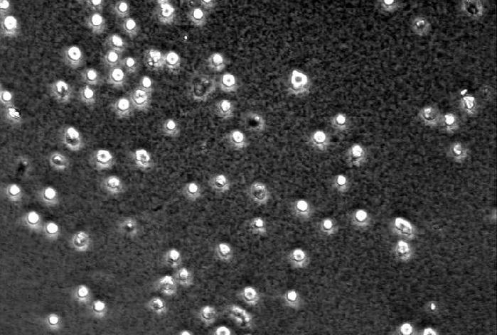
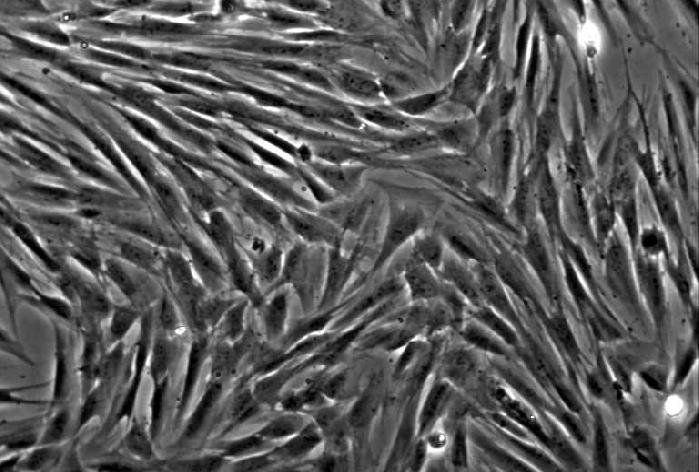
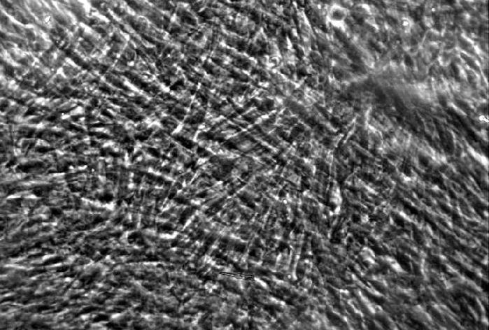
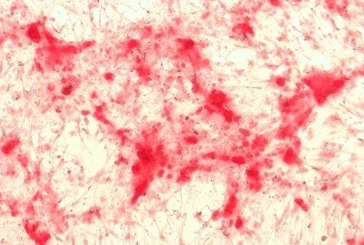
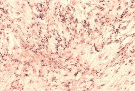
42 Figure A B C D E F Figure 1.


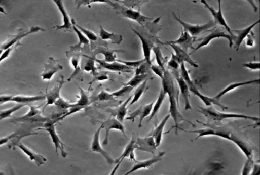
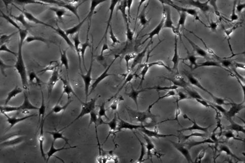
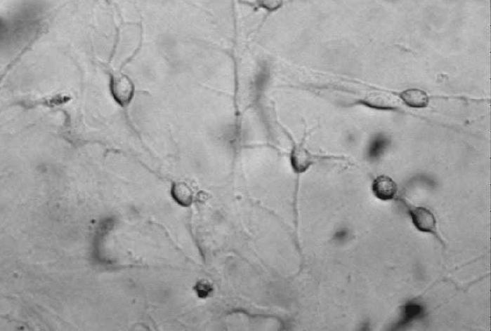
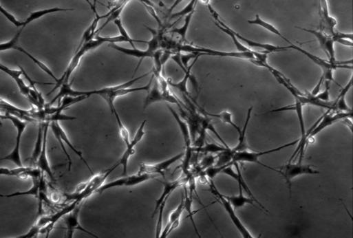
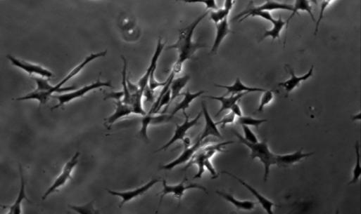
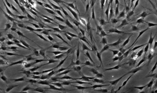
43 Figure A D B C D E F G H Figure 2.
44 Figure 120 Cell viability (% of control) * * * * 0 Figure 3.
 NSE (329 bp) NF-M (333 bp) GFAP (266 bp) β-actin (234 bp) Figure 4.")
45 Figure A B C D E F Vimentin (200 bp) Nestin (220 bp) NGN2 (196 bp) N-tubulin (243 bp) NSE (329 bp) NF-M (333 bp) GFAP (266 bp) β-actin (234 bp) Figure 4.
46 Figure * * Relative normalized quantity A B C D E F * * * * * A B C D E F A B C D E F * * * * * * * * * * A B C D E F * * * * * A B C D E F A B C D E F Figure 5.

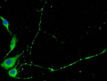
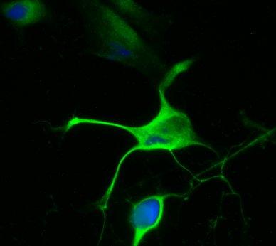
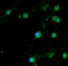
47 Figure A B C D Figure 6.
http://www.geneticliteracyproject.org Nagy Krisztina Semmelweis Egyetem, Orálbiológiai Tanszék
 ŐSSEJTEK http://www.geneticliteracyproject.org 2015. május 6. Nagy Krisztina Semmelweis Egyetem, Orálbiológiai Tanszék Az őssejt definíciója korlátlan önmegújító képesség differenciált utódsejtek létrehozása
Supporting Information
 Supporting Information Cell-free GFP simulations Cell-free simulations of degfp production were consistent with experimental measurements (Fig. S1). Dual emmission GFP was produced under a P70a promoter
Supplementary materials to: Whole-mount single molecule FISH method for zebrafish embryo
 Supplementary materials to: Whole-mount single molecule FISH method for zebrafish embryo Yuma Oka and Thomas N. Sato Supplementary Figure S1. Whole-mount smfish with and without the methanol pretreatment.
Involvement of ER Stress in Dysmyelination of Pelizaeus-Merzbacher Disease with PLP1 Missense Mutations Shown by ipsc-derived Oligodendrocytes
 Stem Cell Reports, Volume 2 Supplemental Information Involvement of ER Stress in Dysmyelination of Pelizaeus-Merzbacher Disease with PLP1 Missense Mutations Shown by ipsc-derived Oligodendrocytes Yuko
A rosszindulatú daganatos halálozás változása 1975 és 2001 között Magyarországon
 A rosszindulatú daganatos halálozás változása és között Eredeti közlemény Gaudi István 1,2, Kásler Miklós 2 1 MTA Számítástechnikai és Automatizálási Kutató Intézete, Budapest 2 Országos Onkológiai Intézet,
Phenotype. Genotype. It is like any other experiment! What is a bioinformatics experiment? Remember the Goal. Infectious Disease Paradigm
 It is like any other experiment! What is a bioinformatics experiment? You need to know your data/input sources You need to understand your methods and their assumptions You need a plan to get from point
Supplementary Table 1. Cystometric parameters in sham-operated wild type and Trpv4 -/- rats during saline infusion and
 WT sham Trpv4 -/- sham Saline 10µM GSK1016709A P value Saline 10µM GSK1016709A P value Number 10 10 8 8 Intercontractile interval (sec) 143 (102 155) 98.4 (71.4 148) 0.01 96 (92 121) 109 (95 123) 0.3 Voided
On The Number Of Slim Semimodular Lattices
 On The Number Of Slim Semimodular Lattices Gábor Czédli, Tamás Dékány, László Ozsvárt, Nóra Szakács, Balázs Udvari Bolyai Institute, University of Szeged Conference on Universal Algebra and Lattice Theory
FAMILY STRUCTURES THROUGH THE LIFE CYCLE
 FAMILY STRUCTURES THROUGH THE LIFE CYCLE István Harcsa Judit Monostori A magyar társadalom 2012-ben: trendek és perspektívák EU összehasonlításban Budapest, 2012 november 22-23 Introduction Factors which
Elektrofiziológiai vizsgálómódszerek alkalmazása a sejtek elektromos tevékenységének kutatásában. A kezdetek 1.
 Elektrofiziológiai vizsgálómódszerek alkalmazása a sejtek elektromos tevékenységének kutatásában A kezdetek 1. 1935-1952 A klasszikus membrán biofizika megteremtése membránpermeabilitás kinetikája, membránfehérjék
Fogbél eredetű felnőtt szöveti őssejtek idegi differenciációja
 Fogbél eredetű felnőtt szöveti őssejtek idegi differenciációja Doktori értekezés Király Marianna Semmelweis Egyetem Klinikai Orvostudományok Doktori Iskola Témavezető: Dr. Varga Gábor, az MTA doktora Hivatalos
Összefoglalás. Summary. Bevezetés
 A talaj kálium ellátottságának vizsgálata módosított Baker-Amacher és,1 M CaCl egyensúlyi kivonószerek alkalmazásával Berényi Sándor Szabó Emese Kremper Rita Loch Jakab Debreceni Egyetem Agrár és Műszaki
Miskolci Egyetem Gazdaságtudományi Kar Üzleti Információgazdálkodási és Módszertani Intézet Nonparametric Tests
 Nonparametric Tests Petra Petrovics Hypothesis Testing Parametric Tests Mean of a population Population proportion Population Standard Deviation Nonparametric Tests Test for Independence Analysis of Variance
2012.11.27. Neuronok előkészítése funkcionális vizsgálatokra. Az alkalmazható technikák előnyei és hátrányai. Neuronok izolálása I
 Neuronok előkészítése funkcionális vizsgálatokra. Az alkalmazható technikák előnyei és hátrányai Sejtszintű elektrofiziológia 1.: csatornák funkcionális Sejtszintű elektrofiziológia 2.: izolált/sejtkultúrában
Miskolci Egyetem Gazdaságtudományi Kar Üzleti Információgazdálkodási és Módszertani Intézet Factor Analysis
 Factor Analysis Factor analysis is a multiple statistical method, which analyzes the correlation relation between data, and it is for data reduction, dimension reduction and to explore the structure. Aim
Gottsegen National Institute of Cardiology. Prof. A. JÁNOSI
 Myocardial Infarction Registry Pilot Study Hungarian Myocardial Infarction Register Gottsegen National Institute of Cardiology Prof. A. JÁNOSI A https://ir.kardio.hu A Web based study with quality assurance
Tudományos Ismeretterjesztő Társulat
 Sample letter number 5. International Culture Festival PO Box 34467 Harrogate HG 45 67F Sonnenbergstraße 11a CH-6005 Luzern Re: Festival May 19, 2009 Dear Ms Atkinson, We are two students from Switzerland
Trinucleotide Repeat Diseases: CRISPR Cas9 PacBio no PCR Sequencing MFMER slide-1
 Trinucleotide Repeat Diseases: CRISPR Cas9 PacBio no PCR Sequencing 2015 MFMER slide-1 Fuch s Eye Disease TCF 4 gene Fuchs occurs in about 4% of the US population. Leads to deteriorating vision without
Expression analysis of PIN genes in root tips and nodules of Lotus japonicus
 Article Expression analysis of PIN genes in root tips and nodules of Lotus japonicus Izabela Sańko-Sawczenko 1, Dominika Dmitruk 1, Barbara Łotocka 1, Elżbieta Różańska 1 and Weronika Czarnocka 1, * 1
Construction of a cube given with its centre and a sideline
 Transformation of a plane of projection Construction of a cube given with its centre and a sideline Exercise. Given the center O and a sideline e of a cube, where e is a vertical line. Construct the projections
Fogbél eredetű felnőtt szöveti őssejtek idegi differenciációja
 Fogbél eredetű felnőtt szöveti őssejtek idegi differenciációja Doktori értekezés Király Marianna Semmelweis Egyetem Klinikai Orvostudományok Doktori Iskola Témavezető: Dr. Varga Gábor, az MTA doktora Hivatalos
Statistical Inference
 Petra Petrovics Statistical Inference 1 st lecture Descriptive Statistics Inferential - it is concerned only with collecting and describing data Population - it is used when tentative conclusions about
A nemi különbségek vizsgálatáról lévén szó, elsődleges volt a nemi hormonok, mint belső környezetbeli különbségeket létrehozó tényezők szerepének
 Kutatási beszámoló Pályázatunk célja annak kiderítése volt, hogy az agyi asztrociták mutatnak-e nemi különbségeket, akár struktura, akár területi megoszlás, akár reaktivitás tekintetében. Alkalmazott megközelítésünk
University of Bristol - Explore Bristol Research
 Al-Salihi, S. A. A., Scott, T. A., Bailey, A. M., & Foster, G. D. (2017). Improved vectors for Agrobacterium mediated genetic manipulation of Hypholoma spp. and other homobasidiomycetes. Journal of Microbiological
Miskolci Egyetem Gazdaságtudományi Kar Üzleti Információgazdálkodási és Módszertani Intézet. Nonparametric Tests. Petra Petrovics.
 Nonparametric Tests Petra Petrovics PhD Student Hypothesis Testing Parametric Tests Mean o a population Population proportion Population Standard Deviation Nonparametric Tests Test or Independence Analysis
Suppl. Materials. Polyhydroxyalkanoate (PHA) Granules Have no Phospholipids. Germany
 Granules Have no Phospholipids. Germany") Suppl. Materials Polyhydroxyalkanoate (PHA) Granules Have no Phospholipids Stephanie Bresan 1, Anna Sznajder 1, Waldemar Hauf 2, Karl Forchhammer 2, Daniel Pfeiffer 3 and Dieter Jendrossek 1 1 Institute
Suppl. Materials Polyhydroxyalkanoate (PHA) Granules Have no Phospholipids Stephanie Bresan 1, Anna Sznajder 1, Waldemar Hauf 2, Karl Forchhammer 2, Daniel Pfeiffer 3 and Dieter Jendrossek 1 1 Institute
Modular Optimization of Hemicellulose-utilizing Pathway in. Corynebacterium glutamicum for Consolidated Bioprocessing of
 [Supplementary materials] Modular Optimization of Hemicellulose-utilizing Pathway in Corynebacterium glutamicum for Consolidated Bioprocessing of Hemicellulosic Biomass Sung Sun Yim 1, Jae Woong Choi 1,
Fogbél eredetű felnőtt szöveti őssejtek idegi differenciációja. Doktori tézis
 Fogbél eredetű felnőtt szöveti őssejtek idegi differenciációja Doktori tézis Király Marianna Semmelweis Egyetem Klinikai Orvostudományok Doktori Iskola Témavezető: Dr. Varga Gábor, az MTA doktora Hivatalos
Correlation & Linear Regression in SPSS
 Petra Petrovics Correlation & Linear Regression in SPSS 4 th seminar Types of dependence association between two nominal data mixed between a nominal and a ratio data correlation among ratio data Correlation
Using the CW-Net in a user defined IP network
 Using the CW-Net in a user defined IP network Data transmission and device control through IP platform CW-Net Basically, CableWorld's CW-Net operates in the 10.123.13.xxx IP address range. User Defined
Nan Wang, Qingming Dong, Jingjing Li, Rohit K. Jangra, Meiyun Fan, Allan R. Brasier, Stanley M. Lemon, Lawrence M. Pfeffer, Kui Li
 Supplemental Material IRF3-dependent and NF- B-independent viral induction of the zinc-finger antiviral protein Nan Wang, Qingming Dong, Jingjing Li, Rohit K. Jangra, Meiyun Fan, Allan R. Brasier, Stanley
Supporting Information
 Supporting Information Paired design of dcas9 as a systematic platform for the detection of featured nucleic acid sequences in pathogenic strains Yihao Zhang 1,2,8, Long Qian 4,8, Weijia Wei 1,3,7,8, Yu
A cell-based screening system for RNA Polymerase I inhibitors
 Electronic Supplementary Material (ESI) for MedChemComm. This journal is The Royal Society of Chemistry 2019 Supporting Information A cell-based screening system for RNA Polymerase I inhibitors Xiao Tan,
Szerződéses kutatások/contract research
 Szerződéses kutatások/contract research Év/year Cím/subject (témavezető/principle investigator) Partner Összeg/amount Epitópok predikciója és szintézismódszer kidolgozása/ Epitope prediction and development
A BÜKKI KARSZTVÍZSZINT ÉSZLELŐ RENDSZER KERETÉBEN GYŰJTÖTT HIDROMETEOROLÓGIAI ADATOK ELEMZÉSE
 KARSZTFEJLŐDÉS XIX. Szombathely, 2014. pp. 137-146. A BÜKKI KARSZTVÍZSZINT ÉSZLELŐ RENDSZER KERETÉBEN GYŰJTÖTT HIDROMETEOROLÓGIAI ADATOK ELEMZÉSE ANALYSIS OF HYDROMETEOROLIGYCAL DATA OF BÜKK WATER LEVEL
Miskolci Egyetem Gazdaságtudományi Kar Üzleti Információgazdálkodási és Módszertani Intézet. Hypothesis Testing. Petra Petrovics.
 Hypothesis Testing Petra Petrovics PhD Student Inference from the Sample to the Population Estimation Hypothesis Testing Estimation: how can we determine the value of an unknown parameter of a population
Kezdőlap > Termékek > Szabályozó rendszerek > EASYLAB és TCU-LON-II szabályozó rendszer LABCONTROL > Érzékelő rendszerek > Típus DS-TRD-01
 Típus DS-TRD FOR EASYLAB FUME CUPBOARD CONTROLLERS Sash distance sensor for the variable, demand-based control of extract air flows in fume cupboards Sash distance measurement For fume cupboards with vertical
KÉPI INFORMÁCIÓK KEZELHETŐSÉGE. Forczek Erzsébet SZTE ÁOK Orvosi Informatikai Intézet. Összefoglaló
 KÉPI INFORMÁCIÓK KEZELHETŐSÉGE Forczek Erzsébet SZTE ÁOK Orvosi Informatikai Intézet Összefoglaló Tanórákon és az önálló tanulás részeként is, az informatika világában a rendelkezésünkre álló óriási mennyiségű
Akce platí od do Do objednávky uveďte kód akce CLON16.
 Média Lonza se slevou 25-30% Seznam produktů se slevou, konkrétní cenovou nabídku žádejte na eastport@eastport.cz nebo tel. 721 867 038. Akce platí od 1.10.2016 do 31.12. 2016. Do objednávky uveďte kód
First experiences with Gd fuel assemblies in. Tamás Parkó, Botond Beliczai AER Symposium 2009.09.21 25.
 First experiences with Gd fuel assemblies in the Paks NPP Tams Parkó, Botond Beliczai AER Symposium 2009.09.21 25. Introduction From 2006 we increased the heat power of our units by 8% For reaching this
TIOLKARBAMÁT TÍPUSÚ NÖVÉNYVÉDŐ SZER HATÓANYAGOK ÉS SZÁRMAZÉKAIK KÉMIAI OXIDÁLHATÓSÁGÁNAK VIZSGÁLATA I
 Műszaki Földtudományi Közlemények, 83. kötet, 1. szám (2012), pp. 137 146. TIOLKARBAMÁT TÍPUSÚ NÖVÉNYVÉDŐ SZER HATÓANYAGOK ÉS SZÁRMAZÉKAIK KÉMIAI OXIDÁLHATÓSÁGÁNAK VIZSGÁLATA I. S-ETIL-N,N-DI-N-PROPIL-TIOLKARBAMÁT
SOLiD Technology. library preparation & Sequencing Chemistry (sequencing by ligation!) Imaging and analysis. Application specific sample preparation
 Imaging and analysis. Application specific sample preparation") SOLiD Technology Application specific sample preparation Application specific data analysis library preparation & emulsion PCR Sequencing Chemistry (sequencing by ligation!) Imaging and analysis SOLiD
SOLiD Technology Application specific sample preparation Application specific data analysis library preparation & emulsion PCR Sequencing Chemistry (sequencing by ligation!) Imaging and analysis SOLiD
Statistical Dependence
 Statistical Dependence Petra Petrovics Statistical Dependence Deinition: Statistical dependence exists when the value o some variable is dependent upon or aected by the value o some other variable. Independent
Sejtek - őssejtek dióhéjban. 2014. február. Sarkadi Balázs, MTA-TTK Molekuláris Farmakológiai Intézet - SE Kutatócsoport, Budapest
 Sejtek - őssejtek dióhéjban 2014. február Sarkadi Balázs, MTA-TTK Molekuláris Farmakológiai Intézet - SE Kutatócsoport, Budapest A legtöbb sejtünk osztódik, differenciálódik, elpusztul... vérsejtek Vannak
Computer Architecture
 Computer Architecture Locality-aware programming 2016. április 27. Budapest Gábor Horváth associate professor BUTE Department of Telecommunications ghorvath@hit.bme.hu Számítógép Architektúrák Horváth
2 kultúra. Zétényi Tamás. www.nytud.hu/depts/tlp/quantification zetenyi@nytud.hu
 2 kultúra Zétényi Tamás www.nytud.hu/depts/tlp/quantification zetenyi@nytud.hu MTA Nyelvtudományi Intézet - 'Elmélet és kísérlet a nyelvészetben' Budapest, 2014. február 25 agenda: mit csinálunk? mit veszünk
ELEKTRONIKAI ALAPISMERETEK ANGOL NYELVEN
 ÉRETTSÉGI VIZSGA 200. május 4. ELEKTRONIKAI ALAPISMERETEK ANGOL NYELVEN KÖZÉPSZINTŰ ÍRÁSBELI VIZSGA 200. május 4. 8:00 Az írásbeli vizsga időtartama: 80 perc Pótlapok száma Tisztázati Piszkozati OKTATÁSI
Mapping Sequencing Reads to a Reference Genome
 Mapping Sequencing Reads to a Reference Genome High Throughput Sequencing RN Example applications: Sequencing a genome (DN) Sequencing a transcriptome and gene expression studies (RN) ChIP (chromatin immunoprecipitation)
Cluster Analysis. Potyó László
 Cluster Analysis Potyó László What is Cluster Analysis? Cluster: a collection of data objects Similar to one another within the same cluster Dissimilar to the objects in other clusters Cluster analysis
Az orvosi biotechnológiai mesterképzés megfeleltetése az Európai Unió új társadalmi kihívásainak a Pécsi Tudományegyetemen és a Debreceni Egyetemen
 Az orvosi biotechnológiai mesterképzés megfeleltetése az Európai Unió új társadalmi kihívásainak a Pécsi Tudományegyetemen és a Debreceni Egyetemen Azonosító szám: TÁMOP-4.1.2-08/1/A-2009-0011 Az orvosi
SAJTÓKÖZLEMÉNY Budapest 2011. július 13.
 SAJTÓKÖZLEMÉNY Budapest 2011. július 13. A MinDig TV a legdinamikusabban bıvülı televíziós szolgáltatás Magyarországon 2011 elsı öt hónapjában - A MinDig TV Extra a vezeték nélküli digitális televíziós
CELLULÁRIS SZÍV- ELEKTROFIZIOLÓGIAI MÉRÉSI TECHNIKÁK. Dr. Virág László
 CELLULÁRIS SZÍV- ELEKTROFIZIOLÓGIAI MÉRÉSI TECHNIKÁK Dr. Virág László Intracelluláris mikroelektród technika Voltage clamp technika Patch clamp technika Membrane potentials and excitation of impaled single
Flowering time. Col C24 Cvi C24xCol C24xCvi ColxCvi
 Flowering time Rosette leaf number 50 45 40 35 30 25 20 15 10 5 0 Col C24 Cvi C24xCol C24xCvi ColxCvi Figure S1. Flowering time in three F 1 hybrids and their parental lines as measured by leaf number
Humán szubmandibuláris nyálmirigy-eredetű primer sejtek és immortalizált sejtvonal differenciálódásának in vitro vizsgálata
 Humán szubmandibuláris nyálmirigy-eredetű primer sejtek és immortalizált sejtvonal differenciálódásának in vitro vizsgálata Doktori tézisek Szlávik Vanda Semmelweis Egyetem Molekuláris Orvostudományok
Computational Neuroscience
 Computational Neuroscience Zoltán Somogyvári senior research fellow KFKI Research Institute for Particle and Nuclear Physics Supporting materials: http://www.kfki.hu/~soma/bscs/ BSCS 2010 Lengyel Máté:
4-42 ELECTRONICS WX210 - WX240
 4-42 ELECTRONICS WX210 - WX240 PCS 40000499-en Fig. 8 WX210 - WX240 ELECTRONICS 4-43 PCS COMPONENTS 40000471-en Load-limit regulator Legend Fig. 1 Fig. 2 1 Power supply 2 PWM1 output, proportional valve
A polikomb fehérje, Rybp kulcsfontosságú az egér embrionális őssejtek neurális differenciációjához
 A polikomb fehérje, Rybp kulcsfontosságú az egér embrionális őssejtek neurális differenciációjához Ph.D. értekezés tézisei Kovács Gergő Témavezető: Dr. Pirity Melinda MTA-Szegedi Biológiai Kutatóközpont,
HALLGATÓI KÉRDŐÍV ÉS TESZT ÉRTÉKELÉSE
 HALLGATÓI KÉRDŐÍV ÉS TESZT ÉRTÉKELÉSE EVALUATION OF STUDENT QUESTIONNAIRE AND TEST Daragó László, Dinyáné Szabó Marianna, Sára Zoltán, Jávor András Semmelweis Egyetem, Egészségügyi Informatikai Fejlesztő
Hasznos és kártevő rovarok monitorozása innovatív szenzorokkal (LIFE13 ENV/HU/001092)
") Hasznos és kártevő rovarok monitorozása innovatív szenzorokkal (LIFE13 ENV/HU/001092) www.zoolog.hu Dr. Dombos Miklós Tudományos főmunkatárs MTA ATK TAKI Innovative Real-time Monitoring and Pest control
Hasznos és kártevő rovarok monitorozása innovatív szenzorokkal (LIFE13 ENV/HU/001092) www.zoolog.hu Dr. Dombos Miklós Tudományos főmunkatárs MTA ATK TAKI Innovative Real-time Monitoring and Pest control
STUDENT LOGBOOK. 1 week general practice course for the 6 th year medical students SEMMELWEIS EGYETEM. Name of the student:
 STUDENT LOGBOOK 1 week general practice course for the 6 th year medical students Name of the student: Dates of the practice course: Name of the tutor: Address of the family practice: Tel: Please read
ANNEX V / V, MELLÉKLET
 ANNEX V / V, MELLÉKLET VETERINARY CERTIFICATE EMBRYOS OF DOMESTIC ANIMALS OF THE BOVINE SPECIES FOR IMPORTS COLLECTED OR PRODUCED BEFORE 1 st JANUARY 2006 ÁLLAT-EGÉSZSÉGÜGYI BIZONYÍTVÁNY 2006. JANUÁR 1-JE
ELEKTRONIKAI ALAPISMERETEK ANGOL NYELVEN
 ÉRETTSÉGI VIZSGA 2008. május 26. ELEKTRONIKAI ALAPISMERETEK ANGOL NYELVEN KÖZÉPSZINTŰ ÍRÁSBELI VIZSGA 2008. május 26. 8:00 Az írásbeli vizsga időtartama: 180 perc Pótlapok száma Tisztázati Piszkozati OKTATÁSI
A évi fizikai Nobel-díj
 A 2012. évi fizikai Nobel-díj "for ground-breaking experimental methods that enable measuring and manipulation of individual quantum systems" Serge Haroche David Wineland Ecole Normale Superieure, Párizs
Márkaépítés a YouTube-on
 Márkaépítés a YouTube-on Tv+ Adj hozzá YouTube-ot, Google Ground, 2016 Március 7. Bíró Pál, Google - YouTube 9,000,000 INTERNETTEL BÍRÓ ESZKÖZÖK VOLUMENE GLOBÁLISAN WEARABLES OKOS TV 8,000,000 7,000,000
Nevezze meg a jelölt csontot latinul! Name the bone marked! Nevezze meg a jelölt csont típusát! What is the type of the bone marked?
 1 Nevezze meg a jelölt csontot latinul! Name the bone marked! 2 Nevezze meg a jelölt csont típusát! What is the type of the bone marked? 3 Milyen csontállomány található a jelölt csont belsejében? What
IES TM Evaluating Light Source Color Rendition
 IES TM-30-15 Evaluating Light Source Color Rendition "Original" "CRI = 80" Desaturated "CRI = 80" Saturated More metrics Color Fidelity Color Discrimination Color Preference Metrics/Measures R f (IES TM-30-15)
EPILEPSY TREATMENT: VAGUS NERVE STIMULATION. Sakoun Phommavongsa November 12, 2013
 EPILEPSY TREATMENT: VAGUS NERVE STIMULATION Sakoun Phommavongsa November 12, 2013 WHAT IS EPILEPSY? A chronic neurological disorder characterized by having two or more unprovoked seizures Affects nearly
A STRATÉGIAALKOTÁS FOLYAMATA
 BUDAPESTI CORVINUS EGYETEM VÁLLALATGAZDASÁGTAN INTÉZET VERSENYKÉPESSÉG KUTATÓ KÖZPONT Szabó Zsolt Roland: A STRATÉGIAALKOTÁS FOLYAMATA VERSENYBEN A VILÁGGAL 2004 2006 GAZDASÁGI VERSENYKÉPESSÉGÜNK VÁLLALATI
Animal welfare, etológia és tartástechnológia
 Animal welfare, etológia és tartástechnológia Animal welfare, ethology and housing systems Volume 9 Issue 3 Különszám/Special Issue Gödöllő 2013 69 TEJ ALAPÚ HIGÍTÓK ALKALMAZÁSÁNAK LEHETŐSÉGE NYÚL SPERMA
4 vana, vanb, vanc1, vanc2
 77 1) 2) 2) 3) 4) 5) 1) 2) 3) 4) 5) 16 12 7 17 4 8 2003 1 2004 7 3 Enterococcus 5 Enterococcus faecalis 1 Enterococcus avium 1 Enterococcus faecium 2 5 PCR E. faecium 1 E-test MIC 256 mg/ml vana MRSA MRSA
Hibridspecifikus tápanyag-és vízhasznosítás kukoricánál csernozjom talajon
 Hibridspecifikus tápanyag-és vízhasznosítás kukoricánál csernozjom talajon Karancsi Lajos Gábor Debreceni Egyetem Agrár és Gazdálkodástudományok Centruma Mezőgazdaság-, Élelmiszertudományi és Környezetgazdálkodási
ELEKTRONIKAI ALAPISMERETEK ANGOL NYELVEN
 ÉRETTSÉGI VIZSGA 2012. május 25. ELEKTRONIKAI ALAPISMERETEK ANGOL NYELVEN KÖZÉPSZINTŰ ÍRÁSBELI VIZSGA 2012. május 25. 8:00 Az írásbeli vizsga időtartama: 180 perc Pótlapok száma Tisztázati Piszkozati NEMZETI
Whole mount in situ hybridization, sectioning, and immunostaining Ribonuclease protection assay (RPA) Collagen gel tube formation assay
 Collagen gel tube formation assay") Whole mount in situ hybridization, sectioning, and immunostaining In situ hybridization for tie-1 and lncrna tie-1as and sectioning of ISH stained embryos were performed according to previously published
Whole mount in situ hybridization, sectioning, and immunostaining In situ hybridization for tie-1 and lncrna tie-1as and sectioning of ISH stained embryos were performed according to previously published
PIACI HIRDETMÉNY / MARKET NOTICE
 PIACI HIRDETMÉNY / MARKET NOTICE HUPX DAM Másnapi Aukció / HUPX DAM Day-Ahead Auction Iktatási szám / Notice #: Dátum / Of: 18/11/2014 HUPX-MN-DAM-2014-0023 Tárgy / Subject: Változások a HUPX másnapi piac
Jelitai Márta OTKA pályázat (F 68940) zárójelentés (2007 07.01-2010 06.30) Idegi ős/progenitor sejtek fejlődésének fiziológiai jellemzése
 zárójelentés (2007 07.01-2010 06.30) Idegi ős/progenitor sejtek fejlődésének fiziológiai jellemzése") Jelitai Márta OTKA pályázat (F 68940) zárójelentés (2007 07.01-2010 06.30) Idegi ős/progenitor sejtek fejlődésének fiziológiai jellemzése Alulírott Jelitai Márta, Fiatal Kutatói kategóriában (F 68940)
Jelitai Márta OTKA pályázat (F 68940) zárójelentés (2007 07.01-2010 06.30) Idegi ős/progenitor sejtek fejlődésének fiziológiai jellemzése Alulírott Jelitai Márta, Fiatal Kutatói kategóriában (F 68940)
klorid ioncsatorna az ABC (ATP Binding Casette) fehérjecsaládba tartozik, amelyek általánosságban részt vesznek a gyógyszerek olyan alapvetı
 fehérjecsaládba tartozik, amelyek általánosságban részt vesznek a gyógyszerek olyan alapvetı") Szegedi Tudományegyetem Farmakológiai és Farmakoterápiai Intézet Igazgató: Prof. Dr. Varró András 6720 Szeged, Dóm tér 12., Tel.: (62) 545-682 Fax: (62) 545-680 e-mail: varro.andras@med.u-szeged.hu Opponensi
Szegedi Tudományegyetem Farmakológiai és Farmakoterápiai Intézet Igazgató: Prof. Dr. Varró András 6720 Szeged, Dóm tér 12., Tel.: (62) 545-682 Fax: (62) 545-680 e-mail: varro.andras@med.u-szeged.hu Opponensi
Őssejtkezelés kardiovaszkuláris kórképekben
 Őssejtkezelés kardiovaszkuláris kórképekben Papp Zoltán Debreceni Egyetem Kardiológiai Intézet Klinikai Fiziológiai Tanszék Megmenthető a károsodott szív őssejtekkel? Funkcionális változások az öregedő
AZ ERDÕ NÖVEKEDÉSÉNEK VIZSGÁLATA TÉRINFORMATIKAI ÉS FOTOGRAMMETRIAI MÓDSZEREKKEL KARSZTOS MINTATERÜLETEN
 Tájökológiai Lapok 5 (2): 287 293. (2007) 287 AZ ERDÕ NÖVEKEDÉSÉNEK VIZSGÁLATA TÉRINFORMATIKAI ÉS FOTOGRAMMETRIAI MÓDSZEREKKEL KARSZTOS MINTATERÜLETEN ZBORAY Zoltán Honvédelmi Minisztérium Térképészeti
Néhány folyóiratkereső rendszer felsorolása és példa segítségével vázlatos bemutatása Sasvári Péter
 Néhány folyóiratkereső rendszer felsorolása és példa segítségével vázlatos bemutatása Sasvári Péter DOI: http://doi.org/10.13140/rg.2.2.28994.22721 A tudományos közlemények írása minden szakma művelésének
Performance Modeling of Intelligent Car Parking Systems
 Performance Modeling of Intelligent Car Parking Systems Károly Farkas Gábor Horváth András Mészáros Miklós Telek Technical University of Budapest, Hungary EPEW 2014, Florence, Italy Outline Intelligent
TÁMOPͲ4.2.2.AͲ11/1/KONVͲ2012Ͳ0029
 AUTOTECH Jármipari anyagfejlesztések: célzott alapkutatás az alakíthatóság, hkezelés és hegeszthetség témaköreiben TÁMOP4.2.2.A11/1/KONV20120029 www.autotech.unimiskolc.hu ANYAGSZERKEZETTANI ÉS ANYAGTECHNOLÓGIAI
EN United in diversity EN A8-0206/419. Amendment
 22.3.2019 A8-0206/419 419 Article 2 paragraph 4 point a point i (i) the identity of the road transport operator; (i) the identity of the road transport operator by means of its intra-community tax identification
Manuscript Title: Identification of a thermostable fungal lytic polysaccharide monooxygenase and
 1 2 3 4 5 Journal name: Applied Microbiology and Biotechnology Manuscript Title: Identification of a thermostable fungal lytic polysaccharide monooxygenase and evaluation of its effect on lignocellulosic
Környezetben részlegesen lebomló műanyag fóliák degradációjának nyomon követése
 Környezetben részlegesen lebomló műanyag fóliák degradációjának nyomon követése Rétháti Gabriella 1, Pogácsás Krisztina 1, Heffner Tamás 2, Simon Barbara 1, Czinkota Imre 1, Tolner László 1, Kelemen Ottó
A TÓGAZDASÁGI HALTERMELÉS SZERKEZETÉNEK ELEMZÉSE. SZATHMÁRI LÁSZLÓ d r.- TENK ANTAL dr. ÖSSZEFOGLALÁS
 A TÓGAZDASÁGI HALTERMELÉS SZERKEZETÉNEK ELEMZÉSE SZATHMÁRI LÁSZLÓ d r.- TENK ANTAL dr. ÖSSZEFOGLALÁS A hazai tógazdasági haltermelés a 90-es évek közepén tapasztalt mélypontról elmozdult és az utóbbi három
A modern e-learning lehetőségei a tűzoltók oktatásának fejlesztésében. Dicse Jenő üzletfejlesztési igazgató
 A modern e-learning lehetőségei a tűzoltók oktatásának fejlesztésében Dicse Jenő üzletfejlesztési igazgató How to apply modern e-learning to improve the training of firefighters Jenő Dicse Director of
Erdészettudományi Közlemények
 Erdészettudományi Közlemények 2. évfolyam 1. szám 2012 73 80 oldal AZ EZÜSTHÁRS FATERMÉSI TÁBLÁJÁNAK MÓDOSÍTÁSA Peszlen Roland József és Veperdi Gábor Nyugat-magyarországi Egyetem, Erdőmérnöki Kar, Erdővagyon-gazdálkodási
A felszíni ektoderma differenciációja
 A felszíni ektoderma differenciációja - elkülönülése - kialakulása az egyedfejlődés során - szerkezete és a felnőttkori őssejtek -interfollikuláris epidermis -szőrtüsző -faggyúmirigy - a bőrpótlás (klinikai)
Sejt- és szövetkultúra
 Sejt- és szövetkultúra Subline Innenteil 14pt TC Tested Tested Cryo Performance Sterile/ DNA-/ DNase-/ RNase-/ Pyrogen-free/ non-cytotoxic CE/ IVD/ Sterile/ Pyrogen-free/ non-cytotoxic/ non-mutagenic Come
Új temékek az UD- GenoMed Kft. kínálatában!
 Új temékek az UD- GenoMed Kft. kínálatában! Szolgáltatásaink: Medical Genomic Technologies Kft. Betegtoborzás és biobanking Bioinformatika o Adatelemzés/adatbányászás o Integrált adatbázis készítés Sejtvonal
Az orvosi biotechnológiai mesterképzés megfeleltetése az Európai Unió új társadalmi kihívásainak a Pécsi Tudományegyetemen és a Debreceni Egyetemen
 Az orvosi biotechnológiai mesterképzés megfeleltetése az Európai Unió új társadalmi kihívásainak a Pécsi Tudományegyetemen és a Debreceni Egyetemen Azonosító szám: Az orvosi biotechnológiai mesterképzés
FORGÁCS ANNA 1 LISÁNYI ENDRÉNÉ BEKE JUDIT 2
 FORGÁCS ANNA 1 LISÁNYI ENDRÉNÉ BEKE JUDIT 2 Hátrányos-e az új tagállamok számára a KAP támogatások disztribúciója? Can the CAP fund distribution system be considered unfair to the new Member States? A
7 th Iron Smelting Symposium 2010, Holland
 7 th Iron Smelting Symposium 2010, Holland Október 13-17 között került megrendezésre a Hollandiai Alphen aan den Rijn városában található Archeon Skanzenben a 7. Vasolvasztó Szimpózium. Az öt napos rendezvényen
Animal welfare, etológia és tartástechnológia
 Animal welfare, etológia és tartástechnológia Animal welfare, ethology and housing systems Volume 5 Issue 4 Különszám Gödöllı 2009 459 MORFOLÓGIAI ÉS GENETIKAI VIZSGÁLATOK MAGYARORSZÁGI TÖRPEHARCSÁKON
Supplementary Figure 1
 Supplementary Figure 1 Plot of delta-afe of sequence variants detected between resistant and susceptible pool over the genome sequence of the WB42 line of B. vulgaris ssp. maritima. The delta-afe values
Cashback 2015 Deposit Promotion teljes szabályzat
 Cashback 2015 Deposit Promotion teljes szabályzat 1. Definitions 1. Definíciók: a) Account Client s trading account or any other accounts and/or registers maintained for Számla Az ügyfél kereskedési számlája
Hogyan szűrjük a röntgensugarat?
 Hogyan szűrjük a röntgensugarat? (A röntgencsőegység állandó szűrésének nemzetközi szabványáról) Porubszky Tamás OSSKI Munkahelyi Sugáregészségügyi Osztály E-mail: porubszky@osski.hu 1 IEC 60522 (IEC 522):
Expansion of Red Deer and afforestation in Hungary
 Expansion of Red Deer and afforestation in Hungary László Szemethy, Róbert Lehoczki, Krisztián Katona, Norbert Bleier, Sándor Csányi www.vmi.szie.hu Background and importance large herbivores are overpopulated
Influence of geogas seepage on indoor radon. István Csige Sándor Csegzi Sándor Gyila
 VII. Magyar Radon Fórum és Radon a környezetben Nemzetközi workshop Veszprém, 2013. május 16-17. Influence of geogas seepage on indoor radon István Csige Sándor Csegzi Sándor Gyila Debrecen Marosvásárhely
Correlation & Linear Regression in SPSS
 Correlation & Linear Regression in SPSS Types of dependence association between two nominal data mixed between a nominal and a ratio data correlation among ratio data Exercise 1 - Correlation File / Open